. 2024 Feb 18;14(1):3997.
doi: 10.1038/s41598-024-54228-0.

Bioactive glass-ceramics containing fluorapatite, xonotlite, cuspidine and wollastonite form apatite faster than their corresponding glasses

Affiliations

Bioactive glass-ceramics containing fluorapatite, xonotlite, cuspidine and wollastonite form apatite faster than their corresponding glasses

Gloria Kirste et al. Sci Rep. .

Abstract

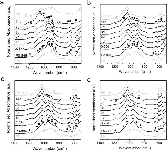
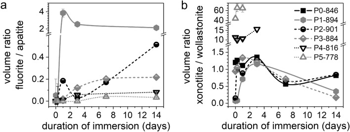
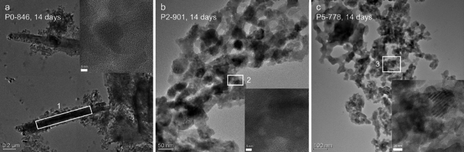
Crystallisation of bioactive glasses has been claimed to negatively affect the ion release from bioactive glasses. Here, we compare ion release and mineralisation in Tris-HCl buffer solution for a series of glass-ceramics and their parent glasses in the system SiO2-CaO-P2O5-CaF2. Time-resolved X-ray diffraction analysis of glass-ceramic degradation, including quantification of crystal fractions by full pattern refinement, show that the glass-ceramics precipitated apatite faster than the corresponding glasses, in agreement with faster ion release from the glass-ceramics. Imaging by transmission electron microscopy and X-ray nano-computed tomography suggest that this accelerated degradation may be caused by the presence of nano-sized channels along the internal crystal/glassy matrix interfaces. In addition, the presence of crystalline fluorapatite in the glass-ceramics facilitated apatite nucleation and crystallisation during immersion. These results suggest that the popular view of bioactive glass crystallisation being a disadvantage for degradation, apatite formation and, subsequently, bioactivity may depend on the actual system study and, thus, has to be reconsidered.


Conflict of interest statement

The authors declare no competing interests.

Figures

Figure 1
Figure 1
Changes in the composition of parent glasses (due to fluoride loss during melting) and GC (due to incongruent crystallisation; concentrations estimated for different degrees of crystallinity; for calculation details see Supplementary File) compared to the nominal glass composition: (a) CaF2, (b) P2O5, (c) CaO and (d) SiO2 content; (e) network connectivity (NC; calculated using Eq. 1).
Figure 2
Figure 2
Characterisation of buffer solutions after immersion: (a) pH, relative (b) Ca and (c) Si ion concentrations and (d) absolute P concentrations of glasses and corresponding GC. (Ion concentrations in b and c have been normalised to the concentration of the respective ion in the untreated sample. Lines are drawn as visual guides only).
Figure 3
Figure 3
FTIR spectra at different time points of immersion for (a) P0 and P0-846, (b) P2 and P2-901, (c) P3 and P3-884, (d) P5 and P5-778. Parent glasses for comparison plotted in grey, labelling of vibration bands: (circle) amorphous Si–O, (star) cuspidine, (filled square) wollastonite, (diamond) xonotlite, (asterik) apatite/phosphate, (square) carbonate.
Figure 4
Figure 4
Volume fractions of cuspidine, xonotlite, wollastonite, apatite and fluorite within the crystalline share of the GC at different time points of immersion, calculated by full pattern refinement for (a) P0-846, (b) P2-901, (c) P3-884 and (d) P5-778. (Lines are drawn as a visual guide only, original diffraction patterns depicted in Suppl. Figure 1.)
Figure 5
Figure 5
Evolution of volumetric ratios among crystalline phases in GC during immersion: (a) Fluorite-to-apatite and (b) xonotlite-to-wollastonite. (For samples P4-816 and P5-778, the xonotlite-to-wollastonite ratio at later time points could not be calculated because of very low wollastonite concentrations.)
Figure 6
Figure 6
TEM micrographs at different magnifications of the GC powders of (a) P0-846, (b) P2-901 and (c) P5-778 after 14 days immersed in Tris buffer. Insets display details of crystalline areas, showing the crystalline planes.
Figure 7
Figure 7
Nano-CT images of (a) GC sample P0-846 at 14 days of immersion in Tris buffer and (b) P0 glass under the same conditions, as discussed previously. Volumetric reconstructions of the samples with a virtual cut (centre and bottom). The phase with the brightest contrast is highlighted in green.

References

    1. Hench LL, Day DE, Höland W, Rheinberger VM. Glass and medicine. Int. J. Appl. Glass Sci. 2010;1:104–117. doi: 10.1111/j.2041-1294.2010.00001.x. - DOI
    1. Hench LL, Paschall HA. Direct chemical bond of bioactive glass-ceramic materials to bone and muscle. J. Biomed. Mater. Res. 1973;7:25–42. doi: 10.1002/jbm.820070304. - DOI - PubMed
    1. Jones JR, Boccaccini AR. Cellular Ceramics: Structure, Manufacturing, Processing and Applications. Wiley; 2005. pp. 547–570.
    1. Baino F, Novajra G, Vitale-Brovarone C. Bioceramics and scaffolds: A winning combination for tissue engineering. Front. Bioeng. Biotechnol. 2015;3:202. doi: 10.3389/fbioe.2015.00202. - DOI - PMC - PubMed
    1. Jones JR. Review of bioactive glass: From Hench to hybrids. Acta Biomater. 2013;9:4457–4486. doi: 10.1016/j.actbio.2012.08.023. - DOI - PubMed