

Characteristics of Vascular Access Cannulation Complications in End Stage Kidney Disease Patients in West Java from 2018 to 2022: A Retrospective Observational Study
- PMID: 38370010
- PMCID: PMC10870994
- DOI: 10.2147/IJNRD.S440467
Characteristics of Vascular Access Cannulation Complications in End Stage Kidney Disease Patients in West Java from 2018 to 2022: A Retrospective Observational Study
Abstract
Background: End-stage kidney disease (ESKD) is associated with a tremendous financial burden. Data in Indonesia shows an increasing number of patients with ESKD taking hemodialysis as a routine procedure every year. Establishment and maintenance of vascular access are important in the management of ESKD. Vascular complications that often arise due to hemodialysis are common and one of the main reasons for hospitalization. Cannulation complications ranged from minor hematomas to acute bleeding from pseudoaneurysms that required emergency surgical procedures. This study aims to assess the different clinicopathological characteristics of ESKD patients with vascular access cannulation complications and the surgical management related to the complications.
Materials and methods: This research is a retrospective observational study. The research subjects in this study were ESKD patients in the vascular and endovascular surgery division of the tertiary hospital in West Java, Indonesia. There were 121 study subjects. Clinicopathological characteristics of vascular cannulation complications and surgical management are extracted from the medical record.
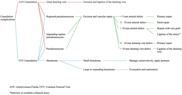
Results: Three major vascular complications were ruptured pseudoaneurysms 64/121 (52.9%), impending rupture pseudoaneurysms 28/121 (23.1%), and pseudoaneurysms 21/121 (17.4%). Common surgical procedures were ligation of the draining vein 47/121 (38.8%), arterial primary repair 28/121 (23.1%), and arterial patch repair 18/121 (14.9%). There was a significant relationship between symptoms of bleeding in ruptured pseudoaneurysms and bulging masses in pseudoaneurysms (p = 0.001). There was a significant relationship between the diameter of the vascular mass, vascular defect size, and hematoma and the type of surgical procedure taken (p < 0.010).
Conclusion: Ruptured, impending rupture, and pseudoaneurysms are major complications of vascular access in ESKD patients, and there was a significant relationship between the carried-out surgical procedure and the size of the vascular mass, defect, and hematoma.
Keywords: arteriovenous fistula; end-stage kidney disease; hemodialysis; pseudoaneurysm; vascular access.
© 2024 Djajakusumah et al.
Conflict of interest statement
The authors affirm that they have no known financial or interpersonal conflicts that would have appeared to have an impact on the study presented.
Figures


Similar articles
-
Endovascular Stent Graft Repair is an Effective and Safe Alternative Therapy for Arteriovenous Graft Pseudoaneurysms.Eur J Vasc Endovasc Surg. 2016 Nov;52(5):682-688. doi: 10.1016/j.ejvs.2016.07.019. Epub 2016 Sep 2. Eur J Vasc Endovasc Surg. 2016. PMID: 27592733
-
Endovascular repair of symptomatic hemodialysis access graft pseudoaneurysms.J Vasc Access. 2014 Jan-Feb;15(1):5-11. doi: 10.5301/jva.5000161. Epub 2013 Aug 9. J Vasc Access. 2014. PMID: 23934930
-
Stent graft treatment for hemodialysis access aneurysms.J Vasc Surg. 2011 Oct;54(4):1088-94. doi: 10.1016/j.jvs.2011.03.252. Epub 2011 Jun 12. J Vasc Surg. 2011. PMID: 21658886
-
Surgical Treatment Options of Subclavian Artery Pseudoaneurysms: A Case Report and Litterature Review.Rev Port Cir Cardiotorac Vasc. 2017 Jul-Dec;24(3-4):105-106. Rev Port Cir Cardiotorac Vasc. 2017. PMID: 29701339 Review.
-
The role of interventional radiology in management of patients with end-stage renal disease.Eur J Radiol. 2003 May;46(2):96-114. doi: 10.1016/s0720-048x(03)00074-3. Eur J Radiol. 2003. PMID: 12714226 Review.
Cited by
-
Types of Vascular Access and Associated Clinical Outcomes in Dialysis Patients.Cureus. 2025 May 7;17(5):e83691. doi: 10.7759/cureus.83691. eCollection 2025 May. Cureus. 2025. PMID: 40486410 Free PMC article.
References
-
- Afiatin A, Agustian D, Wahyudi K, Riono P, Roesli RM. Survival analysis of chronic kidney disease patients with hemodialysis in West Java. Indonesia, year 2007–2018. Majalah Kedokteran Bandung. 2020;52(3):172–179. doi:10.15395/mkb.v52n3.2124 - DOI
LinkOut - more resources
Full Text Sources
