

MIHIC: a multiplex IHC histopathological image classification dataset for lung cancer immune microenvironment quantification
- PMID: 38370413
- PMCID: PMC10869447
- DOI: 10.3389/fimmu.2024.1334348
MIHIC: a multiplex IHC histopathological image classification dataset for lung cancer immune microenvironment quantification
Abstract
Background: Immunohistochemistry (IHC) is a widely used laboratory technique for cancer diagnosis, which selectively binds specific antibodies to target proteins in tissue samples and then makes the bound proteins visible through chemical staining. Deep learning approaches have the potential to be employed in quantifying tumor immune micro-environment (TIME) in digitized IHC histological slides. However, it lacks of publicly available IHC datasets explicitly collected for the in-depth TIME analysis.
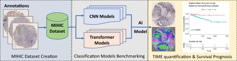
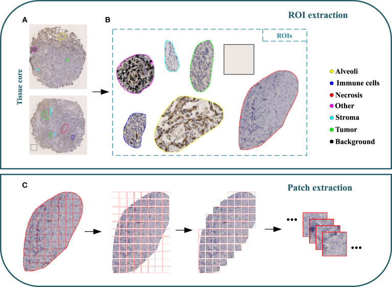
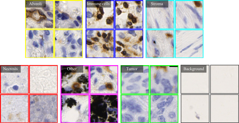
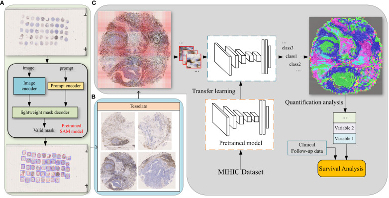
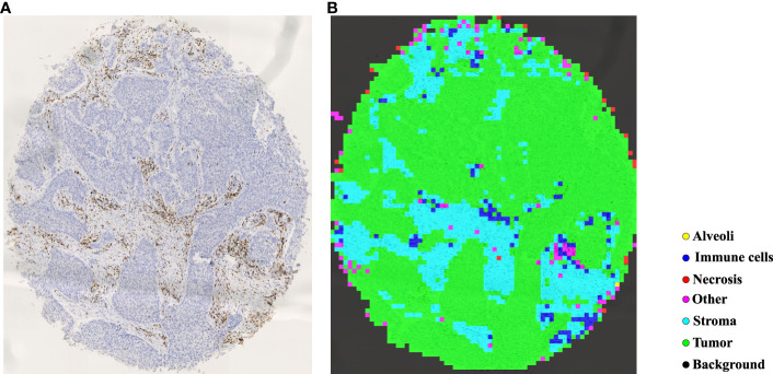
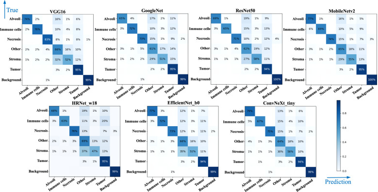
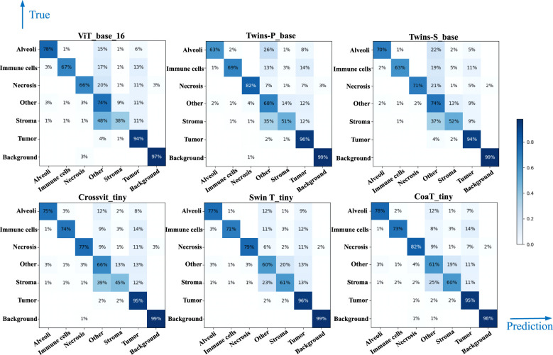
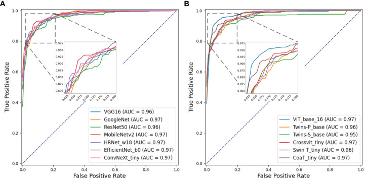
Method: In this paper, a notable Multiplex IHC Histopathological Image Classification (MIHIC) dataset is created based on manual annotations by pathologists, which is publicly available for exploring deep learning models to quantify variables associated with the TIME in lung cancer. The MIHIC dataset comprises of totally 309,698 multiplex IHC stained histological image patches, encompassing seven distinct tissue types: Alveoli, Immune cells, Necrosis, Stroma, Tumor, Other and Background. By using the MIHIC dataset, we conduct a series of experiments that utilize both convolutional neural networks (CNNs) and transformer models to benchmark IHC stained histological image classifications. We finally quantify lung cancer immune microenvironment variables by using the top-performing model on tissue microarray (TMA) cores, which are subsequently used to predict patients' survival outcomes.
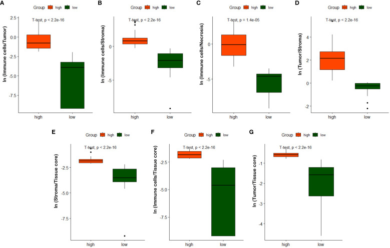
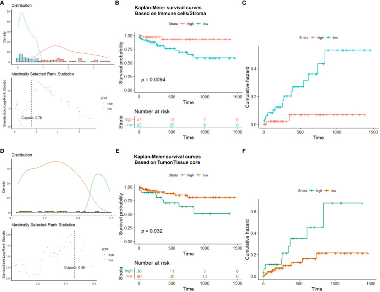
Result: Experiments show that transformer models tend to provide slightly better performances than CNN models in histological image classifications, although both types of models provide the highest accuracy of 0.811 on the testing dataset in MIHIC. The automatically quantified TIME variables, which reflect proportions of immune cells over stroma and tumor over tissue core, show prognostic value for overall survival of lung cancer patients.
Conclusion: To the best of our knowledge, MIHIC is the first publicly available lung cancer IHC histopathological dataset that includes images with 12 different IHC stains, meticulously annotated by multiple pathologists across 7 distinct categories. This dataset holds significant potential for researchers to explore novel techniques for quantifying the TIME and advancing our understanding of the interactions between the immune system and tumors.
Keywords: database; image classification; immunohistochemical image; lung cancer; transformer models.
Copyright © 2024 Wang, Qiu, Wang, Wang, Jin, Cong, Zhang and Xu.
Conflict of interest statement
The authors declare that the research was conducted in the absence of any commercial or financial relationships that could be construed as a potential conflict of interest.
Figures
Comment on
-
Predicting survival from colorectal cancer histology slides using deep learning: A retrospective multicenter study.PLoS Med. 2019 Jan 24;16(1):e1002730. doi: 10.1371/journal.pmed.1002730. eCollection 2019 Jan. PLoS Med. 2019. PMID: 30677016 Free PMC article.
References
Publication types
MeSH terms
LinkOut - more resources
Full Text Sources
Medical