

Case Reports
doi: 10.1093/jscr/rjae058.
eCollection 2024 Feb.
Slipped capital femoral epiphysis in a 5-year-old boy with cerebral palsy on valproic acid and levetiracetam for epilepsy: a case report
Affiliations
- PMID: 38370596
- PMCID: PMC10871696
- DOI: 10.1093/jscr/rjae058
Item in Clipboard
Case Reports
Slipped capital femoral epiphysis in a 5-year-old boy with cerebral palsy on valproic acid and levetiracetam for epilepsy: a case report
J Surg Case Rep.
.
Abstract
This study presents a rare case of unilateral slipped capital femoral epiphysis treated surgically in a 5-year-old boy with cerebral palsy who was born at 27 weeks' gestation and developed grade III intraventricular haemorrhage and periventricular leucomalacia and was on antiepileptic drugs, including valproic acid and levetiracetam for >3 years. The patient had no history of endocrine, renal, and significant familial diseases.
Keywords: cerebral palsy; hip pain; levetiracetam; slipped capital femoral epiphysis; valproic acid.
Published by Oxford University Press and JSCR Publishing Ltd. © The Author(s) 2024.
Conflict of interest statement
None declared.
Figures
Pelvic anterior–posterior radiograph showing coxa valga deformity in the left hip.

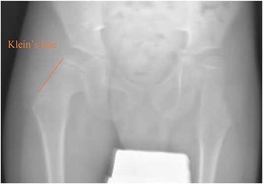
Pelvic anterior–posterior radiograph showing SCFE in the right hip, with Klein’s line not intersecting the capital femoral epiphysis
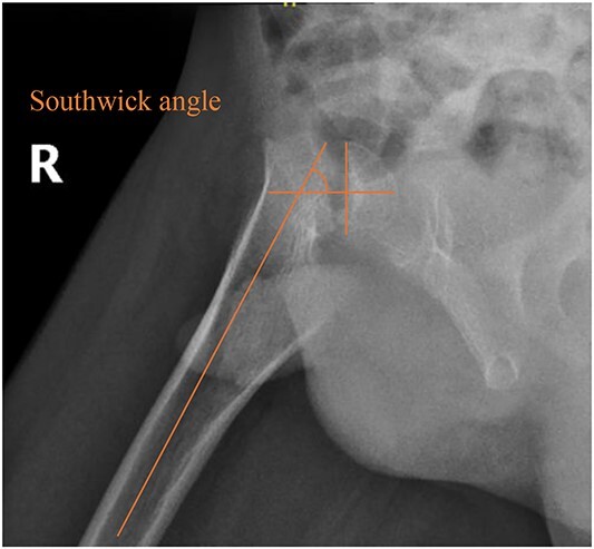
Pelvic frog-leg lateral view radiograph showing Southwick’s slip angle 50° in the right hip.
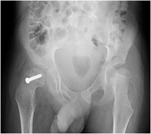
Pelvic anterior–posterior radiograph immediately after in situ fixation with single cannulate screw.

Six weeks following post-operative fixation: (A) pelvic anterior–posterior radiograph and (B) pelvic frog-leg lateral view radiograph.
Pelvic anterior–posterior radiograph, 3 months following post-operative fixation.
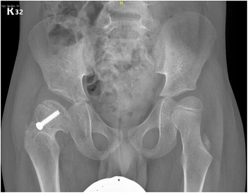
Pelvic anterior–posterior radiograph, 15 months following post-operative fixation.
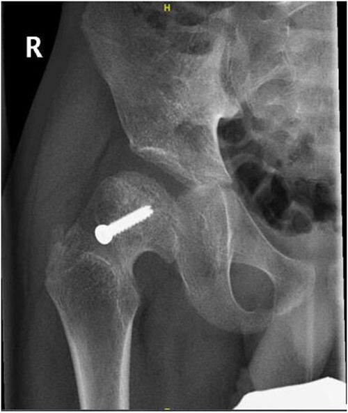
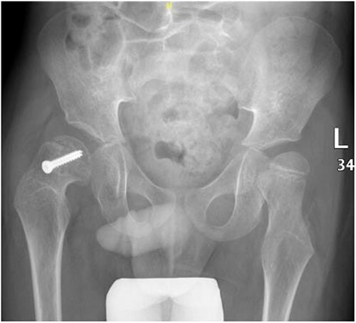
Right hip anterior–posterior radiograph, 36 months following post-operative fixation.
References
Publication types
LinkOut - more resources
Full Text Sources
