[Preprint]. 2024 Apr 15:2024.02.11.579846.
doi: 10.1101/2024.02.11.579846.

Single-cell and Spatial Transcriptomics Identified Fatty Acid-binding Proteins Controlling Endothelial Glycolytic and Arterial Programming in Pulmonary Hypertension

Affiliations

Single-cell and Spatial Transcriptomics Identified Fatty Acid-binding Proteins Controlling Endothelial Glycolytic and Arterial Programming in Pulmonary Hypertension

Bin Liu et al. bioRxiv. .

Update in

Abstract

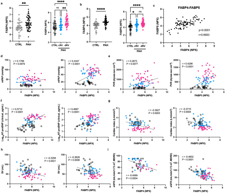
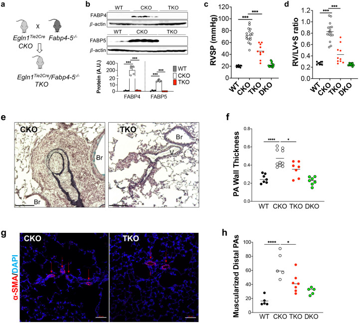
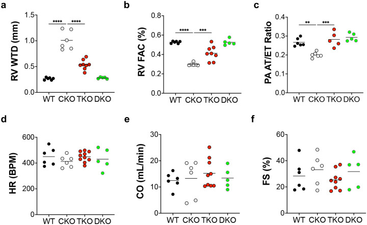
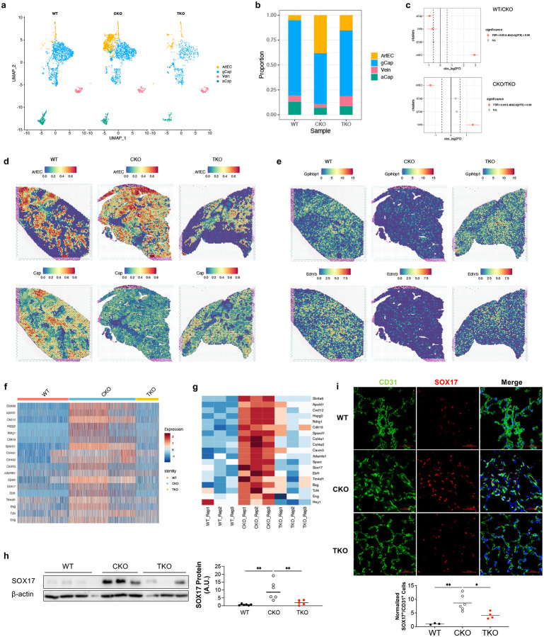
Pulmonary arterial hypertension (PAH) is a devastating disease characterized by obliterative vascular remodeling and persistent increase of vascular resistance, leading to right heart failure and premature death. Understanding the cellular and molecular mechanisms will help develop novel therapeutic approaches for PAH patients. Single-cell RNA sequencing (scRNAseq) analysis found that both FABP4 and FABP5 were highly induced in endothelial cells (ECs) of Egln1 Tie2Cre (CKO) mice, which was also observed in pulmonary arterial ECs (PAECs) from idiopathic PAH (IPAH) patients, and in whole lungs of pulmonary hypertension (PH) rats. Plasma levels of FABP4/5 were upregulated in IPAH patients and directly correlated with severity of hemodynamics and biochemical parameters using plasma proteome analysis. Genetic deletion of both Fabp4 and 5 in CKO mice (Egln1 Tie2Cre /Fabp4-5 -/- ,TKO) caused a reduction of right ventricular systolic pressure (RVSP) and RV hypertrophy, attenuated pulmonary vascular remodeling and prevented the right heart failure assessed by echocardiography, hemodynamic and histological analysis. Employing bulk RNA-seq and scRNA-seq, and spatial transcriptomic analysis, we showed that Fabp4/5 deletion also inhibited EC glycolysis and distal arterial programming, reduced ROS and HIF-2α expression in PH lungs. Thus, PH causes aberrant expression of FABP4/5 in pulmonary ECs which leads to enhanced ECs glycolysis and distal arterial programming, contributing to the accumulation of arterial ECs and vascular remodeling and exacerbating the disease.

Keywords: arterial; glycolysis; hypoxia; lipid metabolism; right heart failure.


Figures

Figure 1.
Figure 1.. Upregulation of FABP4 and 5 in the PAH patients and PH rodents.
(a) Single-cell RNA sequencing analysis showed that FABP4 and 5 were upregulated in the lung ECs of Egln1Tie2Cre (CKO) mice compared to WT mice. Wilcoxon Rank Sum Test, **** adjusted P < 0.0001; ns=no significance. (b) Representative images of immunostaining against FABP4 and 5 showed that FABP4 and 5 were upregulated in the lung ECs of Egln1Tie2Cre mice compared to WT mice. Lung tissues were co-stained with anti-FABP4 or anti-FABP5 and anti-CD31 (marker for ECs). Nuclei were counterstained with DAPI. FABP4 and 5 expression was upregulated in pulmonary vascular ECs in CKO mice. V, vessel. More than 3 lungs from each group of mice were checked. (c) Representative Western Blotting demonstrating upregulation of FABP4 and 5 protein expression in the lung lysate isolated from CKO lungs compared with WT mice. (d) Representative Western Blotting showing an increase of FABP4 and 5 protein expression in the lung lysate isolated from Sugen5416/hypoxia (SuHx) -exposed lungs or monocrotaline (MCT) -exposed lungs compared with basal rats. (e, f) QRT-PCR analysis and Western Blotting demonstrating an upregulation of FABP4 and 5 in the isolated PAECs from IPAH patients compared with healthy donor lungs. Each sample represents PAECs from individual patients or healthy controls (n=3). t test (c, d, e). *P<0.05; **P<0.01; Scale bar, 50 μm.
Figure 2.
Figure 2.. Upregulation of the plasma FABP4 and 5 in patients with PAH.
(a, b) Plasma protein NPX levels of the FABP4 (a) and 5 (b) in donors (CTRL) and patients with PAH from Canada cohorts. Scatter dot plots show individual values (n). According to contemporary Cardiac Index (CI) measured by right heart catheterization at the time the blood sample was drawn. Patients with PAH were categorized as compensated RV (cRV, CI> 2.2 L/min/m2) and decompensated RV (dRV, CI ≤ 2.2 L/min/m2). CTRL N=28, cRV N= 31, dRV N=29. (c) Plasma protein NPX level of FABP4 were correlated with plasma protein NPX level of FABP5 in the human subjects including both donors and PAH patients. (d–i) Pearson’s correlation coefficient of the levels of FABP4 and 5 and hemodynamics/biomedical parameters of PAH patients with associated P value is shown in each graph. mPAP, mean pulmonary arterial pressure; PVR, pulmonary vascular resistance; SV, strove volume; eGFR, estimating glomerular filtration rate. t test (left panels of a, b). ANOVA followed by Turkey post hoc analysis was used for statistical analysis (right panels of a, b). Pearson’s correlation test (c-i). **P<0.01; ****P<0.0001. ns=not significant.
Figure 3.
Figure 3.. FABP4 and 5 deletions protected from Egln1-deficiency-induced PH in mice.
(a) A diagram showing the strategy for generation of FABP4 and 5 double knockouts in Egln1Tie2Cre mice (TKO). (b) Representative Western blotting demonstrating diminished FABP4 and 5 protein expression in lung lysate isolated from TKO lungs compared with CKO lungs. (c) Dramatic reduction in RVSP was seen in TKO mice compared with CKO mice. Bars represent the mean. (d) Marked inhibition of RV hypertrophy in TKO mice compared with CKO mice. (e, f) Representative images from Russel-Movat pentachrome staining and quantification of wall thickness of PA exhibited reduced thickness in the intima, media, and adventitia layers of tissue of TKO mice compared with CKO mice. Occlusive lesions were diminished in TKO mice compared with CKO mice. Br, bronchus; V, vessel. (g) Exemplary images showed a reduction in muscularization of distal pulmonary vessels in TKO mice. Lung sections were subjected to immunostaining using anti-smooth muscle α-actin (SMA). (h) Muscularization quantification was performed by tallying SMA-positive vessels across 40 fields (at ×20 magnification) within every lung section. The results were presented as the mean value along with the standard deviation (mean ± SD) based on a sample size of 5 to 10 mice per group. ANOVA followed by Turkey post hoc analysis was used for statistical analysis (b, c, d, f and h). *P<0.05; ***P<0.001, and ****P<0.0001.
Figure 4.
Figure 4.. Genetical deletion of Fabp4–5 improved right heart function in PH mice
(a) Representative echocardiography images revealed a reduction of RV wall thickness during diastole (RVWTD) in TKO mice compared with CKO mice. (b) Improved RV fraction area change (RVFAC), indicating enhancing RV contractility, in TKO mice compared with CKO mice. (c) An increased ratio of pulmonary artery acceleration time to ejection time (PA AT/ET) in TKO mice compared with CKO mice. (d–f) There is no significant change of heart rate, cardiac output and left ventricular fractional shorting. ANOVA followed by Turkey post hoc analysis was used for statistical analysis (a, b, c). Significance levels were denoted as **P<0.01, ***P<0.001, and ****P<0.0001.
Figure 5.
Figure 5.. FABP4–5 regulated genes related to PH and glycolysis
(a) Principal component analysis showed that FABP4–5 deletion normalized the altered gene signature by Egln1 deficiency in mice. (b) A representative heatmap showed that Fabp4–5 deletion altered the overall gene signature of CKO lungs toward to WT lungs. (c) Venn diagrams indicated FABP4–5 controlled the expression of the dysregulated genes in CKO mice. (d) The Kyoto Encyclopedia of Genes and Genomes (KEGG) pathway analysis of the genes upregulated in CKO lungs and normalized by FABP4–5 deletion. (e) Gene Set Enrichment Analysis (GESA) analysis showed the enrichment of Hypoxia and Glycolysis pathways based on the gene sets regulated by FABP4/5. (f) A panel of representative genes related to PH pathogenesis and glycolysis were normalized by FABP4–5 deletion in CKO mice. (g) qPCR analysis confirmed the PH causing genes and glycolysis related genes were altered in CKO lungs and normalized in the TKO mice by RNA-seq data. (h) Western blotting showing FABP4–5 deletion reduced the protein level of glycolytic genes in CKO mice. ANOVA followed by Turkey post hoc analysis was used for statistical analysis (g and h). *P<0.05; **P<0.01; ***P<0.001, and ****P<0.0001.
Figure 6.
Figure 6.. Single-cell transcriptomics analysis on FABP4–5 regulated PH development.
(a, b) ScRNA-seq analysis demonstrated that FABP4–5 deletion partially normalized the alteration of EC subpopulations in CKO mice. (c) Venn diagrams indicated FABP4–5 controlled the expression of the dysregulated genes in ECs. (d) The Kyoto Encyclopedia of Genes and Genomes (KEGG) pathway analysis of the genes upregulated in CKO ECs and normalized by FABP4–5 deletion in ECs. (e). A heatmap analysis based on the scRNA-seq analysis on ECs population showed that the glycolytic genes were upregulated in CKO lungs and normalized in TKO ECs. (f) The glycolytic score calculation showed that glycolysis is controlled by FABP4–5 in ECs.
Figure 7.
Figure 7.. Single-cell and spatial transcriptomics analysis identified induction of distal arterialization by FABP4–5 in PH.
(a, b) A representative UMAP and a cellular proportion plot showing the EC subpopulation change between groups. Upregulation of arterial EC proportion in CKO lungs was inhibited in TKO lungs. (c) Statistical analysis showed that an increase in arterial ECs and a reduction of aCap in CKO lungs were rescued in TKO lungs. (d) Integration of scRNAseq and Visium spatial data revealed increased arterial ECs and decreased gCap ECs in the distal capillary bed of CKO mice, which was normalized in the TKO mice. The same WT and CKO lungs were used in our previous publication. The visualization shows the predicted spatial distribution of arterial ECs and capillary ECs within the lungs. (e) A spatial Plot showed that both gCap marker Gpihbpi and aCap marker Car4 were reduced in the CKO lungs and restored in the TKO lungs. (f) A heatmap showing a panel of representative genes related to arterial EC markers were increased in the CKO ECs and normalized in TKO ECs based on the scRNAseq data. (g) A heatmap based on the Bulk RNAseq data showing the rescue of arterial gene programming in TKO lungs compared to CKO lungs. (h) Western blotting demonstrated the classical arterial marker SOX17 was increased in the CKO lungs and inhibited in the TKO lungs. (i) Immunostaining against SOX17 demonstrated the increase in distal arterial ECs in CKO lungs, which was rescued in the TKO lungs. ANOVA followed by Turkey post hoc analysis was used for statistical analysis (h and i). *P<0.05; **P<0.01.
Figure 8.
Figure 8.. Overexpression of FABP4–5 induced ECs dysfunction
(A) Overexpression of FABP4 and 5 fused with Flag tag in hPAECs using lentiviruses mediated gene overexpression. (B, C) FABP4–5 overexpression promoted ECs glycolysis and proliferation assessed by BrdU incorporation assay. (D) Glycolytic inhibitor 2-DG treatment inhibited FABP4–5 induced PAEC proliferation. (E) FABP4–5 deletion in vivo reduced lung PAECs proliferation in TKO mice compared with CKO mice. (F and G) Overexpression of FABP4–5 promoted fatty acid oxidation (FAO) in hPAECs in the presence of palmitic acid. The double arrows label the OCR generation contributed by FAO. (H) Both CPT1α inhibitor Etomoxir (ETO, 20 μM) and DNA synthesis inhibitor (5-FU, 20 μM) blocked FABP4–5 induced ECs proliferation. (I and J) A representative seahorse data showing upregulation of FAO in PAECs isolated from IPAH patients compared to control donors. Six lines of control or IPAH patients PAECs were tested. (C, D, H) For BrdU assay, each dot represents a biological replicate. The experiments were performed at least three times. Student t test (D). ANOVA followed by Turkey post hoc analysis was used for statistical analysis (B, C, E, F, G, H, J). Significance levels were denoted as *P<0.05, **P<0.01, ***P<0.001, and ****P<0.0001.
Figure 9.
Figure 9.. HIF-2α mediated glycolysis induced by FABP4–5 in pulmonary ECs
(A) A diagram showing the predicted transcription factors based on the DEGs and literature. (B) Western blotting demonstrated HIF-2α but not p53 or C-Myc was upregulated in CKO lungs and normalized in TKO lungs. (C) A representative heatmap showed that glycolytic genes were depended on HIF-2α using WT, CKO mice, and Egln1Tie2Cre/Hif2aTie2Cre (EH2) mice. (D) HIF-2α knockdown inhibited FABP4–5 induced endothelial proliferation. (E) Nitrative stress assessed by protein nitrotyrosine modification was reduced in TKO lungs compared to CKO lungs. (F) Overexpression of FABP4 and 5 promoted mitochondrial ROS levels in HPAECs. (G) A diagram showing our proposed model. Our study addresses a novel role of lung endothelial FABP4–5 controlling PAECs accumulation through increased glycolysis in the pathogenesis of PAH. By facilitating fatty acid uptake and translocation into mitochondria, FABP4–5 promote FAO and ROS generation, which activates HIF-2α signaling to promote endothelial glycolysis. For BrdU assay in (D) and mitochondrial ROS assay in (F), each dot represents a biological replicate. The experiments were performed at least three times. ANOVA followed by Turkey post hoc analysis was used for statistical analysis (B, D, E, F). Significance levels were denoted as *P<0.05, **P<0.01, ***P<0.001, and ****P<0.0001.

References

    1. Stacher E, Graham BB, Hunt JM, Gandjeva A, Groshong SD, McLaughlin VV, Jessup M, Grizzle WE, Aldred MA, Cool CD, Tuder RM. Modern age pathology of pulmonary arterial hypertension. American Journal of Respiratory and Critical Care Medicine. 2012;186:261–272. - PMC - PubMed
    1. Humbert M, Guignabert C, Bonnet S, Dorfmüller P, Klinger JR, Nicolls MR, Olschewski AJ, Pullamsetti SS, Schermuly RT, Stenmark KR, Rabinovitch M. Pathology and pathobiology of pulmonary hypertension: state of the art and research perspectives. The European respiratory journal. 2019;53:1801887. - PMC - PubMed
    1. Thenappan T, Ormiston ML, Ryan JJ, Archer SL. Pulmonary arterial hypertension: Pathogenesis and clinical management. BMJ (Online). 2018;360:j5492. - PMC - PubMed
    1. Paulin R, Michelakis ED. The metabolic theory of pulmonary arterial hypertension. Circulation Research. 2014;115:148–164. - PubMed
    1. Nickel NP, Yuan K, Dorfmuller P, Provencher S, Lai YC, Bonnet S, Austin ED, Koch CD, Morris A, Perros F, Montani D, Zamanian RT, De Jesus Perez VA. Beyond the lungs: Systemic manifestations of pulmonary arterial hypertension. American Journal of Respiratory and Critical Care Medicine. 2020;201:148–157. - PMC - PubMed

Publication types