[Preprint]. 2024 May 22:2024.02.08.576722.
doi: 10.1101/2024.02.08.576722.

Mosaic sarbecovirus nanoparticles elicit cross-reactive responses in pre-vaccinated animals

Affiliations

Mosaic sarbecovirus nanoparticles elicit cross-reactive responses in pre-vaccinated animals

Alexander A Cohen et al. bioRxiv. .

Update in

  • Mosaic sarbecovirus nanoparticles elicit cross-reactive responses in pre-vaccinated animals.
    Cohen AA, Keeffe JR, Schiepers A, Dross SE, Greaney AJ, Rorick AV, Gao H, Gnanapragasam PNP, Fan C, West AP Jr, Ramsingh AI, Erasmus JH, Pata JD, Muramatsu H, Pardi N, Lin PJC, Baxter S, Cruz R, Quintanar-Audelo M, Robb E, Serrano-Amatriain C, Magneschi L, Fotheringham IG, Fuller DH, Victora GD, Bjorkman PJ. Cohen AA, et al. Cell. 2024 Oct 3;187(20):5554-5571.e19. doi: 10.1016/j.cell.2024.07.052. Epub 2024 Aug 27. Cell. 2024. PMID: 39197450 Free PMC article.

Abstract

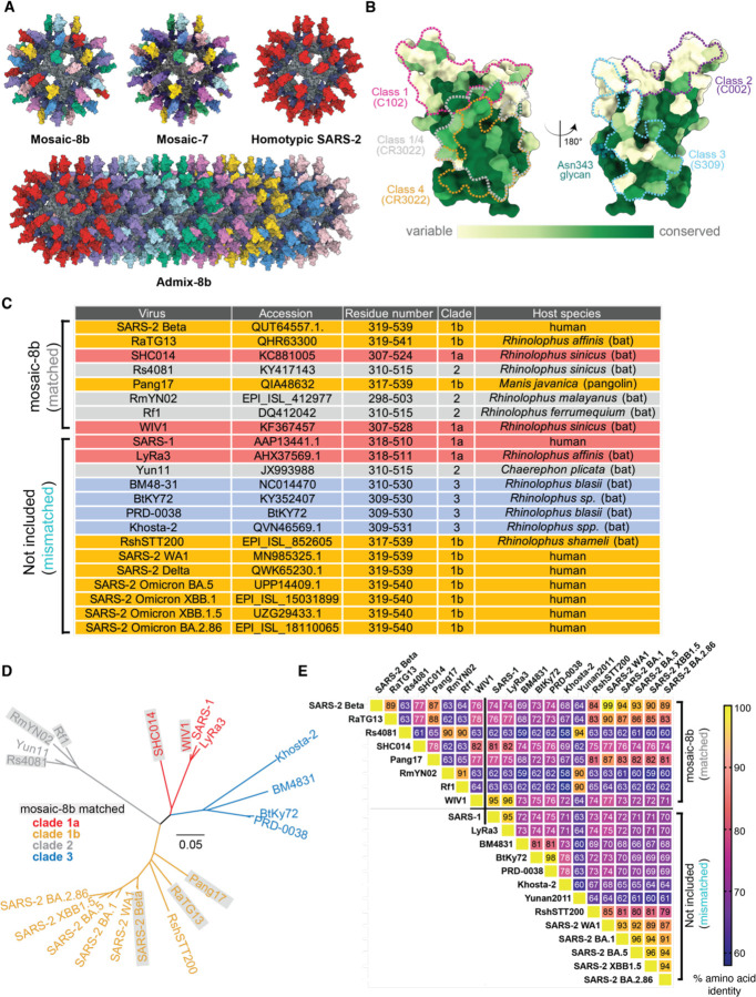
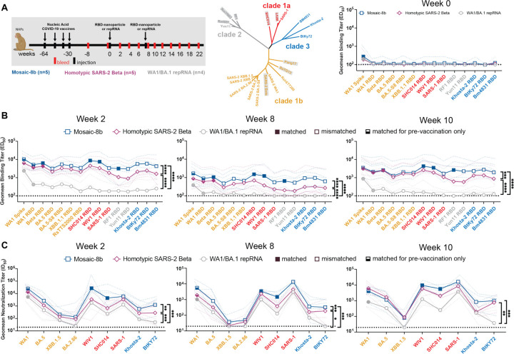
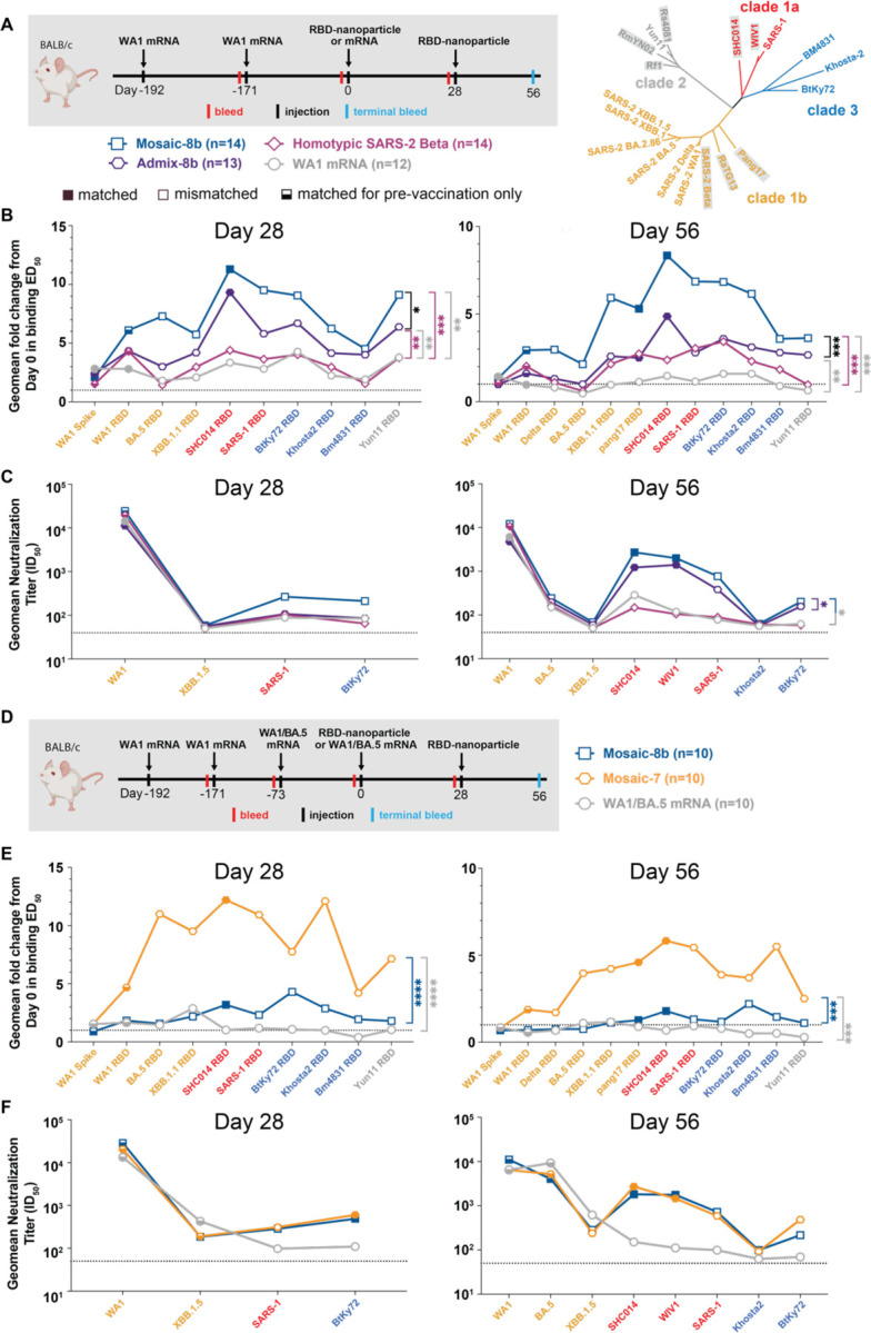
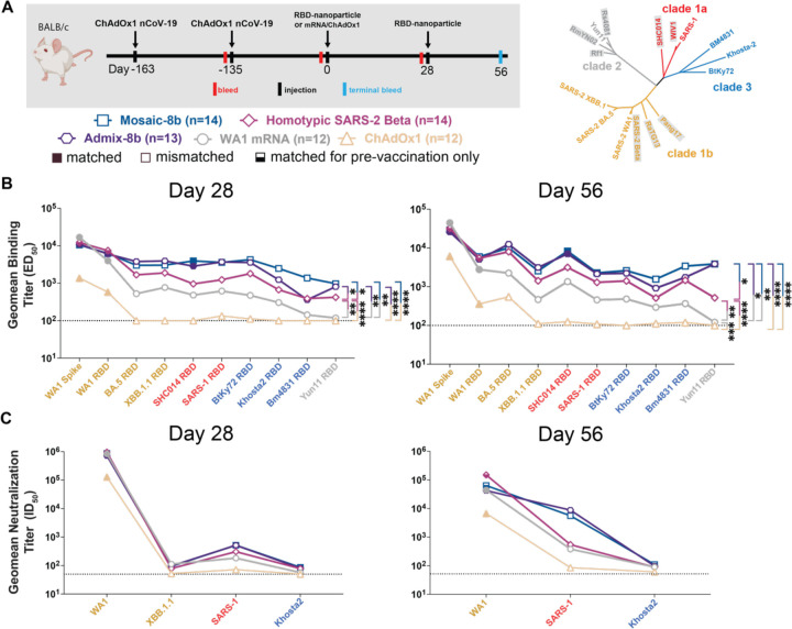
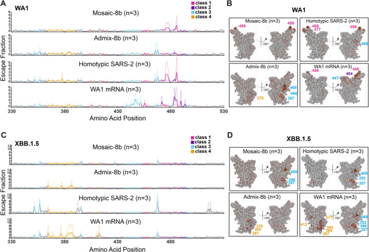
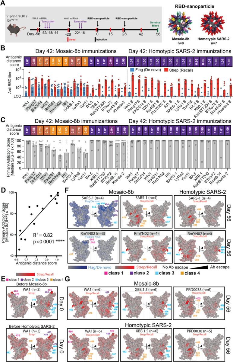
Immunization with mosaic-8b [60-mer nanoparticles presenting 8 SARS-like betacoronavirus (sarbecovirus) receptor-binding domains (RBDs)] elicits more broadly cross-reactive antibodies than homotypic SARS-CoV-2 RBD-only nanoparticles and protects against sarbecoviruses. To investigate original antigenic sin (OAS) effects on mosaic-8b efficacy, we evaluated effects of prior COVID-19 vaccinations in non-human primates and mice on anti-sarbecovirus responses elicited by mosaic-8b, admix-8b (8 homotypics), or homotypic SARS-CoV-2 immunizations, finding greatest cross-reactivity for mosaic-8b. As demonstrated by molecular fate-mapping in which antibodies from specific cohorts of B cells are differentially detected, B cells primed by WA1 spike mRNA-LNP dominated antibody responses after RBD-nanoparticle boosting. While mosaic-8b- and homotypic-nanoparticles boosted cross-reactive antibodies, de novo antibodies were predominantly induced by mosaic-8b, and these were specific for variant RBDs with increased identity to RBDs on mosaic-8b. These results inform OAS mechanisms and support using mosaic-8b to protect COVID-19 vaccinated/infected humans against as-yet-unknown SARS-CoV-2 variants and animal sarbecoviruses with human spillover potential.

Keywords: Antibody; Immune Imprinting; Macaque and Mouse Models; Mosaic-8b RBD-nanoparticle; Original Antigenic Sin; Primary Addiction; RBD; SARS-CoV-2; Sarbecovirus; Vaccination.


Conflict of interest statement

DECLARATION OF INTERESTS P.J.B. and A.A.C. are inventors on a US patent application (17/523,813) filed by the California Institute of Technology that covers the mosaic nanoparticles described in this work. A.I.R. and J.D.P. are inventors on a US patent (11,780,888) that covers the chimeric sequence of RBD fused to the HA2 stem of influenza hemagglutinin. A.J.G. is an inventor on a Fred Hutchinson Cancer Center-optioned technology related to DMS of the RBD of SARS-CoV-2 spike protein. L.M., S.B., R.C., C.S-A., and I.G.F. are inventors on U.S. Patent Applications (16/952,983) and (17/651,476) filed by Ingenza Ltd. that cover Bacillus and Pichia strains established to manufacture endotoxin-free vaccine products. J.H.E. is an employee of HDT Bio that provided the repRNA-LION vaccine. D.H.F. is a co-founder of Orlance, Inc. that is developing gene gun delivery of DNA and repRNA vaccines. S.B., R.C., M.Q.-A., E.R., C.S.-A., L.M., and I.G.F. are employees of Ingenza, LTD. P.J.B. and G.D.V. are scientific advisors for Vaccine Company, Inc, and P.J.B. is a scientific advisor for Vir Biotechnology. N.P. is named on patents describing the use of nucleoside-modified mRNA in lipid nanoparticles as a vaccine platform. N.P. served on the mRNA strategic advisory board of Sanofi Pasteur in 2022 and the mRNA technology advisory board of Pfizer in 2023 and is a member of the Scientific Advisory Board of AldexChem and Bionet-Asia. P.J.C.L. is an employee of Acuitas Therapeutics, a company developing LNP delivery systems for RNA therapeutics.

Figures

Figure 1
Figure 1. RBDs used to make nanoparticles and for assays, related to Figure S1, Table S1.
(A) Models of mosaic-8b, mosaic-7 (mosaic-8b without SARS-2 RBD), homotypic SARS-2, and admix-8b RBD-nanoparticles constructed using coordinates of an RBD (PDB 7BZ5), SpyCatcher (PDB 4MLI), and i3-01 nanoparticle (PDB 7B3Y). (B) Sequence conservation determined using the ConSurf Database of the 16 sarbecovirus RBDs used to make nanoparticles and/or for assays shown on two views of an RBD surface (PDB 7BZ5). Class 1, 2, 3, 4, and 1/4 epitopes are outlined in different colors using information from Fab-RBD or Fab-spike trimer structures (C102, PDB 7K8M; C002, PDB 7K8T; S309, PDB 7JX3; CR3022, PDB 7LOP; and C118, PDB 7RKV). (C) List of sarbecoviruses from which the RBDs in mosaic-8b and admix-8b were included (matched) or not included (mismatched). Clades were defined as described. (D) Phylogenetic tree of selected sarbecoviruses calculated using a Jukes-Cantor generic distance model using Geneious Prime® 2023.1.2 based on amino acid sequences of RBDs aligned using Clustal Omega. Viruses with RBDs included in mosaic-8b are highlighted in gray rectangles. (E) Amino acid sequence identity matrix of RBDs based on alignments using Clustal Omega.
Figure 2
Figure 2. Mosaic-8b immunizations in previously-vaccinated NHPs elicits cross-reactive Ab responses, related to Figure S2.
Data are shown for ELISA and neutralization analyses for serum samples from weeks 0, 2, 8, and 10. (Data for samples from weeks 4, 12, and 22 are in Figure S2.) Geometric means of ED50 or ID50 values for all animals in each cohort are indicated by symbols connected by thick colored lines; results for individual animals are indicated by dotted colored lines. Mean titers against indicated viral antigens or pseudoviruses were compared pairwise across immunization cohorts by Tukey’s multiple comparison test with the Geisser-Greenhouse correction (as calculated by GraphPad Prism). Significant differences between cohorts linked by vertical lines are indicated by asterisks: p<0.05 = *, p<0.01 = **, p<0.001 = ***, p<0.0001 = ****. (A) Left: Schematic of vaccination/immunization regimen. NHPs were vaccinated at the indicated weeks prior to RBD-nanoparticle or repRNA prime and boost immunizations at week 0 and week 8. Middle: Phylogenetic tree of selected sarbecoviruses calculated using a Jukes-Cantor generic distance model using Geneious Prime® 2023.1.2 based on amino acid sequences of RBDs aligned using Clustal Omega. RBDs included in mosaic-8b are highlighted in gray rectangles. Right: Geometric mean ELISA binding titers at week 0 (after pre-vaccinations but prior to RBD-nanoparticle or repRNA immunizations) against indicated viral antigens. (B) Geometric mean ELISA binding titers at the indicated weeks after immunization with mosaic-8b, homotypic SARS-2, or bivalent WA1/BA.1 repRNA against indicated viral antigens. (C) Geometric mean neutralization titers at the indicated weeks after immunization with mosaic-8b, homotypic SARS-2, or bivalent WA1/BA.1 repRNA against indicated sarbecovirus pseudoviruses.
Figure 3
Figure 3. Mosaic-8b immunizations in mice previously immunized with an mRNA-LNP vaccine elicit cross-reactive Ab responses, related to Figure S3.
For experiments shown schematically in panels A and D, binding responses at day 0 (immediately prior to nanoparticle or other vaccine immunizations) showed significant differences across cohorts in titers elicited by the pre-vaccinations. We therefore applied baseline corrections to account for different mean responses at day 0 prior to immunizations in each of the four groups for data in panel B and each of the three groups for data in panel E (see Methods). Non-baseline corrected binding data for panel E are shown in Figure S3G. Geometric means of fold change in ED50 or geometric means of ID50 values for all animals in each cohort are indicated by symbols connected by thick colored lines. Mean titers against indicated viral antigens or pseudoviruses were compared pairwise across immunization cohorts by Tukey’s multiple comparison test with the Geisser-Greenhouse correction (as calculated by GraphPad Prism). Significant differences between cohorts linked by vertical lines are indicated by asterisks: p<0.05 = *, p<0.01 = **, p<0.001 = ***, p<0.0001 = ****. (A) Left: Schematic of vaccination/immunization regimen for panels B and C. Mice were vaccinated at the indicated days prior to RBD-nanoparticle prime and boost immunizations at days 0 and 28 or mRNA-LNP prime immunization at day 0. Right: Phylogenetic tree of selected sarbecoviruses calculated using a Jukes-Cantor generic distance model using Geneious Prime® 2023.1.2 based on amino acid sequences of RBDs aligned using Clustal Omega. RBDs included in mosaic-8b are highlighted in gray rectangles. (B) Geometric mean fold change ELISA binding titers at the indicated days after immunization with mosaic-8b, admix-8b, homotypic SARS-2, or WA1 mRNA-LNP against indicated viral antigens. (C) Geometric mean neutralization titers at the indicated weeks after immunization with mosaic-8b, admix-8b, homotypic SARS-2, or WA1 mRNA-LNP against indicated sarbecovirus pseudoviruses. (D) Schematic of vaccination/immunization regimen for panels E and F. Mice were vaccinated at the indicated days prior to RBD-nanoparticle prime and boost immunizations at days 0 and 28 or mRNA-LNP prime immunization at day 0. (E) Geometric mean fold change ELISA binding titers at the indicated days after immunization with mosaic-8b, mosaic-7, or WA1/BA.5 mRNA-LNP against indicated viral antigens. Compare with Figure S3G (non-baseline corrected geometric mean ELISA binding titers). (F) Geometric mean neutralization titers at the indicated weeks after immunization with mosaic-8b, mosaic-7, or WA1/BA.5 mRNA-LNP against indicated sarbecovirus pseudoviruses.
Figure 4
Figure 4. Mosaic-8b immunizations in ChAdOx1-vaccinated mice elicit cross-reactive Ab responses, related to Figure S4.
Geometric means of ED50 or ID50 values for all animals in each cohort are indicated by symbols connected by thick colored lines. Mean titers against indicated viral antigens or pseudoviruses were compared pairwise across immunization cohorts by Tukey’s multiple comparison test with the Geisser-Greenhouse correction (as calculated by GraphPad Prism). Significant differences between cohorts linked by vertical lines are indicated by asterisks: p<0.05 = *, p<0.01 = **, p<0.001 = ***, p<0.0001 = ****. (A) Left: Schematic of vaccination/immunization regimen. Mice were vaccinated at the indicated days prior to RBD-nanoparticle prime and boost immunizations at days 0 and 28 or mRNA-LNP or ChAdOx1 prime immunizations at day 0. Right: Phylogenetic tree of selected sarbecoviruses calculated using a Jukes-Cantor generic distance model using Geneious Prime® 2023.1.2 based on amino acid sequences of RBDs aligned using Clustal Omega. RBDs included in mosaic-8b are highlighted in gray rectangles. (B) Geometric mean ELISA binding titers at the indicated days after immunization with mosaic-8b, admix-8b, homotypic SARS-2, WA1 mRNA-LNP, or ChAdOx1 against indicated viral antigens. (C) Geometric mean neutralization titers at the indicated weeks after immunization with mosaic-8b, admix-8b, homotypic SARS-2, WA1 mRNA-LNP, or ChAdOx1 against indicated sarbecovirus pseudoviruses.
Figure 5
Figure 5. Differences in epitope targeting of Abs elicited in ChAdOx1 pre-vaccinated mice by mosaic-8b and admix-8b compared with homotypic SARS-2 and WA1 mRNA-LNP, related to Figure S5.
Sera were derived from ChAdOx1 pre-vaccinated mice that were immunized with either two doses of an RBD-nanoparticle immunogen or one dose of WA1 mRNA-LNP. (A) Line plots for DMS results from sera from ChAdOx1-vaccinated mice that were immunized with the indicated immunogens (immunization schedule in Figure 4A). DMS was conducted using a WA1 RBD library. The x-axis shows the RBD residue number and the y-axis shows the sum of the Ab escape of all mutations at a site (larger numbers indicating more Ab escape). Each line represents one antiserum with heavy lines showing the average across the n=3 sera in each group. Lines are colored differently for RBD epitopes within different classes (color definitions in upper right legend; epitopes defined in Figure 1E; gray for residues not assigned to an epitope). (B) The average site-total Ab escape for the WA1 RBD library for mice immunized with the indicated immunogens after ChAdOx1 vaccination mapped to the surface of the WA1 RBD (PDB 6M0J), with gray indicating no escape, and epitope-specific colors indicating sites with the most escape. (C) Line plots for DMS results from sera from ChAdOx1-vaccinated mice that were immunized with the indicated immunogens (immunization schedule in Figure 4A). DMS was conducted using a XBB.1.5 RBD library. The x-axis shows the RBD residue number and the y-axis shows the sum of the Ab escape of all mutations at a site (larger numbers indicating more Ab escape). Each line represents one antiserum with heavy lines showing the average across the n=3 sera in each group. Lines are colored differently for RBD epitopes within different classes (color definitions in upper right legend; epitopes defined in Figure 1E; gray for residues not assigned to an epitope). (D) The average site-total Ab escape for the XBB.1.5 RBD library for mice immunized with the indicated immunogens after ChAdOx1 vaccination mapped to the surface of the WA1 RBD (PDB 6M0J), with gray indicating no escape, and epitope-specific colors indicating sites with the most escape.
Figure 6
Figure 6. Fate mapping of serum antibodies reveals elicitation of cross-reactive recall responses and strain-specific de novo responses elicited by mosaic-8b nanoparticles in previously vaccinated animals, related to Figure S6.
Antigenic distance score is defined as the ratio of % amino acid sequence identity of a strain to WA1 RBD divided by % sequence identity of that strain to the closest non-self RBD relative on the nanoparticle. The primary addiction index is defined as Strep/(Flag + Strep) x 100. See Figure 1B for RBD epitope classifications. (A) (Left) Schematic of vaccine regimen in S1pr2-IgkTag/Tag immunized mice. Mice were vaccinated with an mRNA-LNP vaccine encoding WA1 spike at the indicated days prior to RBD-nanoparticle immunizations at day 0 and day 28. After each dose of mRNA-LNP vaccine, mice were treated with tamoxifen to switch epitope tags on the Abs produced by B cells activated by the mRNA-LNP vaccinations from Flag to Strep, so that Ab responses elicited by RBD-nanoparticle boosting can be separated into recall (Strep+) and de novo (Flag+) responses. (Right) Models of RBD-nanoparticles that were used for immunizations at days 0 and 28. (B) Comparison of the Flag+/de novo (blue) and Strep+/recall (red) anti-RBD titers shown at day 42, two weeks after a second immunization with mosaic-8b or homotypic SARS-2 nanoparticles. For each strain, the antigenic distance score is shown above the plots and colored with a gradient from purple (score=1) to yellow (score=0). Gray shaded strains are matched to mosaic-8b. (C) The primary addiction index for each RBD is plotted for day 42 serum after two doses of mosaic-8b or homotypic SARS-2. The antigenic distance score is shown above the plots and colored with a gradient from purple (score=1) to yellow (score=0). Gray shaded strains are matched to mosaic-8b. (D) Correlation of the primary addiction index with the antigenic distance score. The best-fit line, R2, and p value were calculated by linear regression using Prism. (E) DMS analysis of Strep+ Abs from day 0 (prior to RBD-nanoparticle immunization) serum from n=3 pre-vaccinated mice using an RBD mutant library derived from WA1. Ab binding sites are shaded red according to degree of Strep+ Ab escape on the surface of the WA1 RBD (PDB 6M0J). Locations of residues with high escape scores are indicated on RBD surfaces with gray indicating no escape and shades of red indicating sites with most escape. Residue numbers are color coded according to their RBD epitope classification. (F) DMS analysis using SARS-1 (antigenic distance score = 0.81) and RmYN02 (antigenic distance score = 0.63) RBD mutant libraries of day 56 serum from n=4 or n=3 RBD-nanoparticle immunized mice. Ab binding sites are shaded according to degree of Ab escape, with blue for Flag/de novo responses and red for Strep/recall responses, on the surface of the WA1 RBD (PDB 6M0J). Comparisons are made for Flag/de novo and Strep/recall elicited by mosaic-8b and for Strep/recall elicited by homotypic SARS-2 (Flag/de novo responses after homotypic SARS-2 immunization were weak to undetectable). Gray shaded virus names represent strains that are matched to mosaic-8b. Locations of residues with high escape scores are indicated on RBD surfaces with gray indicating no escape and darker shades indicating sites with most escape. Residue numbers are color coded according to their RBD epitope classification. (G) DMS analysis of the Strep/recall compartment of day 56 serum from n=5 or n=6 mosaic-8b or homotypic SARS-2 immunized mice using RBD mutant libraries derived from strains with antigenic distance scores of 1.01 (WA1), 1.01 (XBB.1.5), and 0.94 (PRD008). Ab binding sites are shaded red according to degree of Strep/Recall Ab escape on the surface of the WA1 RBD (PDB 6M0J). Locations of residues with high escape scores are indicated on RBD surfaces with gray indicating no escape and darker shades indicating sites with most escape. Residue numbers are color coded according to their RBD epitope classification.

References

    1. Menachery V. D., Yount B. L., Debbink K., Agnihothram S., Gralinski L. E., Plante J. A., Graham R. L., Scobey T., Ge X.-Y., Donaldson E. F., Randell S. H., Lanzavecchia A., Marasco W. A., Shi Z.-L. & Baric R. S. (2015). A SARS-like cluster of circulating bat coronaviruses shows potential for human emergence. Nature Medicine 21, 1508–1513. - PMC - PubMed
    1. Menachery V. D., Yount B. L., Sims A. C., Debbink K., Agnihothram S. S., Gralinski L. E., Graham R. L., Scobey T., Plante J. A., Royal S. R., Swanstrom J., Sheahan T. P., Pickles R. J., Corti D., Randell S. H., Lanzavecchia A., Marasco W. A. & Baric R. S. (2016). SARS-like WIV1-CoV poised for human emergence. Proceedings of the National Academy of Sciences 113, 3048–3053. - PMC - PubMed
    1. Zhou H., Ji J., Chen X., Bi Y., Li J., Wang Q., Hu T., Song H., Zhao R., Chen Y., Cui M., Zhang Y., Hughes A. C., Holmes E. C. & Shi W. (2021). Identification of novel bat coronaviruses sheds light on the evolutionary origins of SARS-CoV-2 and related viruses. Cell 184, 4380–4391 e4314. - PMC - PubMed
    1. Davis H. E., McCorkell L., Vogel J. M. & Topol E. J. (2023). Long COVID: major findings, mechanisms and recommendations. Nature Reviews Microbiology 21, 133–146. - PMC - PubMed
    1. Planas D., Bruel T., Grzelak L., Guivel-Benhassine F., Staropoli I., Porrot F., Planchais C., Buchrieser J., Rajah M. M., Bishop E., Albert M., Donati F., Prot M., Behillil S., Enouf V., Maquart M., Smati-Lafarge M., Varon E., Schortgen F., Yahyaoui L., Gonzalez M., De Seze J., Pere H., Veyer D., Seve A., Simon-Loriere E., Fafi-Kremer S., Stefic K., Mouquet H., Hocqueloux L., van der Werf S., Prazuck T. & Schwartz O. (2021). Sensitivity of infectious SARS-CoV-2 B.1.1.7 and B.1.351 variants to neutralizing antibodies. Nat Med 27, 917–924. - PubMed

Publication types