. 2024 Jan 26;57(3):1050-1071.
doi: 10.1021/acs.macromol.3c02600. eCollection 2024 Feb 13.

Solid-Phase Synthesis as a Tool to Create Exactly Defined, Branched Polymer Vectors for Cell Membrane Targeting

Affiliations

Solid-Phase Synthesis as a Tool to Create Exactly Defined, Branched Polymer Vectors for Cell Membrane Targeting

Johanna K Elter et al. Macromolecules. .

Abstract

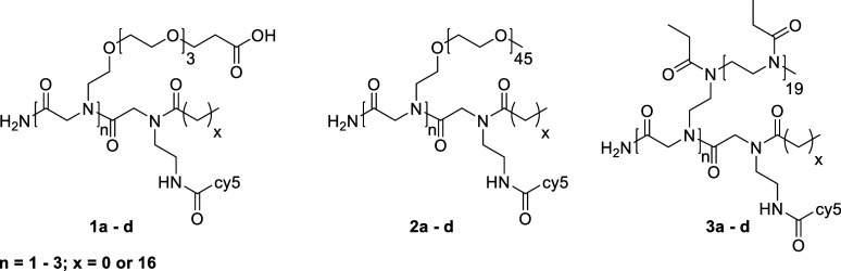
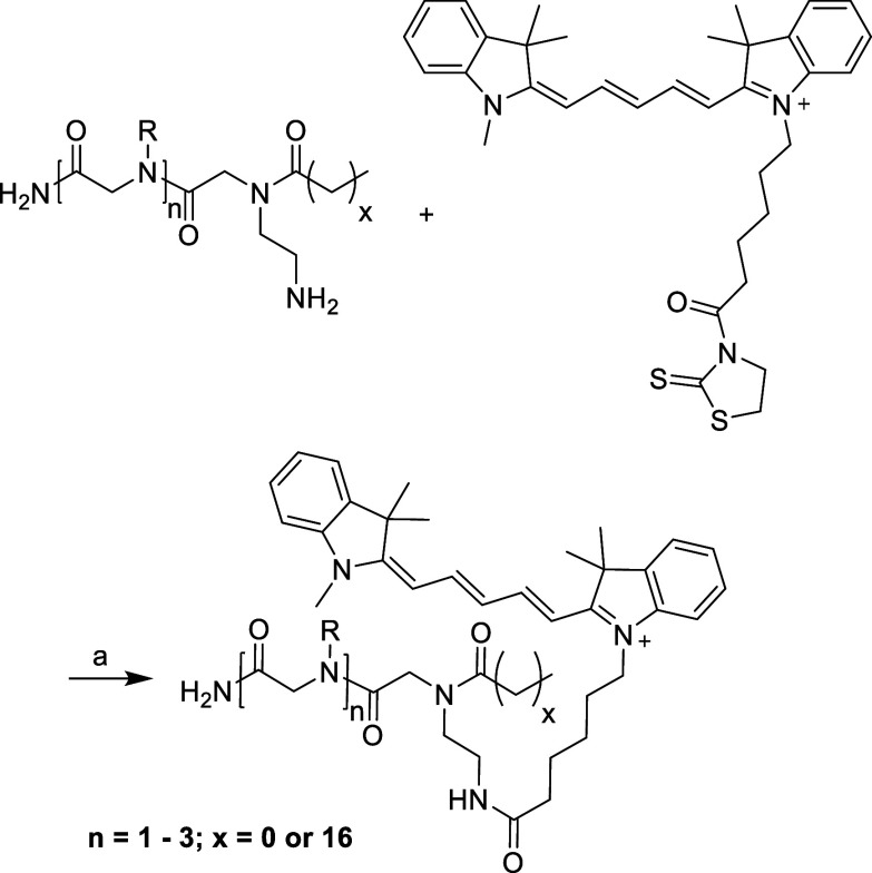
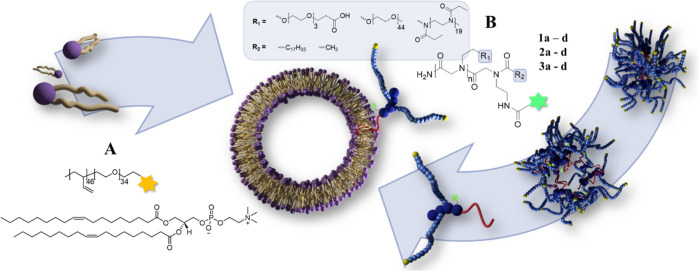
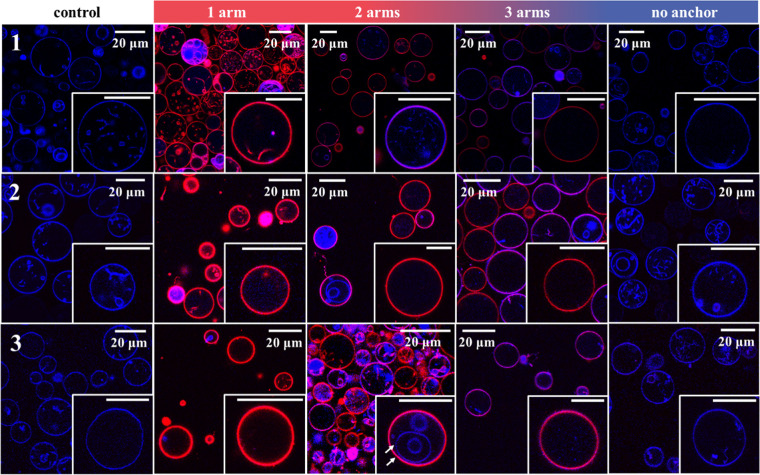
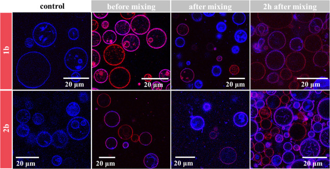
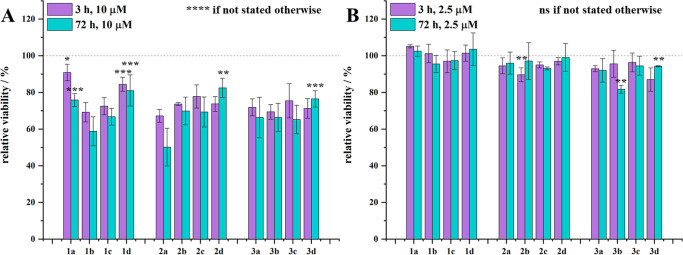
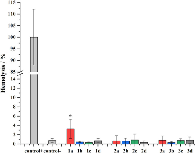
Modern drug formulations often require, besides the active drug molecule, auxiliaries to enhance their pharmacological properties. Tailor-made, biocompatible polymers covalently connected to the drug molecule can fulfill this function by increasing its solubility, reducing its toxicity, and guiding it to a specific target. If targeting membrane-bound proteins, localization of the drug close to the cell membrane and its target is beneficial to increase drug efficiency and residence time. In this study, we present the synthesis of highly defined, branched polymeric structures with membrane-binding properties. One to three hydrophilic poly(ethylene oxide) or poly(2-ethyloxazoline) side chains were connected via a peptoid backbone using a two-step iterative protocol for solid-phase peptoid synthesis. Additional groups, e.g., a hydrophobic anchor for membrane attachment, were introduced. Due to the nature of solid-phase synthesis, the number and order of the side chains and additional units can be precisely defined. The method proved to be versatile for the generation of multifunctional, branched polymeric structures of molecular weights up to approximately 7000 g mol-1. The behavior of all compounds towards biological membranes and cells was investigated using liposomes as cell membrane models, HEK293 and U251-MG cell lines, and red blood cells, thereby demonstrating their potential value as drug auxiliaries with cell membrane affinity.


Conflict of interest statement

The authors declare no competing financial interest.

Figures

Figure 1
Figure 1
Final compounds based on functional OEG (1a–1d), mPEG (2a–2d), and mPEtOx (3a–3d).
Figure 2
Figure 2
1H NMR spectra of compounds 1a–1d (A), 2a–2d, and (C) 3a–3d (E), HPLC traces of compounds 1a–1d (B), and GPC traces of compounds 2a–2d (D) and 3a–3d (F). 1H NMR spectra were recorded before freeze-drying for biological studies and can therefore contain solvent residues (e.g., EtOH). The 1H NMR spectra of compounds 2a–2d were recorded before the attachment of cy5. An exemplary 1H NMR spectrum after attachment of cy5 can be found in the Supporting Information (Figure S1).
Scheme 1
Scheme 1. Polymerization of 2-Ethyloxazoline for the Generation of Polymer Fragments to Connect on the Solid Phase
(a): MeTos, ACN, 100 °C, 4h, then NaN3 (5 equiv), 100 °C, 18 h; (b): Pd/C (10 wt %), Et3SiH, MeOH, rt, 1 h, or PPh3, THF, 0 °C to rt, 6 h, then H2O, rt, 18 h n ≈ 20.
Scheme 2
Scheme 2. Connection of PEG or PEtOx Fragments on a Peptoid Backbone via Solid-Phase Synthesis
(a): 20% piperidine in DMF, rt, 2 min, then 15 min; (b): bromoacetic acid, DIC, rt, 1 h; (c): OEG-NH2 or PEG-NH2 or PetOx-NH2-fragment, DMF, rt, 3 h–1 d; (d): steps b and c are repeated 0–2 times; (e): step b is repeated, then N-Boc-ethylenediamine, DMF, rt, 3 h; (f): stearic acid, DIC, DMF, 40 °C, 18 h; (g): TFA/H2O 95:5, rt, 1 h.
Scheme 3
Scheme 3. Attachment of a cy5-Dye to the Structures Obtained from Solid-Phase Synthesis
(a): peptoid, cy5-2-mercaptothiazoline (cy5-TT, 3 equiv), diisopropylethylamine (DIPEA), DCM, rt, 18 h, dark.
Figure 3
Figure 3
UV/vis absorption spectra of compounds 1a–1d (A), 2a–2d (B), and 3a–3d (C), compared to the free dye in solution (cy5-COOH was used for the measurements, as the direct precursor, cy5-TT, decomposes to cy5-COOH in water). The concentration of the compounds was set to 10 μg mL–1 to obtain reasonable absorbance values.
Figure 4
Figure 4
Fluorescence excitation and emission maps for compounds 1a–1d (A), 2a–2d (B), and 3a–3d (C). Excitation maps were measured at a detection wavelength of 660 nm, while emission spectra were measured at an excitation wavelength of 638 nm. The concentration of the compounds was set to 10 μg mL–1 to obtain reasonable absorbance and emission values. A magnified map for compounds 1a–1c can be found in the Supporting Information (Figure S2).
Scheme 4
Scheme 4. Liposomes were Generated From DOPC and poly(ethylene oxide)-b-1,2-poly(butadiene) (PEO-b-PBD) (A), Purified, and Incubated with Oligopeptoids (B) to Investigate Their Ability to Bind to Lipid Bilayer Membranes
Figure 5
Figure 5
CLSM images of liposomes incubated with oligopeptoids 1a–1d, 2a–2d, and 3a–3d (see numbers on the left) for 3 h. The control samples show liposomes of the same batch without any oligopeptoid added. Scale bars in magnification: 20 μm; blue channel: excitation 405 nm, emission 440–480 nm, atto390; red channel: excitation 635 nm, emission 700–770 nm, cy5.
Figure 6
Figure 6
CLSM images of liposomes incubated with compounds 1b and 2b for 3 h, respectively. Nonincubated liposomes (control) were added, and images were taken directly after mixing both solutions and 2 h after mixing both solutions. Blue channel: excitation 405 nm, emission 440–480 nm, atto390; red channel: excitation 635 nm, emission 700–770 nm, cy5.
Figure 7
Figure 7
Relative metabolic activity of the HEK293 cells, normalized to the respective untreated control (3 or 72 h) in each assay, as determined by probing the ATP production of the cells via a luminescent-based assay. At compound concentrations of 10 μM, a significant decrease of metabolic activity was detected (A, p < 0.05 to 0.0001), while no significant changes were observed for compound concentrations of 2.5 μM for most samples after 3 and 72 h, respectively (B). Error bars represent ± SD (n = 3). Statistical significance was probed via one-way ANOVA, as described in Section 2.4.6. As described later, the concentration of 1a was lower due to the low solubility of the compound in aqueous environments (2 and 0.5 μM, respectively).
Figure 8
Figure 8
Hemolysis of human RBCs caused by all investigated compounds at a concentration of 10 μM. control+: 1% Triton X-100; control-: PBS. Compound 1a showed low hemolytic activity (p < 0.05). No significant differences were found comparing the negative control and the other samples. Error bars represent ± SD (n = 3). Statistical significance was probed via one-way ANOVA, as described in Section 2.4.6.
Figure 9
Figure 9
Increase of MFI with increasing concentration for selected samples (A) and comparison of binding affinity of all samples at 2.5 μM (B, corrected from 0.5 μM for compound 1a). Significance levels are displayed for each sample with hydrophobic anchor (1a–1c, 2a–2c, and 3a–3c) in comparison to the corresponding sample without hydrophobic anchor (1d, 2d, and 3d), and the lowest determined significance level of the control sample (untreated cells) in comparison to all other samples. Error bars represent ± SD (n = 3). Statistical significance was probed via one-way ANOVA, as described in Section 2.4.6.
Figure 10
Figure 10
Live-cell CLSM images of HEK293 cells treated with compounds 1b (with hydrophobic anchor) and 1d (without hydrophobic anchor), recorded at different times after a 20 min incubation of the cells with the respective compounds. Cell nuclei were stained with Hoechst 34580. Blue channel: Hoechst 34580, laser wavelength 405 nm; red channel: cy5, laser wavelength 639 nm. Laser power for cy5 was set to 3%.
Figure 11
Figure 11
Live-cell CLSM images of U251-MG cells treated with compounds 1b, 2b, and 3b at 37 °C and at 4 °C, respectively. Images were recorded 15 min after incubation at 37 °C. Cell nuclei were stained with Hoechst 34580. Blue channel: Hoechst 34580, laser wavelength 405 nm; red channel: cy5, laser wavelength 639 nm. Laser power for cy5 was set to 2.4%.
Figure 12
Figure 12
Live-cell CLSM images of U251-MG cells treated either solely with compound 1b or with prior preincubation with inhibitors MDC or Pitstop 2. Images were recorded 15 min after incubation at 37 °C. Cell nuclei were stained with Hoechst 34580. Blue channel: Hoechst 34580, laser wavelength 405 nm; red channel: cy5, laser wavelength 639 nm. Laser power for cy5 was set to 2.4%.
Figure 13
Figure 13
Live-cell CLSM images of U251-MG cells treated with compound 1b in the presence of MitoSpy Orange CMTMRos and LysoTracker Green DND-26, respectively. Cells were incubated with the mitochondria or lysosome staining reagent for 30 min, followed by incubation with compound 1b for 20 min. Cell nuclei were stained with Hoechst 34580. Imaging was performed at 37 °C using lasers with a wavelength of 405 nm for Hoechst 34580, 488 nm for LysoTracker, 561 nm for MitoSpy, and 639 nm for cy5. Laser power for cy5 was adjusted according to the respective dyes used for colocalization.

Similar articles

Cited by

References

    1. Allen T. M.; Cullis P. R. Drug Delivery Systems: Entering the Mainstream. Science 2004, 303 (5665), 1818–1822. 10.1126/science.1095833. - DOI - PubMed
    1. Yang Y.; Wang S.; Ma P.; Jiang Y.; Cheng K.; Yu Y.; Jiang N.; Miao H.; Tang Q.; Liu F.; Zha Y.; Li N. Drug conjugate-based anticancer therapy - Current status and perspectives. Cancer Lett. 2023, 552, 215969.10.1016/j.canlet.2022.215969. - DOI - PubMed
    1. Large D. E.; Soucy J. R.; Hebert J.; Auguste D. T. Advances in Receptor-Mediated, Tumor-Targeted Drug Delivery. Adv. Ther. 2019, 2 (1), 1800091.10.1002/adtp.201800091. - DOI - PMC - PubMed
    1. Nishiyama N.; Okazaki S.; Cabral H.; Miyamoto M.; Kato Y.; Sugiyama Y.; Nishio K.; Matsumura Y.; Kataoka K. Novel Cisplatin-Incorporated Polymeric Micelles Can Eradicate Solid Tumors in Mice. Cancer Res. 2003, 63 (24), 8977–8983. - PubMed
    1. Wais U.; Jackson A. W.; He T.; Zhang H. Nanoformulation and encapsulation approaches for poorly water-soluble drug nanoparticles. Nanoscale 2016, 8 (4), 1746–1769. 10.1039/C5NR07161E. - DOI - PubMed

LinkOut - more resources