. 2024 Jan 17;16(1):e52434.
doi: 10.7759/cureus.52434. eCollection 2024 Jan.

Axillary Lymphadenectomy: Safe Dissection Through a Correct Technique

Affiliations

Axillary Lymphadenectomy: Safe Dissection Through a Correct Technique

Iulian M Slavu et al. Cureus. .

Abstract

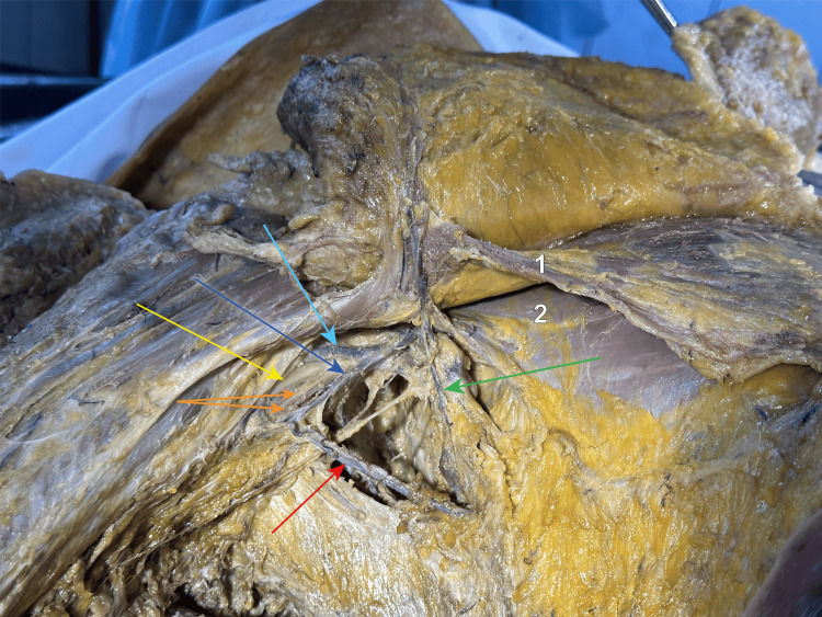
The primary treatment of breast cancer in sentinel-positive ganglia includes axillary lymphatic nodal dissection. The LAD (lymphatic axillary dissection) has decreased in overall numbers but due to the increasing incidence of breast cancer, it is practised on a daily basis, even though there is a myriad of complications such as numbness of the upper limb and chest wall, movement restriction of the upper limb, and chronic pain which appear due to trauma to the nerves which pass through the axilla. However, the utility in the overall survival or DFS (disease-free survival) of the patient is unquestionable. In our study, through the dissection of cadavers, we exposed the vital structures and the anatomical relations of this region. We aimed to offer a map or technique for the surgeon to follow to decrease the overall morbidity of this procedure.

Keywords: breast and endocrine surgery; breast conservation therapy; breast disease; en bloc breast implant removal; thoracic and breast oncology - areas of interest.


Conflict of interest statement

The authors have declared that no competing interests exist.

Figures

Figure 1
Figure 1. Long thoracic nerve on the medial wall of the axilla after the skin was removed
We can observe the trajectory of the long thoracic nerve after the skin and subcutaneous tissue were removed which courses on the medial wall of the axilla, anterior to the serratus muscle (dotted blue line). It is enveloped in a sheath which offers protection against trauma and lesions, thus making this region a good place to start the dissection. Yellow star - lateral thoracic wall covered by the serratus muscle. Red star – large pectoral muscle.
Figure 2
Figure 2. The right axilla is viewed from inferior and lateral
We can observe three intercostobrachial nerves, indicated by the red arrows. They provide sensitive innervation to the skin of the axilla, as well as to the skin on the upper medial aspect of the arm. The yellow arrow points towards the basillic vein ending into the axillary vein. 1 - pectoralis major muscle; 2 - pectoralis minor muscle; Red arrows - three intercostobrachial nerves; Yellow arrow - ending point of the basillic vein.
Figure 3
Figure 3. Close-up view of the axilla from inferior to superior
We dissected into the axilla, sacrificing the anastomosis between the lateral thoracic and the axillary veins, up to the angle between the serratus anterior and the latissimus dorsi muscles, where we identified and individualised the thoracodorsal neurovascular bundle, showed by the green arrow, that runs between the two muscles. The thoracodorsal vein goes posterior to the nerve and underneath the intercostobrachial nerves and drains into the axillary vein (dark blue arrow). The purple arrow points towards the long thoracic nerve, running anterior to the thoracodorsal neurovascular bundle, along with the lateral thoracic artery and vein. The light blue arrow shows the cephalic vein, running in the deltopectoral groove between the deltoid (1) and pectoralis major (2) muscles. The orange arrows show the medial anterbachial and brachial cutaneous nerves and the yellow one indicates the median nerve. 1 - deltoid muscle; 2 - pectoralis major muscle; 3 - pectoralis minor muscle; 4 - serratus anterior muscle; Light blue arrow - cephalic vein in the deltopectoral groove; Yellow arrow - median nerve; Orange arrows - medial brachial and antebrachial cutaneous nerves; Dark blue arrow - axillary vein; Green arrow - thoracodorsal neurovascular bundle; Purple arrow - long thoracic nerve.
Figure 4
Figure 4. View of the axilla apex after the adipose and lymphatic tissues were removed
The axilla after we continued the dissection towards its apex, where we entered by resecting the humeral insertion of the pectoralis minor muscle (2, folded medially). Here we can observe the lateral (red arrow) and medial (green arrow) bundles of the brachial plexus, flanking the axillary artery (red dotted lines follow its pathway), from which a lateral and a medial root emerge and form the median nerve (yellow arrow). Two collateral nerves of the brachial plexus emerge from the lateral and the medial bundles: the lateral (dark blue) and medial (light blue) pectoral nerves. They form an anastomosis that is located anterior and medial to the axillary artery, via the ansa pectoralis, outlined by the yellow lines. 1 - pectoralis major muscle, folded supero-medially; 2 - pectoralis minor muscle, folded medially; 3 - axillary vein; 4 - serratus anterior muscle; Red dotted lines - axillary artery; Red arrow - lateral bundle of the brachial plexus; Green arrow - medial bundle of the brachial plexus; Yellow arrow - median nerve; Dark blue arrows - lateral pectoral nerve; Light blue arrows - medial pectoral nerve; Yellow lines - ansa pectoralis
Figure 5
Figure 5. Overview of the axilla, location and surrounding elements
We can observe venous anastomoses at the level of the right axilla. Indicated by the dark blue arrow, the axillary vein is the most medial element of the main neurovascular bundle of the axilla and the ending point of the axillary lymphadenectomy that includes the Berg stations I and II. Here we can observe the basilic vein, shown by the light blue arrow, that enters the axilla and ends into the axillary vein. The green arrow points towards the lateral thoracic vein, which lies on the lateral thoracic wall and ends into the axillary vein. The red arrow indicates an anastomotic branch between the lateral thoracic and the axillary veins, running on the base of the axilla. The existence of this branch demonstrates that the venous drainage at the level of the axilla is network-shaped, with numerous collateral drainage pathways that ensure a proper venous flow from the upper limb and from the lateral thoracic wall. The yellow arrow shows the median nerve, while the two orange ones show the medial cut. 1 - pectoralis major muscle; 2 - pectoralis minor muscle; Dark blue arrow - axillary vein; Light blue arrow - basillic vein; Green arrow - lateral thoracic vein; Red arrow - anastomosis between the lateral thoracic and the axillary vein; Yellow arrow - median nerve; Orange arrows - medial brachial and antebrachial cutaneous nerves.

References

    1. Breast cancer-epidemiology, risk factors, classification, prognostic markers, and current treatment strategies-an updated review. Łukasiewicz S, Czeczelewski M, Forma A, Baj J, Sitarz R, Stanisławek A. Cancers (Basel) 2021;13 - PMC - PubMed
    1. The potential of breast cancer screening in Europe. Zielonke N, Kregting LM, Heijnsdijk EA, et al. Int J Cancer. 2021;148:406–418. - PMC - PubMed
    1. Sentinel lymph node methods in breast cancer. Giammarile F, Vidal-Sicart S, Paez D, et al. Semin Nucl Med. 2022;52:551–560. - PubMed
    1. Controversies in axillary treatment of breast cancer patients and metastatic sentinel lymph node. Novoa AG, Nebril BA. J Cancer Sci Ther. 2016;8:66–68.
    1. The decline of axillary lymph node dissection in breast cancer. Evolution of its indication over the last 20 years. García-Novoa A, Acea-Nebril B, Casal-Beloy I, et al. Cir Esp (Engl Ed) 2019;97:222–229. - PubMed

LinkOut - more resources