

Guide to serial synchrotron crystallography
- PMID: 38371325
- PMCID: PMC10869752
- DOI: 10.1016/j.crstbi.2024.100131
Guide to serial synchrotron crystallography
Abstract
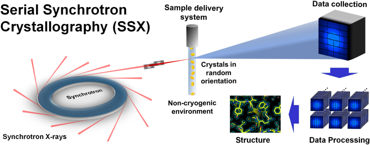
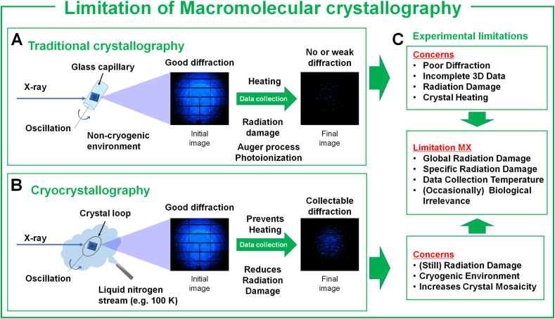
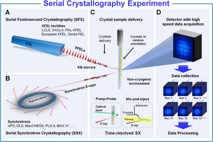
Serial crystallography (SX) is an emerging technique that can be used to determine the noncryogenic crystal structure of macromolecules while minimizing radiation damage. Applying SX using pump-probe or mix-and-inject techniques enables the observation of time-resolved molecular reactions and dynamics in macromolecules. After the successful demonstration of the SX experimental technique with structure determination in serial femtosecond crystallography using an X-ray free electron laser, this method was adapted to the synchrotron, leading to the development of serial synchrotron crystallography (SSX). SSX offers new opportunities for researchers to leverage SX techniques, contributing to the advancement of structural biology and offering a deeper understanding of the structure and function of macromolecules. This review covers the background and advantages of SSX and its experimental approach. It also discusses important considerations when conducting SSX experiments.
Keywords: Radiation damage; Room temperature; Serial crystallography; Serial synchrotron crystallography; Time-resolved study; X-ray crystallography.
© 2024 The Author.
Conflict of interest statement
The authors declare that they have no known competing financial interests or personal relationships that could have appeared to influence the work reported in this paper.
Figures
Similar articles
-
Sample Delivery Media for Serial Crystallography.Int J Mol Sci. 2019 Mar 4;20(5):1094. doi: 10.3390/ijms20051094. Int J Mol Sci. 2019. PMID: 30836596 Free PMC article. Review.
-
A simple goniometer-compatible flow cell for serial synchrotron X-ray crystallography.J Appl Crystallogr. 2023 Mar 9;56(Pt 2):449-460. doi: 10.1107/S1600576723001036. eCollection 2023 Apr 1. J Appl Crystallogr. 2023. PMID: 37032973 Free PMC article.
-
Lard Injection Matrix for Serial Crystallography.Int J Mol Sci. 2020 Aug 19;21(17):5977. doi: 10.3390/ijms21175977. Int J Mol Sci. 2020. PMID: 32825186 Free PMC article.
-
High-viscosity injector-based pink-beam serial crystallography of microcrystals at a synchrotron radiation source.IUCrJ. 2019 Apr 5;6(Pt 3):412-425. doi: 10.1107/S205225251900263X. eCollection 2019 May 1. IUCrJ. 2019. PMID: 31098022 Free PMC article.
-
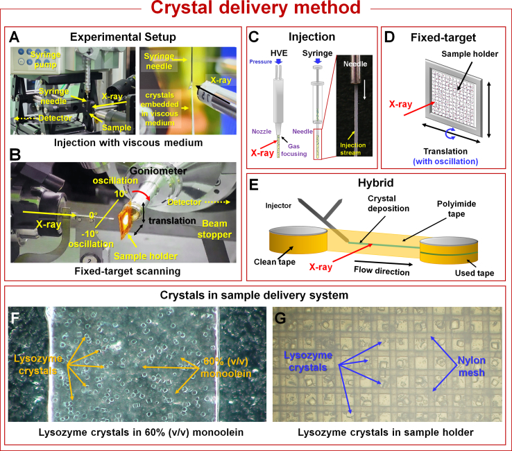
A guide to sample delivery systems for serial crystallography.FEBS J. 2019 Nov;286(22):4402-4417. doi: 10.1111/febs.15099. Epub 2019 Nov 6. FEBS J. 2019. PMID: 31618529 Review.
Cited by
-
Status of macromolecular crystallography beamlines at SSRF.J Synchrotron Radiat. 2025 Jan 1;32(Pt 1):234-245. doi: 10.1107/S1600577524011597. Epub 2025 Jan 1. J Synchrotron Radiat. 2025. PMID: 39705252 Free PMC article.
-
Effect of crystal-to-detector distance shift on data processing in serial crystallography.PLoS One. 2025 Jun 26;20(6):e0327019. doi: 10.1371/journal.pone.0327019. eCollection 2025. PLoS One. 2025. PMID: 40569973 Free PMC article.
-
Novel polymer fixed-target microfluidic platforms with an ultra-thin moisture barrier for serial macromolecular crystallography.bioRxiv [Preprint]. 2025 Jul 18:2025.07.13.663488. doi: 10.1101/2025.07.13.663488. bioRxiv. 2025. PMID: 40791522 Free PMC article. Preprint.
-
Scalable fabrication of an array-type fixed-target device for automated room temperature X-ray protein crystallography.Sci Rep. 2025 Jan 2;15(1):334. doi: 10.1038/s41598-024-83341-3. Sci Rep. 2025. PMID: 39747265 Free PMC article.
References
Publication types
LinkOut - more resources
Full Text Sources
