

Model-based correction of rapid thermal confounds in fluorescence neuroimaging of targeted perturbation
- PMID: 38371339
- PMCID: PMC10871046
- DOI: 10.1117/1.NPh.11.1.014413
Model-based correction of rapid thermal confounds in fluorescence neuroimaging of targeted perturbation
Abstract
Significance: An array of techniques for targeted neuromodulation is emerging, with high potential in brain research and therapy. Calcium imaging or other forms of functional fluorescence imaging are central solutions for monitoring cortical neural responses to targeted neuromodulation, but often are confounded by thermal effects that are inter-mixed with neural responses.
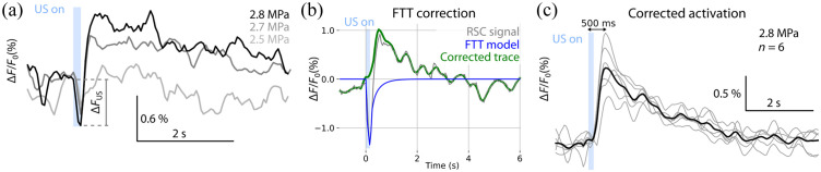
Aim: Here, we develop and demonstrate a method for effectively suppressing fluorescent thermal transients from calcium responses.
Approach: We use high precision phased-array 3 MHz focused ultrasound delivery integrated with fiberscope-based widefield fluorescence to monitor cortex-wide calcium changes. Our approach for detecting the neural activation first takes advantage of the high inter-hemispheric correlation of resting state dynamics and then removes the ultrasound-induced thermal effect by subtracting its simulated spatio-temporal signature from the processed profile.
Results: The focused -sized ultrasound stimulus triggered rapid localized activation events dominated by transient thermal responses produced by ultrasound. By employing bioheat equation to model the ultrasound heat deposition, we can recover putative neural responses to ultrasound.
Conclusions: The developed method for canceling transient thermal fluorescence quenching could also find applications with optical stimulation techniques to monitor thermal effects and disentangle them from neural responses. This approach may help deepen our understanding of the mechanisms and macroscopic effects of ultrasound neuromodulation, further paving the way for tailoring the stimulation regimes toward specific applications.
Keywords: calcium imaging; mouse brain; neuroimaging; thermal effects; ultrasound neuromodulation.
© 2024 The Authors.
Figures


Similar articles
-
High-resolution fluorescence-guided transcranial ultrasound mapping in the live mouse brain.Sci Adv. 2021 Dec 10;7(50):eabi5464. doi: 10.1126/sciadv.abi5464. Epub 2021 Dec 8. Sci Adv. 2021. PMID: 34878843 Free PMC article.
-
A review of the bioeffects of low-intensity focused ultrasound and the benefits of a cellular approach.Front Physiol. 2022 Nov 10;13:1047324. doi: 10.3389/fphys.2022.1047324. eCollection 2022. Front Physiol. 2022. PMID: 36439246 Free PMC article. Review.
-
Endocavitary thermal therapy by MRI-guided phased-array contact ultrasound: experimental and numerical studies on the multi-input single-output PID temperature controller's convergence and stability.Med Phys. 2009 Oct;36(10):4726-41. doi: 10.1118/1.3215534. Med Phys. 2009. PMID: 19928104
-
Implantable acousto-optic window for monitoring ultrasound-mediated neuromodulation in vivo.Neurophotonics. 2022 Jul;9(3):032203. doi: 10.1117/1.NPh.9.3.032203. Epub 2022 Jul 20. Neurophotonics. 2022. PMID: 35874142 Free PMC article.
-
Photoacoustic: A Versatile Nongenetic Method for High-Precision Neuromodulation.Acc Chem Res. 2024 Jun 4;57(11):1595-1607. doi: 10.1021/acs.accounts.4c00119. Epub 2024 May 17. Acc Chem Res. 2024. PMID: 38759211 Free PMC article. Review.
Cited by
-
Special Section Guest Editorial: Frontiers in Neurophotonics.Neurophotonics. 2024 Jan;11(1):014401. doi: 10.1117/1.NPh.11.1.014401. Epub 2024 Mar 28. Neurophotonics. 2024. PMID: 38550388 Free PMC article.
-
Holographic transcranial ultrasound neuromodulation enhances stimulation efficacy by cooperatively recruiting distributed brain circuits.Nat Biomed Eng. 2025 Jul 7. doi: 10.1038/s41551-025-01449-x. Online ahead of print. Nat Biomed Eng. 2025. PMID: 40624336
-
Two-photon imaging of excitatory and inhibitory neural response to infrared neural stimulation.Neurophotonics. 2024 Apr;11(2):025003. doi: 10.1117/1.NPh.11.2.025003. Epub 2024 May 24. Neurophotonics. 2024. PMID: 38800606 Free PMC article.
-
Infrared neuroglial modulation of spinal locomotor networks.Sci Rep. 2024 Sep 27;14(1):22282. doi: 10.1038/s41598-024-73577-4. Sci Rep. 2024. PMID: 39333287 Free PMC article.
References
Grants and funding
LinkOut - more resources
Full Text Sources
Miscellaneous
