Changes in lumbar muscle diffusion tensor indices with age
- PMID: 38371493
- PMCID: PMC10873271
- DOI: 10.1093/bjro/tzae002
Changes in lumbar muscle diffusion tensor indices with age
Abstract
Objective: To investigate differences in diffusion tensor imaging (DTI) parameters and proton density fat fraction (PDFF) in the spinal muscles of younger and older adult males.
Methods: Twelve younger (19-30 years) and 12 older (61-81years) healthy, physically active male participants underwent T1W, T2W, Dixon and DTI of the lumbar spine. The eigenvalues (λ1, λ2, and λ3), fractional anisotropy (FA), and mean diffusivity (MD) from the DTI together with the PDFF were determined in the multifidus, medial and lateral erector spinae (ESmed, ESlat), and quadratus lumborum (QL) muscles. A two-way ANOVA was used to investigate differences with age and muscle and t-tests for differences in individual muscles with age.
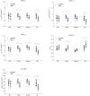
Results: The ANOVA gave significant differences with age for all DTI parameters and the PDFF (P < .01) and with muscle (P < .01) for all DTI parameters except for λ1 and for the PDFF. The mean of the eigenvalues and MD were lower and the FA higher in the older age group with differences reaching statistical significance for all DTI measures for ESlat and QL (P < .01) but only in ESmed for λ3 and MD (P < .05).
Conclusions: Differences in DTI parameters of muscle with age result from changes in both in the intra- and extra-cellular space and cannot be uniquely explained in terms of fibre length and diameter.
Advances in knowledge: Previous studies looking at age have used small groups with uneven age spacing. Our study uses two well defined and separated age groups.
Keywords: DTI; ageing; fat fraction; muscle.
© The Author(s) 2024. Published by Oxford University Press on behalf of the British Institute of Radiology.
Conflict of interest statement
None declared.
Figures


References
-
- Boutin RD, Yao L, Canter RJ, Lenchik L.. Sarcopenia: current concepts and imaging implications. AJR Am J Roentgenol. 2015;205(3):W255-W266. - PubMed
-
- Rosenberg IH. Sarcopenia: origins and clinical relevance. J Nutr. 1997;127(5 Suppl):990S-991S. - PubMed
-
- Janssen I, Heymsfield S, Ross R.. Low relative skeletal muscle mass (sarcopenia) in older persons is associated with functional impairment and physical disability. J Am Geriatr Soc. 2002;50(5):889-896. - PubMed
-
- Oh J, Jung J, Ko Y.. Can diffusion tensor imaging and tractography represent cross-sectional area of lumbar multifidus in patients with lumbar spine disease? Muscle Nerve. 2018;57(2):200-205. - PubMed
LinkOut - more resources
Full Text Sources

