Review
. 2024 May;121(5):895-911.
doi: 10.1111/mmi.15241. Epub 2024 Feb 19.

Bacteriophage lambda site-specific recombination

Affiliations
Review

Bacteriophage lambda site-specific recombination

Gregory D Van Duyne et al. Mol Microbiol. 2024 May.

Abstract

The site-specific recombination pathway of bacteriophage λ encompasses isoenergetic but highly directional and tightly regulated integrative and excisive reactions that integrate and excise the vial chromosome into and out of the bacterial chromosome. The reactions require 240 bp of phage DNA and 21 bp of bacterial DNA comprising 16 protein binding sites that are differentially used in each pathway by the phage-encoded Int and Xis proteins and the host-encoded integration host factor and factor for inversion stimulation proteins. Structures of higher-order protein-DNA complexes of the four-way Holliday junction recombination intermediates provided clarifying insights into the mechanisms, directionality, and regulation of these two pathways, which are tightly linked to the physiology of the bacterial host cell. Here we review our current understanding of the mechanisms responsible for regulating and executing λ site-specific recombination, with an emphasis on key studies completed over the last decade.

Keywords: DNA bending; Holliday junction; bacteriophage lambda; integration; site‐specific recombination; tyrosine recombinase; viral excision; viral integration.


Figures

Figure 1.
Figure 1.
Life styles of bacteriophage lambda. During infection, the phage particle binds to a bacterial host cell and injects its linear DNA genome into the cell. Host factors circularize and supercoil the lambda DNA. Depending upon regulatory signals from the phage and the bacterial cell the phage proceeds down the lytic or lysogenic path. In the former the phage DNA is transcribed, replicated, and packaged into new phage particles that are released from the lysed cell. In the lysogenic pathway the phage DNA is integrated into the host chromosome at a specific site using the phage-encoded integrase (Int) enzyme and the host integration host factor (IHF) protein. Lambda can then undergo lysogenic growth, where almost all of the viral genes are repressed and the prophage is replicated along with the host chromosome. During lysogenic growth signals from the environment and/or the host cause induction of the prophage and excision from the host chromosome (not shown in this figure). The excision reaction is catalyzed by Int, IHF, and the phage-encoded Xis protein. Transcription of lambda genes to produce new phage particles and to lyse the cell represent the lytic phase of growth. The phage DNA is drawn in red in this figure.
Figure 2.
Figure 2.
Site-specific integration of λ DNA into and excision out of the bacterial chromosome. Integration occurs between the phage attP site (C’-O-C) and the bacterial attB site (B-O-B’). The hybrid sites C’-O-B and C-O-B’ that are generated are called attL and attR, respectively. During excision, recombination between attL and attR sites results in free phage DNA containing attP and the unaltered bacterial chromosome. Additional binding sites for Int, IHF, and Xis located on the attP “arms” that flank C and C’ are not shown here. The full attP site is 240 bp in length, vs. the minimal 21 bp core attB site.
Figure 3.
Figure 3.
Generation of transducing phage λ. Inefficient and rare integrations at a degenerate mimic of the canonical attB site (called a secondary att sites; attA in this figure) positions the λ prophage adjacent to E. coli genes X and Y. A rare illegitimate recombination, involving sequences with very little homology, between a site on the bacterial chromosome and a site in a nonessential region of the viral chromosome results in the formation of a viral chromosome linked to an adjacent segment of bacterial DNA, in this case gene X. This new transuding phage lacks the A’OC site and some adjacent nonessential viral sequences. The resulting phage can be used to transfer gene X into new host cells.
Figure 4.
Figure 4.
The protein binding sites that comprise complete att DNA sites. Integrative recombination between supercoiled attP and linear attB requires Int and the host-encoded IHF and gives rise to an integrated phage chromosome bounded by attL and attR. Excisive recombination between attL and attR to regenerate attP and attB additionally requires the phage-encoded Xis protein (which inhibits integrative recombination) and is stimulated by the host-encoded Fis protein. Both reactions proceed through the stepwise cleavage and strand exchange pathway shown in Figure 8. Integration and excision use different subsets of protein binding sites in the P and P′ arms, as indicated by the filled boxes: Int arm-type P1, P2, P′1, P′2, and P′3 (light green); IHF, H1, H2, and H′ (yellow); Xis, X1, X1.5, and X2 (gold); and Fis (pink). The four core-type Int binding sites, C, C′, B, and B′ (dark green boxes) are each bound by the core-binding and catalytic domains. The integration reaction requires four Int proteins bridging between core and arm binding sites, as shown in Figures 9 & 10. The excision reaction requires four Int proteins binding to core sites, but only three bridges to arm sites are required.
Figure 5.
Figure 5.
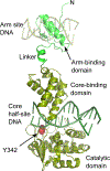
Structure of heterobivalent Int bound to arm- and core-type DNA sites. The conserved catalytic tyrosine responsible for cleavage of the core sites is shown in red and is poised to cleave at the edges of the overlap region of the core site. Based on pdb entry 1Z1G (Biswas et al., 2005). The N-terminal arm-binding domain of Int binds to one of the five arm-type DNA sites located outside of the core DNA region while the C-terminal core-binding and catalytic domains form a clamp around each core half-site sequence. The Int protein thus forms intra- and inter-molecular bridges between the core- and arm-type sites facilitated by the accessory DNA bending proteins (as shown in Figures 8, 9, & 10).
Figure 6.
Figure 6.
Structure of integration host factor (IHF) bound to H’ DNA site. IHF is a tightly associated heterodimer composed of α and β subunits. β-hairpins from each subunit create sharp kinks in the DNA separated by 9-bp. The result is an overall bend in the DNA of ~160°. The H’ sequence (one of the three IHF binding sites, as shown in Figure 8) provides both specific IHF interactions and non-specific conformational flexibility in the DNA. Modified from pdb entry 1IHF (Rice et al., 1996).
Figure 7.
Figure 7.
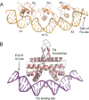
Structures of Xis and Fis bound to DNA. A) Three Xis molecules bind cooperatively to successive sites (named X1, X1.5, and X2) to form a mini-filament on DNA. Xis binding results in a smooth bend in the DNA. Based on pdb entry 2IEF (Abbani et al., 2007). B) Fis binds as a homodimer to its DNA binding site, resulting in a bend similar in magnitude to that seen with Xis. Based on pdb entry 3IV5 (Stella, Cascio and Johnson, 2010). In the λ P-arm, the Fis and Xis binding sites are adjacent, allowing for synergy between Fis binding and recruitment of the first Xis subunit at X2 (see Fig 8).
Figure 8.
Figure 8.
Biochemistry of recombination. Isoenergetic cleaving, recombining, and resealing DNA during recombination proceeds by two pairs of sequential single-strand DNA exchanges shown here as steps i->iii and steps iii->v. The phosphodiester linkages to be cleaved (indicated by solid circles) are staggered by seven base pairs that are identical in the substrate and product sites; they are referred to as the ‘overlap’ region (O). Viral and bacterial DNAs are colored as in Figures 1–3. (i) The attP (C’OC) and attB (BOB’) sites are aligned anti-parallel with respect to their identical overlap sequences. (ii) The Int subunits bound to the C and B core sites cleave the DNA to form covalent 3’-phosphotyrosine linkages, exposing free 5’-hydroxyl groups. The liberated DNA strands can melt away from their complementary strands and migrate to the identical complementary strand of the partner substrate. (iii) The 5’-OH groups attack the phosphotyrosine linkages at C’ and B to generate new DNA strands and form the four-way DNA (HJ) intermediate. (iv) A similar pair of single strand cleavage and exchange steps is executed by the Int subunits at C’ and B’ on the other sides of the overlap regions. (v) After resealing the nicked DNA strands following strand exchange, the HJ is resolved to recombinant products, attL (C’OB) and attR (COB’), and all four DNA strands have new junction sequences. In both integrative and excisive recombination λ Int always initiates cleavage at the C and B sites, which means that excision is not simply the reverse of integration, i.e., they are two distinct reactions.
Figure 9.
Figure 9.
Model for how DNA-bending proteins facilitate an Int bridge. A protein such as IHF can bring arm-binding and core-binding sites into proximity so that a molecule of Int can interact with both sites using its N-terminal arm-binding domain and its C-terminal core-binding and catalytic domains.
Figure 10.
Figure 10.
Schematic mapping of the bridges Int makes between core half-sites and arm-binding sites during integrative and excisive recombination. The P’1 and P’2 arm sites are used in both pathways. The P1 and P’3 sites are used only during integration and the P2 site is used only during excision.
Figure 11.
Figure 11.
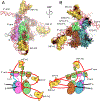
Structure of the excisive Holliday junction complex. Coloring in the structures and in the schematic diagrams below follow the coloring used in Figure 10. Int CTDs are labeled according to the core half-site to which they are bound (B, B’, C, C’). A) View down the ‘top’ of the complex, towards the arm-binding domains (NTDs) of Int. The Int NTDs binding to the P’1, P’2, and P2 sites are indicated. The three Xis molecules bound to the P-arm are also indicated. This view highlights the sharp IHF-mediated DNA bends at H’ that delivers the P’1 and P’2 sites into bridging proximity of the core sites. The center of the DNA bend at H’ is approximately 100 Å from the center of the core sites. This view also illustrates the close association between the Int NTDs bound at P’1 and P’2 with Xis bound at the X1 and X1.5 sites. The close contact between Xis1 and the NTD bound at P2 can also be seen. B) Side view showing the bend of the P-arm in the excisive complex. The IHF-mediated bend at H2 turns the P-arm upwards and Xis provides the bend across the top of the complex to position P2 within bridging distance of the core site. The center of the DNA bend at H2 is approximately 120 Å from the center of the core binding sites. This view also provides an unobscured example of an Int bridging interaction, where the protomer bound at the B’ core site bridges to bind the P2 site.
Figure 12.
Figure 12.
A model for the integrative complex. Coloring and labels in the structures and in the schematic diagrams below follow the coloring used in Figures 10 & 11. A) View along the P-arm showing the upward trajectory of the P-arm following the IHF bend at H2. The DNA reverses course at the H1 site where IHF redirects the P-arm downwards towards the Int tetramer. The P-arm crosses over the P’-arm as a result of the bend at H1. B) View from the opposite side of the complex. The P’-arm engages three Int NTDs at the P’1, P’2, and P’3 sites which bridge to the C’, C, and B’ core sites, respectively. The P-arm can be seen crossing over the P’-arm in this orientation, where the P1 binding site is positioned to bind an Int NTD that bridges to the B core site. During the intitial steps of integration, Int will be tightly bound at P1 and P’3 and the respective CTDs of Int will test for the correct attB sequence in the E. coli chromosome. Labels and coloring schemes follow that shown in Figure 11.

Similar articles

Cited by

References

    1. Abbani M. et al. (2007) Structure of the cooperative Xis-DNA complex reveals a micronucleoprotein filament that regulates phage lambda intasome assembly, Proc Natl Acad Sci U S A, 104, 2109–14. - PMC - PubMed
    1. Abremski K. and Gottesman S. (1982) Purification of the bacteriophage lambda xis gene product required for lambda excisive recombination, The Journal of Biological Chemistry, 257, 9658–9662. - PubMed
    1. Aihara H. et al. (2003) A conformational switch controls the DNA cleavage activity of lambda integrase, Mol Cell, 12, 187–98. - PubMed
    1. Ball CA et al. (1992) Dramatic changes in Fis levels upon nutrient upshift in Escherichia coli, Journal of Bacteriology, 174, 8043–8056. - PMC - PubMed
    1. Ball CA and Johnson RC (1991) Efficient excision of phage lambda from the Escherichia coli chromosome requires the Fis protein, Journal of Bacteriology, 173, 4027–4031. - PMC - PubMed

Publication types

MeSH terms