. 2024 Mar 20;481(6):437-460.
doi: 10.1042/BCJ20230406.

Novel modifications of PARP inhibitor veliparib increase PARP1 binding to DNA breaks

Affiliations

Novel modifications of PARP inhibitor veliparib increase PARP1 binding to DNA breaks

Uday Kiran Velagapudi et al. Biochem J. .

Abstract

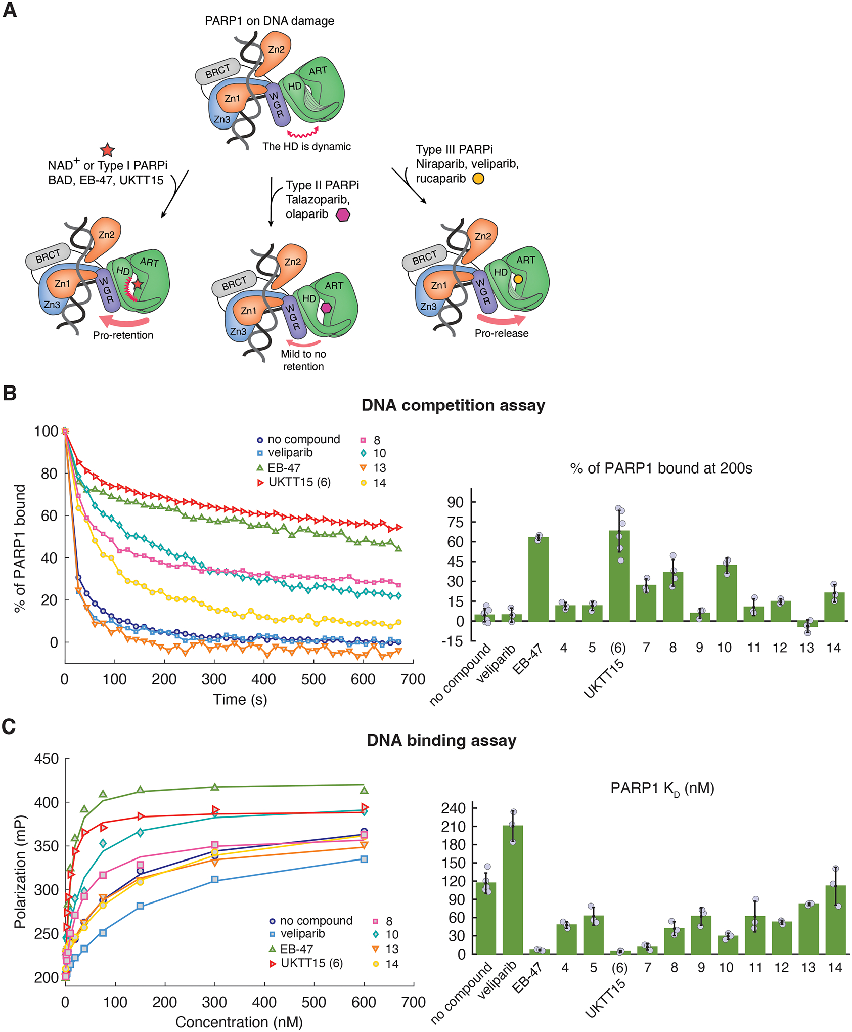
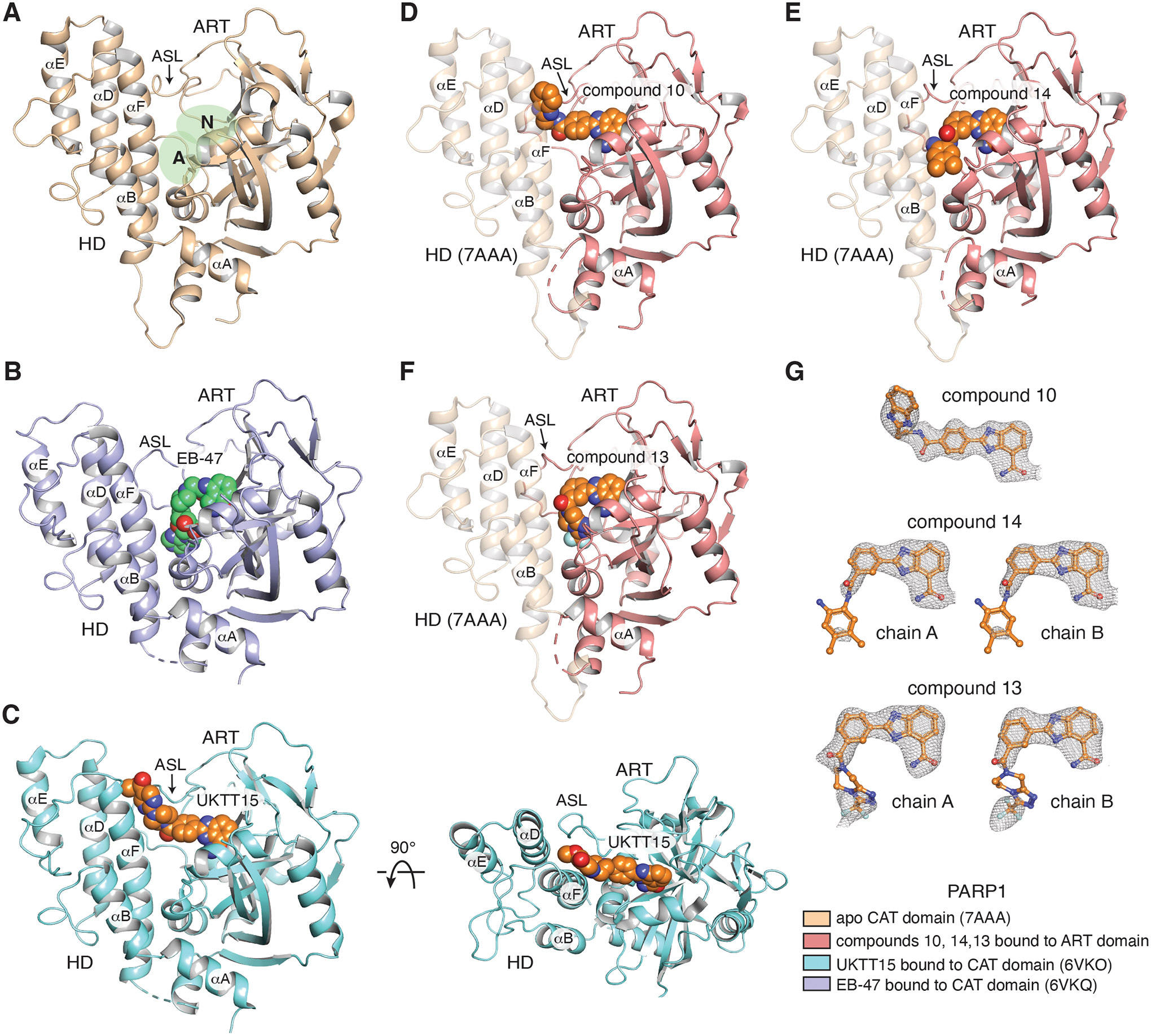
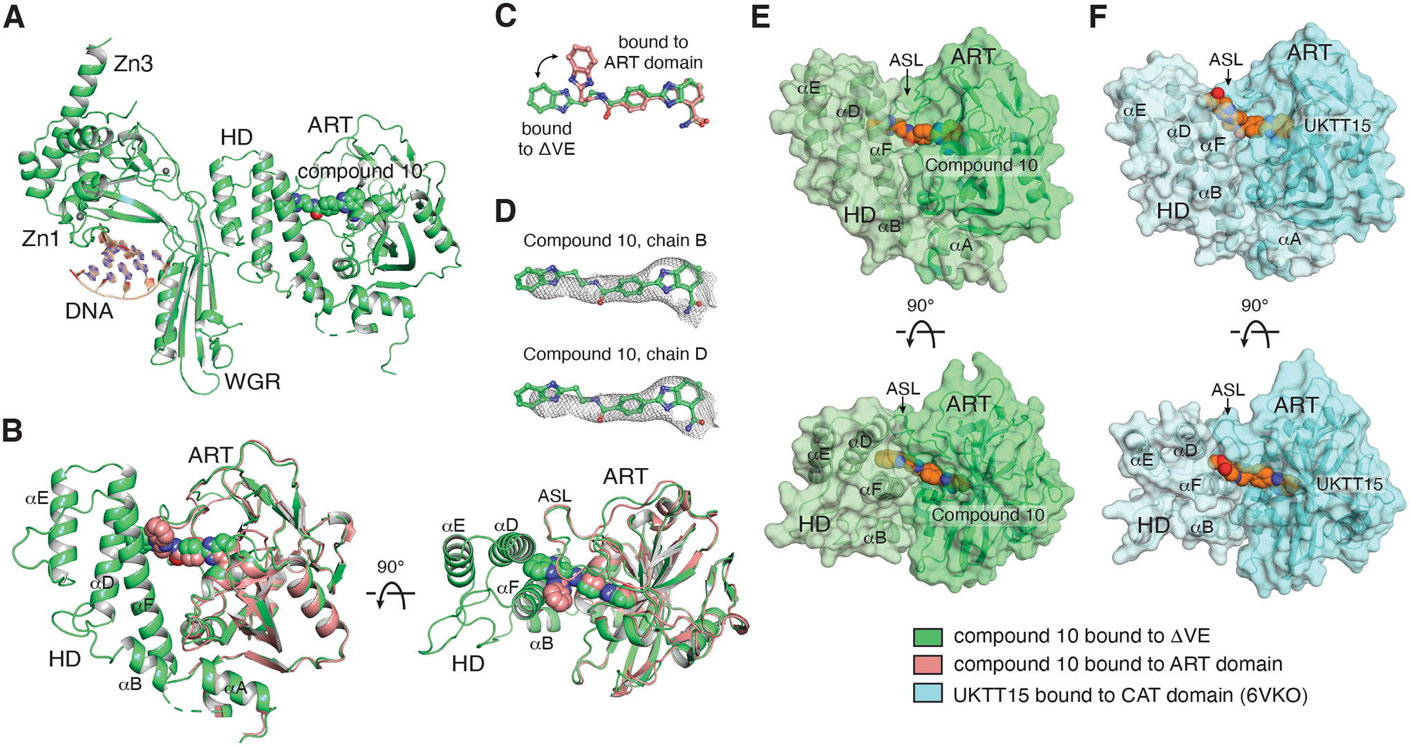
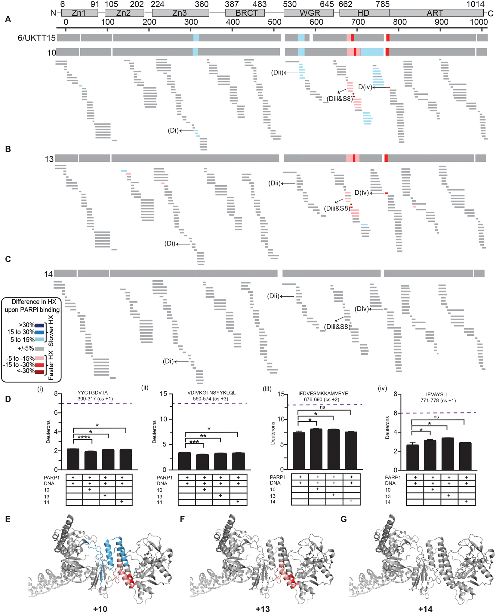
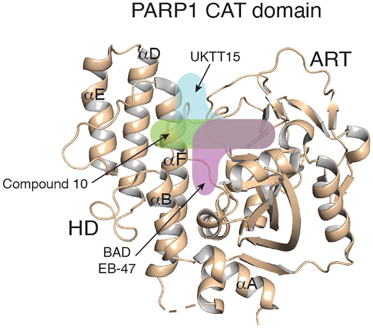
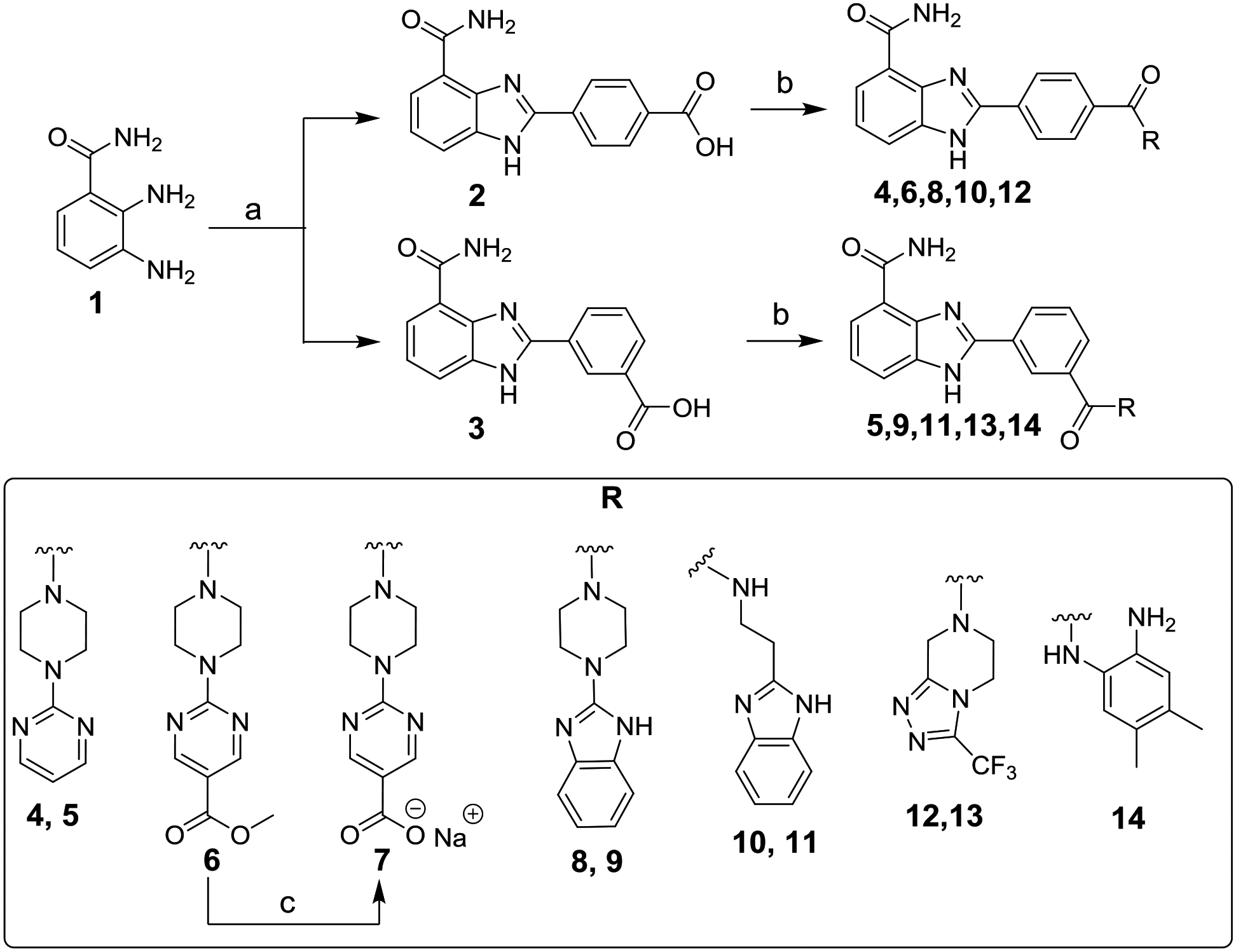
Catalytic poly(ADP-ribose) production by PARP1 is allosterically activated through interaction with DNA breaks, and PARP inhibitor compounds have the potential to influence PARP1 allostery in addition to preventing catalytic activity. Using the benzimidazole-4-carboxamide pharmacophore present in the first generation PARP1 inhibitor veliparib, a series of 11 derivatives was designed, synthesized, and evaluated as allosteric PARP1 inhibitors, with the premise that bulky substituents would engage the regulatory helical domain (HD) and thereby promote PARP1 retention on DNA breaks. We found that core scaffold modifications could indeed increase PARP1 affinity for DNA; however, the bulk of the modification alone was insufficient to trigger PARP1 allosteric retention on DNA breaks. Rather, compounds eliciting PARP1 retention on DNA breaks were found to be rigidly held in a position that interferes with a specific region of the HD domain, a region that is not targeted by current clinical PARP inhibitors. Collectively, these compounds highlight a unique way to trigger PARP1 retention on DNA breaks and open a path to unveil the pharmacological benefits of such inhibitors with novel properties.

Keywords: HXMS; PARP; allosteric regulation; crystallography; veliparib analogs.


Conflict of interest statement

Competing interests

B.E.B., J.M.P., and T.T.T. are co-founders of Hysplex, Inc. with interests in PARP inhibitor development, B.E.B is on the scientific advisory board of Denovicon Therapeutics.

Figures

Figure 1.
Figure 1.. Veliparib core scaffold modifications modulate PARP1 allostery.
(A) PARP1 binding to DNA damage allosterically renders the HD subdomain dynamic and reveals the active site (top). The active site occupancy modulates PARP1 allostery, with substrate NAD+ or Type I inhibitors promoting PARP1 retention to breaks (left). Type II inhibitors mildly or do not promote retention (middle) and type III inhibitors promote PARP1 release from breaks (right). (B) (left panel) Representative FP DNA competition assay that measures PARP1 retention on a fluorescently labeled DNA break in the presence of unlabeled competitor DNA. UKTT15 (6) increases PARP1 retention on DNA damage, similar to EB-47, and in contrast to PARP1 in the absence of inhibitor. Compounds 8, 10, and 14 have a milder effect while compound 13 does not appear to promote PARP1 retention and might even disfavor retention (see Fig. S5 for additional representative curves). (right panel) The percentage of PARP1 still retained at 200 s is plotted. The bars represent the average values from at least three independent experiments, and the individual measurements are plotted. The error values represent the calculated standard deviations. The values are reported in Table 1 and 2. (C) Representative FP DNA binding assay showing that UKTT15 (6) increases PARP1 DNA binding affinity. Again, compounds 10 and 8 mildly increase PARP1 affinity while compounds 13 and 14 show little to no difference (see Fig. S5 for additional representative curves). The lines represent the fit of a 1:1 binding model to the data. (right panel) KD values derived from the FP DNA binding assay are plotted. The bars represent the average of at least three independent experiments. The reported errors are the calculated standard deviations. The values are reported in Table 1 and 2.
Figure 2.
Figure 2.. Crystal structures of target compounds bound to PARP1 ART domain.
(A) Crystal structure of the human PARP1 CAT domain with no inhibitor bound (beige, PDB: 7AAA). The nicotinamide binding pocket (N) and adenosine binding pocket (A) of the active site are highlighted in green. The HD and ART subdomains are labeled. The location of the active site loop (ASL) is labeled and noted with an arrow. Helices of the HD are labeled. (B) Crystal structure of EB-47 (green) bound to PARP1 CAT domain (purple, PDB: 6VKQ). EB-47 contacts both N and A binding pockets of the active site. (C) (left panel) Crystal structure of UKTT15 (6) (orange) bound to PARP1 CAT domain (blue, PDB: 6VKO). UKTT15 (6) does not contact the A binding pocket, but rather occupies a pocket in between helix αF and αD in the HD and the active site loop (ASL) in the ART domain (right panel). (D, E, F) Crystal structures of compounds 10 (D), 14 (E) and 13 (F) (orange) bound to their respective PARP1 ART domain (pink). The semi-transparent HD of PARP1 is represented after having overlayed the structure of PARP1 CAT domain (beige, PDB: 7AAA). (G) Compounds 10, 14 and 13 with a 2FO-FC weighted electron density contoured at 1 σ overlaid on each copy of the compounds found in their respective structures. The color coding for the displayed structures is shown at the bottom right.
Figure 3.
Figure 3.. Compound 10 contacts helices αD and αF of the HD.
(A) The compound 10/ΔVE structure (green) was determined at 3.9 Å (one of the two complexes in the asymmetric unit is shown). (B) The compound 10/ART domain structure (pink) is superposed on the CAT domain of the ΔVE structure, highlighting that the compound contacts helices αD and αF of the HD following a rearrangement of its benzimidazole moiety (panel C). (D) Compound 10 with a 2FO-FC weighted electron density contoured at 0.6 σ overlaid on the compounds from the two catalytic domain protein chains (B and D) found in the asymmetric unit. (E) Two orthogonal views of the surface representation of the CAT domain structure of compound 10 bound to ΔVE (derived from the structure in panel A), highlighting that the compound substituent is buried at the interface of helices αD and αF and the ASL. Compound 10 is drawn with orange carbons. (F) Two orthogonal views of the surface representation of the CAT domain structure of UKTT15 (6) bound to the CAT domain (blue, PDB 6VKO), highlighting that the compound contributes to a groove in between helices αD and αF and the ASL, and that the compound remains accessible to the solvent relative to compound 10 in panel E. UKTT15 (6) is drawn with orange carbons. The color coding for the displayed structures is shown at the bottom right.
Figure 4.
Figure 4.. Hydrogen/deuterium exchange-mass spectrometry (HXMS).
(A) HXMS difference plots between PARP1/DNA complex and PARP1/DNA/10 complex at 100 s. The consensus HX difference plot for the binding of the 10 and UKTT15 (6) is shown on the top for comparison. (B) HXMS difference plots between PARP1/DNA complex and PARP1/DNA/13 complex at 100 s. The consensus HX difference plot for the binding of the 13 is shown on the top. (C) HXMS difference plots between PARP1/DNA complex and PARP1/DNA/14 complex at 100 s. The consensus HX difference plot for the binding of the 14 is shown on the top. For panels A-C, each thin horizontal bar represents a PARP1 peptide. (D) Panels (i-iv) represents the HX of the representative peptides of Zn3, WGR, αB, and αF for PARP1+DNA, and PARP1+DNA+PARPi. An average from three replicates with SD represented by error bars and asterisks indicating P < 0.05 from two-sided t-test between PARP1+DNA and PARP1+DNA+PARPi is shown. Purple dotted line indicates maxD i.e., number of residues minus first two residues (back-exchange within experimental timescale) and minus number of prolines due to no backbone amide hydrogen. Though no major changes in HX were observed for PARP1 in the presence of DNA and compound 14, the number of deuterons between PARP1+DNA complex and PARP1+DNA+compound 14 are statistically different for some peptides (e.g. Di and Dii) indicating that the PARPi is bound to PARP1 and exhibits some mild allostery which classifies as Type II behavior. (E) Consensus HXMS percentage differences with compounds 10 mapped to the crystal structure of PARP1 on DNA damage (PDB 4DQY). (F) Consensus HXMS percentage differences with compound 13 mapped to the crystal structure of PARP1 on DNA damage (PDB 4DQY). (G) Consensus HXMS percentage differences with compound 14 mapped to the crystal structure of PARP1 on DNA damage (PDB 4DQY).
Figure 5.
Figure 5.. Type I inhibitors modulate PARP1 allostery via different binding poses.
The different trajectories of Type I PARPi are indicated by colored and labeled shapes overlaid on the structure of the PARP1 apo CAT domain (beige, PDB 7AAA).
Scheme 1.
Scheme 1.. Synthesis of Target Compounds 4–14.
Reagents and conditions: (a) 4-formylbenzoic acid for 2 or 3-formylbenzoic acid for 3, NH4OAc, DMF, 100 °C, 6 h, 86–89%; (b) appropriate amine, EtN(i-Pr)2, HBTU, DMF, overnight, 23–63%; (c) NaOH, THF/H2O (1:1), rt, overnight, 49%.

Similar articles

Cited by

References

    1. De Vos M, Schreiber V and Dantzer F (2012) The diverse roles and clinical relevance of PARPs in DNA damage repair: current state of the art. Biochem Pharmacol. 84, 137–146. doi: 10.1016/j.bcp.2012.03.018. - DOI - PubMed
    1. Gibson BA and Kraus WL (2012) New insights into the molecular and cellular functions of poly(ADP-ribose) and PARPs. Nat Rev Mol Cell Biol. 13, 411–424. doi: 10.1038/nrm3376. - DOI - PubMed
    1. Ray Chaudhuri A and Nussenzweig A (2017) The multifaceted roles of PARP1 in DNA repair and chromatin remodelling. Nat Rev Mol Cell Biol. 18, 610–621. doi: 10.1038/nrm.2017.53. - DOI - PMC - PubMed
    1. Alemasova EE and Lavrik OI (2019) Poly(ADP-ribosyl)ation by PARP1: reaction mechanism and regulatory proteins. Nucleic Acids Res. 47, 3811–3827. doi: 10.1093/nar/gkz120. - DOI - PMC - PubMed
    1. Bonfiglio JJ, Fontana P, Zhang Q, Colby T, Gibbs-Seymour I, Atanassov I, Bartlett E, Zaja R, Ahel I and Matic I (2017) Serine ADP-Ribosylation Depends on HPF1. Mol Cell. 65, 932–940 e936. doi: 10.1016/j.molcel.2017.01.003. - DOI - PMC - PubMed

Publication types

MeSH terms