. 2024 Apr:306:122496.
doi: 10.1016/j.biomaterials.2024.122496. Epub 2024 Jan 30.

Bioactive nanomaterials kickstart early repair processes and potentiate temporally modulated healing of healthy and diabetic wounds

Affiliations

Bioactive nanomaterials kickstart early repair processes and potentiate temporally modulated healing of healthy and diabetic wounds

Deepanjan Ghosh et al. Biomaterials. 2024 Apr.

Abstract

Slow-healing and chronic wounds represent a major global economic and medical burden, and there is significant unmet need for novel therapies which act to both accelerate wound closure and enhance biomechanical recovery of the skin. Here, we report a new approach in which bioactives that augment early stages of wound healing can kickstart and engender effective wound closure in healthy and diabetic, obese animals, and set the stage for subsequent tissue repair processes. We demonstrate that a nanomaterial dressing made of silk fibroin and gold nanorods (GNR) stimulates a pro-neutrophilic, innate immune, and controlled inflammatory wound transcriptomic response. Further, Silk-GNR, lasered into the wound bed, in combination with exogeneous histamine, accelerates early-stage processes in tissue repair leading to effective wound closure. Silk-GNR and histamine enhanced biomechanical recovery of skin, increased transient neoangiogenesis, myofibroblast activation, epithelial-to-mesenchymal transition (EMT) of keratinocytes and a pro-resolving neutrophilic immune response, which are hitherto unknown activities for these bioactives. Predictive and temporally coordinated delivery of growth factor nanoparticles that modulate later stages of tissue repair further accelerated wound closure in healthy and diabetic, obese animals. Our approach of kickstarting healing by delivering the "right bioactive at the right time" stimulates a multifactorial, pro-reparative response by augmenting endogenous healing and immunoregulatory mechanisms and highlights new targets to promote tissue repair.


Conflict of interest statement

Declaration of competing interest The authors declare the following financial interests/personal relationships which may be considered as potential competing interests: Kaushal Rege reports financial support was provided by National Institutes of Health and Flinn Foundation. Jordan Robin Yaron reports financial support was provided by National Institutes of Health and Flinn Foundation. Kaushal Rege reports a relationship with Synergyan, LLC and Endotat Biotechnologies, LLC that includes: board membership and equity or stocks. Jordan Robin Yaron reports a relationship with Vivo Bioconsulting, LLC and Endotat Biotechnologies, LLC that includes: board membership and equity or stocks. Kaushal Rege and Deepanjan Ghosh have patent #US20210100927A1 pending to Arizona Board of Regents of ASU.

Figures

Fig. 1.
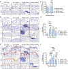
Fig. 1.. Silk-GNR, histamine and their combination accelerate acute wound healing.
(A) Representative wound images of Tegaderm (Teg) + saline (Sal), Silk-GNR + saline, Tegaderm + histamine, and Silk-GNR + histamine wounds on day 0, 4, and 7 post wounding. (B) Daily planimetric measurement of wound areas normalized to day 0 for each mouse. Data shown are mean ± standard error of mean of n = 6 mice per group. Statistical significance was determined using one-way ANOVA with correction for multiple comparisons by Holm-Sidak method. Individual p-values between groups on individual days post wounding are shown using a color-coded heat-map. A change in closure kinetics is seen for silk-GNR, histamine, and the combination of silk-GNR + histamine starting day 5, with the most significant change seen in case of the combination (C) Ultimate tensile strength (UTS) and % recovery of healed skin strength (secondary y-axis) 7-days post wounding. Data shown are mean ± standard error of mean of n = 4 mice per group and the ‘Wound’ group is Tegaderm + saline and ‘Histamine’ is Tegaderm + histamine. Statistical significance was determined using one-way ANOVA with correction for multiple comparisons by Tukey method. Individual p-values between groups are shown in the plot.
Fig. 2.
Fig. 2.. Transcriptomic analyses of wound responses to silk-GNR nanomaterial.
(A) Principal Component Analysis (PCA) of Day 4 wounds treated with Tegaderm (N=3) or silk-GNR (N=2) dressings. (B) K-means clustering heatmap indicating characteristic clusters of genes up- (red) or down- (green) regulated in silk-GNR-treated wounds. (C) Volcano analysis with relevant up- and down-regulated genes indicated. Dots in blue are significant with p<0.05. (D) Selected relevant gene expression responses for silk-GNR which indicate a biomaterial-tissue response of enhanced neutrophil, innate, and inflammatory signaling. (H) Ridgeline GSEA plot of top 20 pathways enriched within the top 1000 differentially expressed genes. Each point on the lines represents a single gene in an indicated pathway. (I) GO pathway enrichment plot of the top 30 pathways significantly enriched among the top 1000 differentially expressed genes.
Fig. 3.
Fig. 3.. Effect of silk-GNR, histamine and their combination modulate innate immune response in wounds.
Representative wound bed micrographs of (A) Arginase-1, (C) iNOS, and (E) Ly-6G-stained IHC fields (20x magnification) on days 2, 4, and 7 post-wounding for all treatments. Scale bar = 200 μm. Quantification of (B) Arginase, (D) iNOS, and (F) Ly-6G positive signals per 20x field. At least four non-overlapping fields were quantified for each group and data shown are mean ± standard error of mean of n = 3 mice per group. Statistical significance was determined using two-way ANOVA with Fisher’s LSD post-hoc analysis. Individual p-values between groups are shown in the plot.
Fig. 4.
Fig. 4.. Enrichment of Ym-1-positive cells in silk-GNR/histamine-treated wounds.
(A) Quantification of % Ym-1 positive signal per 20x field. Data shown are mean ± standard error of mean of n = 3 mice per group. Statistical significance was determined using two-way ANOVA with Fisher’s LSD post-hoc analysis. *p<0.05, **p<0.01. Representative wound bed micrographs of Ym-1 stained IHC field (20x magnification) on day 7 post-wounding are shown (scale bar = 200 μm). (B) Representative IHC serial sections of silk/histamine-treated mouse wounds at 7 days post-wounding stained for neutrophils (Ly6G), N2 neutrophils (Ym-1), N1 neutrophils (CD54), M2 macrophages (Arg-1), and M1 macrophages (iNOS). Scale bar of full field images is 200 μm. Zoomed area is indicated with black box.
Fig. 5.
Fig. 5.. Effect of silk-GNR, histamine and their combination on modulation of EMT in wounds.
(A) Representative micrographs of the epithelial tongue stained for pSTAT3 (20x magnification) at 6 hours and day 2 post wounding for all the groups (scale bar = 200 μm). (B) Quantification of % keratinocytes with positive nuclear pSTAT3 signal per 20x field. (C) Representative micrographs of the epithelial tongue stained for Slug (20x magnification) at 6 hours, day 2, 4, and 7 post wounding for all the groups (scale bar = 200 μm). (D) Quantification of % keratinocytes with positive nuclear Slug signal per 20x field. (E) Representative micrographs of the epithelial tongue stained for E-cadherin (20x magnification) at 6 hours, day 2, 4, and 7 post wounding for all the groups (scale bar = 200 μm). (F) Quantification of number of keratinocytes with loss of E-cadherin signal at the cell boundaries per 20x field. At least four non-overlapping fields were quantified for each group and data shown are mean ± standard error of mean of n = 3 mice per group. Statistical significance was determined using two-way ANOVA with Fisher’s LSD post-hoc analysis. Individual p-values between groups are shown in the plot.
Fig. 6.
Fig. 6.. Effect of silk-GNR, histamine and their combination on myofibroblast response and angiogenesis.
(A) Representative wound bed micrographs of αSMA stained IHC field (10x magnification) on day 4 and 7 post wounding for all the groups (scale bar = 200 μm). (B) Representative wound bed images of CD31 stained IHC field (10x magnification) on day 4 and 7 post wounding for all the groups (scale bar = 200 μm). (C, D) Quantification of αSMA+ and CD31+ pixels per 10x field respectively in the granulation area (wound bed) of wounds treated with Tegaderm (“Teg”) + saline, silk-GNR + saline, Tegaderm + histamine, and silk-GNR + histamine on day 4 and 7 post wounding. At least six non-overlapping fields were quantified for each group and data shown are mean ± standard error of mean of n = 3 mice per group. Statistical significance was determined using two-way ANOVA with Fisher’s LSD post-hoc analysis. Individual p-values between groups are shown in the plot.
Fig. 7.
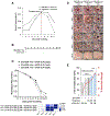
Fig. 7.. Silk-GNR, histamine and their combination promote wound healing in diabetic and obese mice.
(A) Representative wound images of Tegaderm + saline, silk-GNR + saline, Tegaderm + histamine, and silk-GNR + histamine wounds on day 0, 3, 7, and 11 post wounding. (B) Planimetric measurement of wound areas on day 0, 3, 5, 7, 9, and 11 normalized to day 0 for each mouse. Data shown are mean ± standard error, n=4 mice/group. Statistical significance was determined using one-way ANOVA with correction for multiple comparisons by Holm-Sidak method. Individual p-values between groups on individual days post wounding are shown using a color-coded heat-map. (C) Plot showing ultimate tensile strength and % recovery of healed skin strength (secondary y-axis) 11-days post wounding. Data shown are mean ± standard error, n=4 mice/group. Statistical significance was determined using one-way ANOVA with correction for multiple comparisons by Tukey method. Individual p-values between groups are shown in the plot.
Fig. 8.
Fig. 8.. Temporal delivery of second therapeutic along with silk-GNR in diabetic wounds.
(A) Rate-of-healing plot of silk-GNR + histamine and Tegaderm + histamine treated db/db wounds obtained from the healing kinetics in Figure 6B. The day with the maximum healing rate is shown using a red dashed line and indicated as a transition period. Lines connecting the data points are for visualization alone. (B) Experimental schematic indicating on which days silk-GNR + histamine or bFGF-ELP was delivered to wounds. (C) Planimetric measurements of wound area during the healing course normalized to day 0 for each mouse. Data are shown as mean ± standard error, n=3/group. (D) Representative wound images of silk-GNR + histamine treated wounds with bFGF-ELP nanoparticles given on day 0, 3, or 6 post-wounding. (E) Plot showing ultimate tensile strength and % recovery of healed skin strength (secondary y-axis) 7-days post wounding. Data shown are mean ± standard error, n=4 mice/group. Statistical significance was determined using one-way ANOVA with correction for multiple comparisons by Tukey method. Individual p-values between groups are shown in the plot.
Fig. 9.
Fig. 9.. Schematic representation of overarching concepts.
Wounds healing along a continuous spectrum of phases, with hemostasis and inflammation representing the early stages of healing and proliferation and remodeling representing the later stages of healing. Here, we investigated the outcomes of acute (early stage) treatment with a silk-GNR nanomaterial with and without the bioactive immune modulator histamine and found enhanced epidermal EMT and pro-resolution immune responses, particularly in neutrophil populations. Temporally modulated delivery of growth factor nanoparticles (GFNP) in the early-to-late stage transition period further accelerated wound closure and enhanced biomechanical recovery. This study thus demonstrates the principle that rational guidance of treatments can augment healing progress by temporal modulation of therapeutic interventions.

Similar articles

Cited by

References

    1. Sen CK, Gordillo GM, Roy S, Kirsner R, Lambert L, Hunt TK, Gottrup F, Gurtner GC, Longaker MT, Human skin wounds: a major and snowballing threat to public health and the economy, Wound Repair Regen 17(6) (2009) 763–71. - PMC - PubMed
    1. Reinke JM, Sorg H, Wound repair and regeneration, Eur Surg Res 49(1) (2012) 35–43. - PubMed
    1. Sun BK, Siprashvili Z, Khavari PA, Advances in skin grafting and treatment of cutaneous wounds, Science 346(6212) (2014) 941–5. - PubMed
    1. Powers JG, Higham C, Broussard K, Phillips TJ, Wound healing and treating wounds: Chronic wound care and management, Journal of the American Academy of Dermatology 74(4) (2016) 607–625. - PubMed
    1. Armstrong DG, Swerdlow MA, Armstrong AA, Conte MS, Padula WV, Bus SA, Five year mortality and direct costs of care for people with diabetic foot complications are comparable to cancer, J Foot Ankle Res 13(1) (2020) 16. - PMC - PubMed

Publication types