. 2024 Mar 4;21(3):1334-1341.
doi: 10.1021/acs.molpharmaceut.3c01012. Epub 2024 Feb 19.

Large Unilamellar Vesicles of Phosphatidic Acid Reduce the Toxicity of α-Synuclein Fibrils

Affiliations

Large Unilamellar Vesicles of Phosphatidic Acid Reduce the Toxicity of α-Synuclein Fibrils

Abid Ali et al. Mol Pharm. .

Abstract

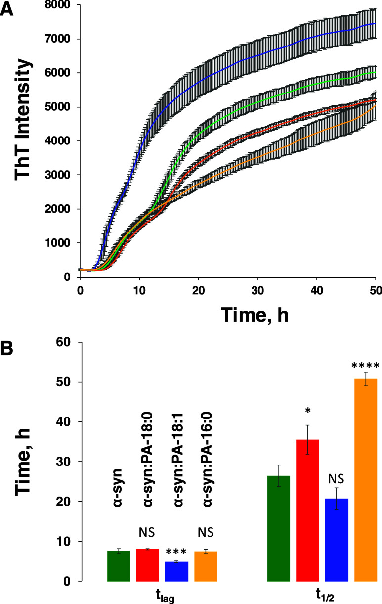
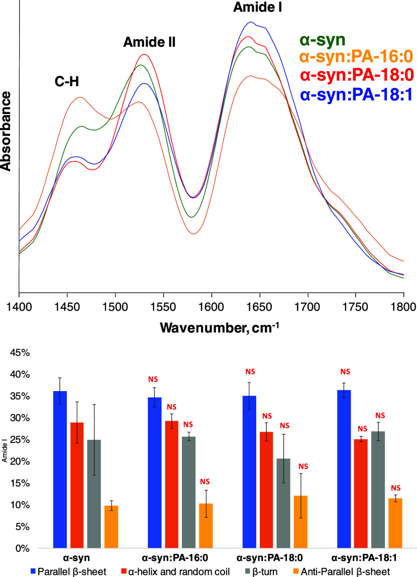
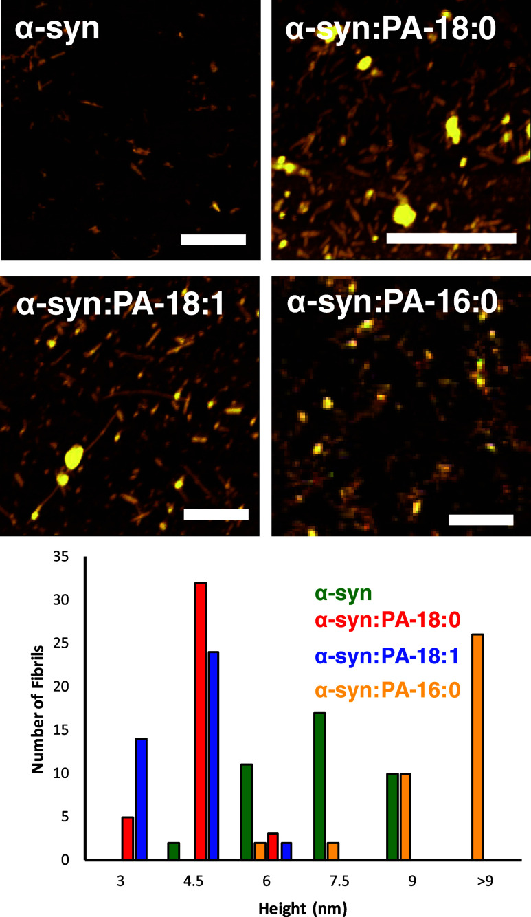
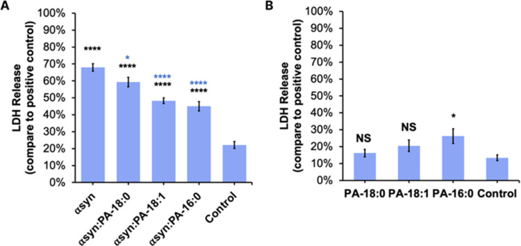
Parkinson's disease (PD) is a severe pathology that is caused by a progressive degeneration of dopaminergic neurons in substantia nigra pars compacta as well as other areas in the brain. These neurodegeneration processes are linked to the abrupt aggregation of α-synuclein (α-syn), a small protein that is abundant at presynaptic nerve termini, where it regulates cell vesicle trafficking. Due to the direct interactions of α-syn with cell membranes, a substantial amount of work was done over the past decade to understand the role of lipids in α-syn aggregation. However, the role of phosphatidic acid (PA), a negatively charged phospholipid with a small polar head, remains unclear. In this study, we examined the effect of PA large unilamellar vesicles (LUVs) on α-syn aggregation. We found that PA LUVs with 16:0, 18:0, and 18:1 FAs drastically reduced the toxicity of α-syn fibrils if were present in a 1:1 molar ratio with the protein. Our results also showed that the presence of these vehicles changed the rate of α-syn aggregation and altered the morphology and secondary structure of α-syn fibrils. These results indicate that PA LUVs can be used as a potential therapeutic strategy to reduce the toxicity of α-syn fibrils formed upon PD.

Keywords: AFM-IR; LDH; fibrils; phosphatidic acid; α-synuclein.


Conflict of interest statement

The authors declare no competing financial interest.

Figures

Figure 1
Figure 1
ThT aggregation kinetics (A) with corresponding tlag and t1/2 (B) of α-syn aggregation in the lipid-free environment (α-syn), as well as in the presence of PA-C18:0 (α-syn:PA-C18:0), PA-C18:1 (α-syn:PA-C18:1), and PA-C16:0 (α-syn:PA-C16:0) at 37 °C. According to one-way ANOVA, * P < 0.05; *** P < 0.001; **** P < 0.0001. NS, nonsignificant differences. Standard deviations of three individual repeats are shown in gray.
Figure 2
Figure 2
IR (top) and CD (bottom) spectra acquired from α-syn, α-syn:PA-C18:0, α-syn:PA-C18:1, and α-syn:PA-C16:0.
Figure 3
Figure 3
Averaged AAFM-IR spectra (top) acquired from α-syn, α-syn:PA-C18:0, α-syn:PA-C18:1, and α-syn:PA-C16:0. A bar graph (bottom) summarizes the distribution of the protein secondary structure in the protein aggregates according to the fitting of the amide I band. Parallel β-sheet (1624 cm–1) in blue, α-helix and random coil (1660 cm–1) in orange, β-turn (1675 cm–1) in gray, and antiparallel β-sheet (1695 cm–1) in yellow.
Figure 4
Figure 4
Length and saturation of FAs in PAs alter the morphology of the α-syn aggregates. AFM images (top) and height histograms (bottom) of α-syn fibrillar aggregates formed in the lipid-free environment (α-syn), as well as in the presence of PA-C18:0 (α-syn:PA-C18:0), PA-C18:1 (α-syn:PA-C18:1), and PA-C16:0 (α-syn:PA-C16:0) formed at 37 °C. Scale bars are 500 nm.
Figure 5
Figure 5
Histograms of LDH assays reveal cell toxicity of (A) α-syn, α-syn:PA-C18:0, α-syn:PA-C18:1, and α-syn:PA-C16:0 and (B) the lipid themselves. Black asterisks (*) show significance level of differences between protein aggregates and the control; blue asterisks show significance level of difference between α-syn and α-syn:PA-C18:1, α-syn:PA-C16:0, and α-syn:PA-C18:0; according to one-way ANOVA, * P < 0.05, **P < 0.01, ***P < 0.001, ****P < 0.0001. NS shows the absence of statistical significance between α-syn and α-syn fibrils formed in the presence of LUVs with different PAs. The results of the LDH assay show no cell toxicity for PA-C18:0 and PA-C18:1, whereas PA-C16:0 was only slightly toxic to N27 rat dopaminergic cells (B). Black asterisks show significant levels of differences between LUVs and the control.

Similar articles

Cited by

References

    1. Tanguy E.; Wang Q. L.; Moine H.; Vitale N. Phosphatidic Acid: From Pleiotropic Functions to Neuronal Pathology. Front. Cell. Neurosci. 2019, 13, 210.3389/fncel.2019.00002. - DOI - PMC - PubMed
    1. Bader M. F.; Vitale N. Phospholipase D in calcium-regulated exocytosis: lessons from chromaffin cells. Biochim. Biophys. Acta, Mol. Cell Biol. Lipids 2009, 1791, 936–941. 10.1016/j.bbalip.2009.02.016. - DOI - PubMed
    1. McMahon H. T.; Gallop J. L. Membrane curvature and mechanisms of dynamic cell membrane remodelling. Nature 2005, 438 (7068), 590–596. 10.1038/nature04396. - DOI - PubMed
    1. Marizzoni M.; Cattaneo A.; Mirabelli P.; Festari C.; Lopizzo N.; Nicolosi V.; Mombelli E.; Mazzelli M.; Luongo D.; Naviglio D.; et al. Short-Chain Fatty Acids and Lipopolysaccharide as Mediators Between Gut Dysbiosis and Amyloid Pathology in Alzheimer’s Disease. J. Alzheimer’s Dis. 2020, 78 (2), 683–697. 10.3233/JAD-200306. - DOI - PubMed
    1. Cecchi C.; Stefani M. The amyloid-cell membrane system. The interplay between the biophysical features of oligomers/fibrils and cell membrane defines amyloid toxicity. Biophys. Chem. 2013, 182, 30–43. 10.1016/j.bpc.2013.06.003. - DOI - PubMed

MeSH terms