. 2024 Feb 19;24(1):60.
doi: 10.1186/s12866-023-03179-z.

What happens to Bifidobacterium adolescentis and Bifidobacterium longum ssp. longum in an experimental environment with eukaryotic cells?

Affiliations

What happens to Bifidobacterium adolescentis and Bifidobacterium longum ssp. longum in an experimental environment with eukaryotic cells?

Dominika Jakubczyk et al. BMC Microbiol. .

Abstract

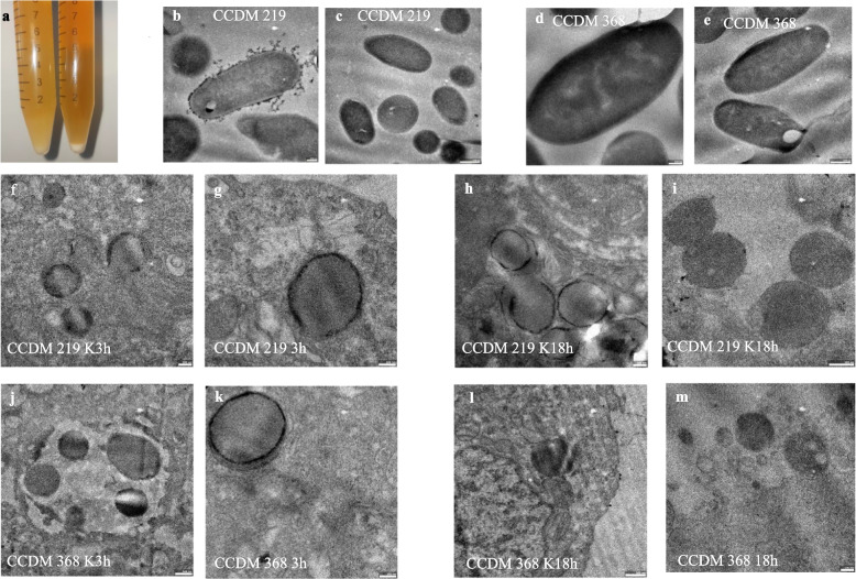
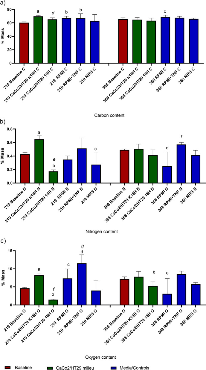
Background: The impact of probiotic strains on host health is widely known. The available studies on the interaction between bacteria and the host are focused on the changes induced by bacteria in the host mainly. The studies determining the changes that occurred in the bacteria cells are in the minority. Within this paper, we determined what happens to the selected Bifidobacterium adolescentis and Bifidobacterium longum ssp. longum in an experimental environment with the intestinal epithelial layer. For this purpose, we tested the bacteria cells' viability, redox activity, membrane potential and enzymatic activity in different environments, including CaCo-2/HT-29 co-culture, cell culture medium, presence of inflammatory inductor (TNF-α) and oxygen.

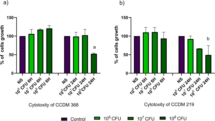
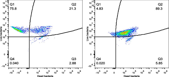
Results: We indicated that the external milieu impacts the viability and vitality of bacteria. Bifidobacterium adolescentis decrease the size of the live population in the cell culture medium with and without TNF-α (p < 0.001 and p < 0.01 respectively). In contrast, Bifidobacterium longum ssp. longum significantly increased survivability in contact with the eukaryotic cells and cell culture medium (p < 0.001). Bifidobacterium adolescentis showed significant changes in membrane potential, which was decreased in the presence of eukaryotic cells (p < 0.01), eukaryotic cells in an inflammatory state (p < 0.01), cell culture medium (p < 0.01) and cell culture medium with TNF-α (p < 0.05). In contrast, Bifidobacterium longum ssp. longum did not modulate membrane potential. Instead, bacteria significantly decreased the redox activity in response to milieus such as eukaryotic cells presence, inflamed eukaryotic cells as well as the culture medium (p < 0.001). The redox activity was significantly different in the cells culture medium vs the presence of eukaryotic cells (p < 0.001). The ability to β-galactosidase production was different for selected strains: Bifidobacterium longum ssp. longum indicated 91.5% of positive cells, whereas Bifidobacterium adolescentis 4.34% only. Both strains significantly reduced the enzyme production in contact with the eukaryotic milieu but not in the cell culture media.

Conclusion: The environmental-induced changes may shape the probiotic properties of bacterial strains. It seems that the knowledge of the sensitivity of bacteria to the external environment may help to select the most promising probiotic strains, reduce research costs, and contribute to greater reproducibility of the obtained probiotic effects.

Keywords: Bifidobacterium; Enzymatic activity; Membrane potential; Probiotic; Redox activity; Viability.


Conflict of interest statement

The authors declare no competing interests.

Figures

Fig. 1
Fig. 1
The cell proliferation analysis of CaCo2/HT29 cells after contact with the selected bacterial strains for. a CCDM 368 impact after 6 h and 24 h CaCo2/HT29 stimulation; b CCDM 219 impact after 6 h and 24 h CaCo2/HT29 stimulation. The statistical analysis was based on the One-way ANOVA test. Significance vs non-stimulated samples: a*, b*. Non-stimulated samples (NS) were marked in purple and stimulated by bacteria in green
Fig. 2
Fig. 2
Morphology of strains CCDM 368 and CCDM 219; a macroscopic appearance of bacterial direct from MRS overnight culture in anaerobic condition, on the right strain CCDM 368, on the left strain CCDM 219; TEM imaging of strain: b-c CCDM 219, d-e CCDM 368 directly from MRS overnight culture in anaerobic condition (baseline); TEM imaging of strain: f-i CCDM 219, j-m CCDM 368 in different environments (in co-culture with CaCo2/HT29 cells); K3h: co-culture CaCo2/HT29 with selected bacteria, without inflammatory state (control), 3 h: CaCo2/HT29 with inflammatory state induced 3 h after bacteria addition, K18h: co-culture CaCo2/HT29 with selected bacteria, without inflammatory state (control), 18 h: CaCo2/HT29 with inflammatory state induced 18 h before bacteria addition
Fig. 3
Fig. 3
EDS analysis for different elements. The baseline samples were presented in claret colour, the eukaryotic environment was presented in green, and the control samples in blue. The analysis of baseline vs samples was based on the One-way ANOVA. The differences between groups K18h vs 18 h and RPMI vs RPMI + TNF-α were analysed by paired t-test. a Carbon contents in bacteria placed in various environments. Significance for strain CCDM 219 versus baseline: a **, b*; K18h vs 18 h: d ***. Significance for strain CCDM 368 versus baseline: c*. b Nitrogen contents in bacteria placed in various environments. Significance for strain CCDM 219 versus baseline: a ***, b ****, c*; K18h vs 18 h: e ****. Significance for strain CCDM 368 versus baseline: d ****, RPMI vs RPMI + TNF-α: f *. c Oxygen contents in bacteria placed in various environments. Significance for strain CCDM 219 versus baseline: a ***, b **, c*, d****; K18h vs 18 h: f ****; RPMI vs RPMI + TNF-α: g*. Significance for strain CCDM 368 versus baseline: e ***, K18h vs 18 h: h *
Fig. 4
Fig. 4
The survival (viability analysis) of Bifidobacterium CCDM 368 strain in different environments. The live population was presented in green, the mixed one in orange, and the dead one in red. The analysis of baseline vs samples was based on the One-way ANOVA. Significance: in live population: a ***, b**; in mixed population: c*, d**, e**** f ***, in dead population: g **
Fig. 5
Fig. 5
Pearson's linear correlation analysis for different groups (baseline, an active inflammatory state model, protection against inflammatory state, and appropriate controls). Interpretation of the correlation coefficient r strength: lack or very weak correlation r = 0–0.3; moderate: 0.3–0.5, strong: 0.5–0.7; very strong: 0.7–1.00)
Fig. 6
Fig. 6
The dots plots examples of the CCDM 219 different states at baseline
Fig. 7
Fig. 7
The survival (viability analysis) of Bifidobacterium CCDM 219 strain in different environments The live population was presented in green, the mixed one in orange, and the dead one in red. The analysis of baseline vs samples was based on the One-way ANOVA test. Significance: in live population: a *, in mixed population: b*, c**
Fig. 8
Fig. 8
Pearson's linear correlation analysis for different groups (baseline, an active inflammatory state model, protection against inflammatory state, and appropriate controls). Interpretation of the correlation coefficient r strength: lack or very weak correlation r = 0–0.3; moderate: 0.3–0.5, strong: 0.5–0.7; very strong: 0.7–1.00)
Fig. 9
Fig. 9
The CCDM 219 and CCDM 368 strains’ redox activity in different environments. The baseline samples were presented in claret, the eukaryotic milieu in green, and the control samples in purple. The analysis of baseline vs samples was based on the One-way ANOVA test. Significance: CCDM 219 baseline vs remaining groups (a****), CCDM 368 baseline vs remaining groups ( b****)
Fig. 10
Fig. 10
The CCDM 219 and CCDM 368 strains membrane potential maintenance. The baseline samples were presented in claret, the eukaryotic milieu in green, and the control samples in purple. The analysis of baseline vs samples was based on the One-way ANOVA test. Significance: a**, b***, c*
Fig. 11
Fig. 11
Percentage of cells which were able to produce β-gal in different environments. The baseline samples were presented in claret, the eukaryotic milieu in green, and the control samples in purple. The analysis of baseline vs samples was based on the One-way ANOVA test. Significance: for CCDM 219: a***, b*, for CCDM 368: c*

References

    1. Arboleya S, Watkins C, Stanton C, Ross RP. Gut Bifidobacteria Populations in Human Health and Aging. Front Microbiol. 2016;7. http://journal.frontiersin.org/Article/.10.3389/fmicb.2016.01204/abstract - PMC - PubMed
    1. Solopova A, Bottacini F, Venturi degli Esposti E, Amaretti A, Raimondi S, Rossi M, et al. Riboflavin Biosynthesis and Overproduction by a Derivative of the Human Gut Commensal Bifidobacterium longum subsp. infantis ATCC 15697. Front Microbiol. 2020;11:573335. doi: 10.3389/fmicb.2020.573335. - DOI - PMC - PubMed
    1. Štofilová J, Kvaková M, Kamlárová A, Hijová E, Bertková I, Guľašová Z. Probiotic-based intervention in the treatment of ulcerative colitis: conventional and new approaches. Biomedicines. 2022;10(9):2236. doi: 10.3390/biomedicines10092236. - DOI - PMC - PubMed
    1. Derwa Y, Gracie DJ, Hamlin PJ, Ford AC. Systematic review with meta-analysis: the efficacy of probiotics in inflammatory bowel disease. Aliment Pharmacol Ther. 2017;46(4):389–400. doi: 10.1111/apt.14203. - DOI - PubMed
    1. Palumbo VD, Romeo M, Gammazza AM, Carini F, Damiani P, Damiano G, et al. The long-term effects of probiotics in the therapy of ulcerative colitis: a clinical study. Biomed Pap Med Fac Univ Palacky Olomouc Czech Repub. 2016;160(3):372–377. doi: 10.5507/bp.2016.044. - DOI - PubMed

Substances

Supplementary concepts

LinkOut - more resources