. 2024 Feb 19;14(1):4055.
doi: 10.1038/s41598-024-54628-2.

Multiple roles for the cytoplasmic C-terminal domains of the yeast cell surface receptors Rgt2 and Snf3 in glucose sensing and signaling

Affiliations

Multiple roles for the cytoplasmic C-terminal domains of the yeast cell surface receptors Rgt2 and Snf3 in glucose sensing and signaling

Jeong-Ho Kim et al. Sci Rep. .

Abstract

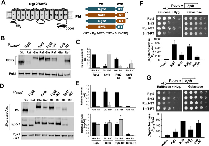
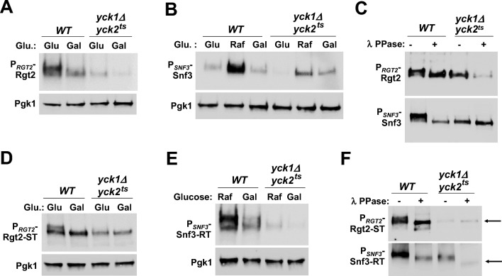
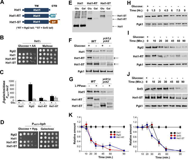
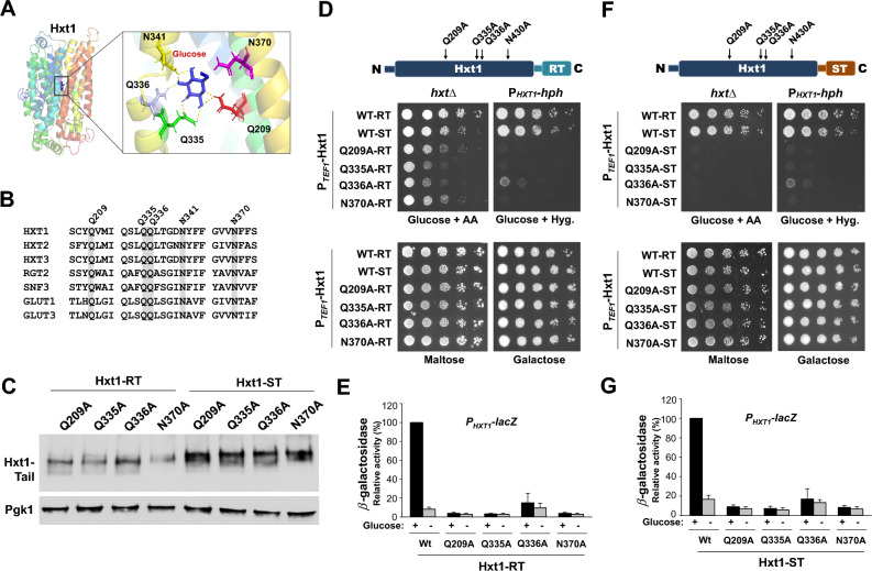
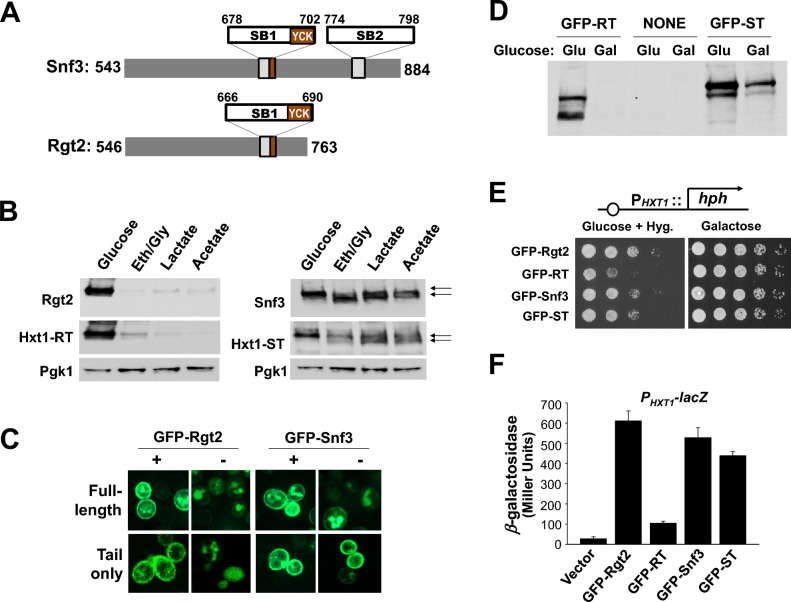
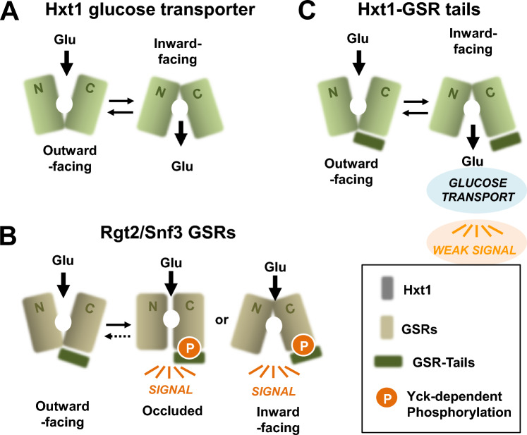
The plasma membrane proteins Rgt2 and Snf3 are glucose sensing receptors (GSRs) that generate an intracellular signal for the induction of gene expression in response to high and low extracellular glucose concentrations, respectively. The GSRs consist of a 12-transmembrane glucose recognition domain and a cytoplasmic C-terminal signaling tail. The GSR tails are dissimilar in length and sequence, but their distinct roles in glucose signal transduction are poorly understood. Here, we show that swapping the tails between Rgt2 and Snf3 does not alter the signaling activity of the GSRs, so long as their tails are phosphorylated in a Yck-dependent manner. Attachment of the GSR tails to Hxt1 converts the transporter into a glucose receptor; however, the tails attached to Hxt1 are not phosphorylated by the Ycks, resulting in only partial signaling. Moreover, in response to non-fermentable carbon substrates, Rgt2 and Hxt1-RT (RT, Rgt2-tail) are efficiently endocytosed, whereas Snf3 and Hxt1-ST (ST, Snf3-tail) are endocytosis-impaired. Thus, the tails are important regulatory domains required for the endocytosis of the Rgt2 and Snf3 glucose sensing receptors triggered by different cellular stimuli. Taken together, these results suggest multiple roles for the tail domains in GSR-mediated glucose sensing and signaling.


Conflict of interest statement

The authors declare no competing interests.

Figures

Figure 1
Figure 1
Swapping the tails between Rgt2 and Snf3 does not alter the signaling specificity of the GSRs. (A) The predicted transmembrane topology model of the Rgt2 and Snf3 GSRs (left) and the schematic representation of the structures of the native and chimeric receptors (right). RT and ST represent the tail domains of Rgt2 and Snf3, respectively. (B) Western blot analysis of protein levels of the wild-type and chimeric GSRs. C-terminally HA-tagged GSRs (RGT2-HA (JKP253, 763aa + 3HA), SNF3-HA (JKP298, 884aa + 3HA), RGT-ST (JKP602, 887aa + 3HA), and SNF3-RT (JKP600, 760aa + 3HA)) were expressed from their native promoters. Wild-type cells (BY4742) were grown in selective SC medium with 2% glucose to mid-log phase (O.D600nm = 1.2–1.5) and equal amounts of cells were shifted to SC medium containing glucose (2%) or raffinose (2%) for 1 h. Membrane fractions were immunoblotted with anti-HA antibody. Pgk1 was used as loading control. (C) Relative band intensities (B) were quantified densitometrically using the NIH ImageJ program. Averages of glucose samples (Rgt2-HA) were set to 1. Results were obtained from at least three independent experiments. (D) Western blot analysis of the wild-type and chimeric GSRs expressed from the constitutive TEF1 promoter in wild-type and rsp5-1 cells. PGK1 was expressed from the wild type cells. Cells were grown and treated as described above (B). The GRS genes expressed were: RGT2 (JKP443), SNF3 (JKP444), RGT2-ST (JKP609), and SNF3-RT (JKP631). (E) Relative band intensities (D) were quantified as described above (C). (F) The PHXT1-hph reporter strains (KLS76) expressing the indicated GSR genes were scored for growth in a SC-2% glucose plate supplemented with 200 µg/ml hygromycin or galactose (2%) plate. The first spot of each row represents a count of ~ 5 × 107 cell/ml, which is diluted 1:10 for each spot thereafter (top). The plates were incubated for 2 days at 30 °C. The rgt2snf3 double mutant (MSY441) was co-transformed the HXT1-lacZ reporter (pBM3212) with plasmids carrying the indicated GSR genes. β-Galactosidase activity was assayed in permeabilized cells and expressed in Miller Units. Values are means for at least three independent experiments (bottom). The GRS genes expressed were: RGT2 (JKP606), SNF3 (JKP607), RGT2-ST (JKP603), and SNF3-RT (JKP604). (G) The PHXT2-hph reporter strains (KLS75) expressing the indicated GSR genes were scored for growth in a SC-2% raffinose (2%)/hygromycin (200 µg/ml) plate or galactose (2%) plate (top). The plates were incubated for 2 days at 30 °C. The rgt2snf3 double mutant (MSY441) was co-transformed the HXT2-lacZ reporter (JKP493) with plasmids carrying the indicated GSR genes. β-Galactosidase activity was assayed as described above (bottom).
Figure 2
Figure 2
The tails of the GSRs are not interchangeable. (A) Western blot analysis of Rgt2-HA protein levels in WT (LRB939) and yck1Δyck2ts (LRB1613) cells. RGT2-HA (JHP253) was expressed from the native promoter. Cells were grown on glucose (2%) or galactose (2%) as described in Fig. 1B, and Pgk1 was used as loading control. The yck1Δyck2ts cells were incubated at 37 °C for 30 min before the precultures were shifted to fresh glucose medium or galactose medium. (B) Western blot analysis of Snf3-HA protein levels in WT and yck1Δyck2ts cells. SNF3-HA (JKP298) was expressed from the native promoter. Cells were grown on glucose (2%), raffinose (2%), or galactose (2%) as described in Fig. 1B, and Pgk1 was used as loading control. (C) WT and yck1Δyck2ts strains expressing RGT2-HA (JKP253) or SNF3-HA (JKP298) were grown on glucose as described in Fig. 1B, and whole cell lysates were immunoprecipitated with agarose-conjugated anti-HA antibody. The precipitates were treated with (+) or without (−) lambda protein phosphatase (10 U) at 30℃ for 30 min and analyzed by Western blotting with anti-HA antibody. (D) Western blot analysis of Rgt2-ST-HA protein levels in WT and yck1Δyck2ts cells. RGT2-ST-HA (JKP602) was expressed from the RGT2 promoter. Cells were grown as described in Fig. 1B, and Pgk1 was used as loading control. (E) Western blot analysis of Snf3-RT-HA protein levels in WT and yck1Δyck2ts cells. SNF3-RT-HA (JKP600) was expressed from the SNF3 promoter. Cells were grown on raffinose (2%) or galactose (2%) as described in Fig. 1B, and Pgk1 was used as loading control. (F) WT and yck1Δyck2ts strains expressing RGT2-ST-HA (JKP602, from the RGT2 promoter) or SNF3-RT-HA (JKP600, from the SNF3 promoter) were grown on glucose as described in Fig. 1B. Whole cell lysates prepared from cells (WT and yck1Δyck2ts) grown on glucose (Rgt2-ST-HA) and on raffinose (Snf3-RT-HA), were immunoprecipitated, treated with (+) or without (−) lambda protein phosphatase, and analyzed by Western blotting with anti-HA antibody, as described above (C). Phosphatase treatment causes different mobility shifts of Rgt2-ST and Snf3-RT on SDS-PAGE (arrows).
Figure 3
Figure 3
The Ycks mediate phosphorylation of the tails of the GSRs but not the tails attached to Hxt1. (A) Schematic representation of the structures of Hxt1 and hybrid transporter/receptor proteins Hxt1-RT (Rgt2 tail) and Hxt1-ST (Snf3 tail). (B) The hxt-null strains (EBY.VW4000, hxtΔ) expressing the indicated genes were scored for growth on SC-medium containing either 2% glucose with Antimycin A (1 μg/ml) or 2% maltose. The genes expressed were: RGT2 (JKP420), HXT1 (JKP504), HXT1-RT (JKP648), and HXT-RT (JKP650). The first spot of each row represents a count of ~ 5 × 107 cell/ml, which is diluted 1:10 for each spot thereafter. The plates were incubated for 2 days at 30 °C. (C) The rgt2snf3 double mutant (MSY441) was co-transformed the HXT1-lacZ reporter (pBM3212) with plasmids carrying the indicated genes. β-Galactosidase activity was assayed in permeabilized cells and expressed in Miller Units, as described in Fig. 1F. (D) The PHXT1-hph reporter strains (KLS76) expressing the indicated genes were scored for growth in a SC-2% glucose plate supplemented with 200 µg/ml hygromycin or galactose (2%) plate, as described in Fig. 1F. The plates were incubated for 2 days at 30 °C. (E) Expression of Hxt1 (570aa + 3HA), Hxt1-RT (788aa + 3HA), and Hxt1-ST (912aa + 3HA) was examined by Western blot analysis, and their respective genes were expressed from the TEF1 promoter (EJ). Wild-type cells (BY4742) were grown in selective SC medium with 2% glucose to mid-log phase and equal amounts of cells were shifted to SC medium containing glucose (2%) or galactose (2%) for 1 h. Membrane fractions were immunoblotted with anti-HA antibody. (F) Western blot analysis of protein levels of Hxt1, Hxt1-RT, and Hxt1-ST in WT (LRB939) and yck1Δyck2ts (LRB1613) cells. Cells were grown as described above (E), and Pgk1 was used as loading control. The glucose-dependent phosphorylation of Hxt1-RT and Hxt1-ST in the yck1Δyck2ts strain was indicated by arrows. (G) WT and yck1Δyck2ts strains expressing HXT1, HXT1-RT, and HXT1-ST were grown on glucose as described above (E), and whole cell lysates were immunoprecipitated, treated with (+) or without (−) lambda protein phosphatase, and analyzed by Western blotting with anti-HA antibody, as described in Fig. 1C. (HJ) Wild-type cells (BY4742) expressing the indicated genes were grown in SC-2% glucose (+) medium to mid-log phase and shifted to 2% galactose (−) medium for times as indicated. Membrane fractions were immunoblotted with anti-HA antibody, and Pgk1 was used as loading control. (K) The intensity of each band on the blot (HJ) was quantified by densitometric scanning, as described in Fig. 1C.
Figure 4
Figure 4
The Hxt1 TM-domain of the Hxt1-RT and Hxt1-ST hybrid proteins generates a signal in response to glucose. (A) Hxt1 homology model was generated using I-TASSER and visualized using PyMol, as we described previously. Docking of glucose to the predicted glucose binding site in Hxt1 was visualized using AutoDock Vina. (B) Clustal Omega was used for sequence alignment of yeast glucose transporters (Hxt1, Hxt2, and Hxt3), yeast GSRs (Rgt2 and Snf3), and human glucose transporters (GLUT1 and GLUT3). (C) Western blot analysis of Hxt1-RT and Hxt1-ST proteins with substitutions in residues Q209A (JKP609/JKP684), Q335A (JKP661/JKP686), Q336A (JKP663/JKP687), or N370A (JKP665/JKP 689). Yeast cells expressing the indicated HXT1-TAIL constructs were grown on glucose as described in Fig. 1B, and membrane fractions were immunoblotted with anti-HA antibody. Pgk1 was used as loading control. (D, F) Schematic representation of the structures of the Hxt1-RT (D) and Hxt1-ST (F) with the locations of the substitutions (top). The hxt-null strains (BSY.VW4000, hxtΔ) expressing the indicated genes were scored for growth on SC-medium containing either 2% glucose with Antimycin A (1 μg/ml) or 2% maltose. The PHXT1-hph reporter strains (KLS76) expressing the indicated genes were scored for growth in a SC-2% glucose plate supplemented with 200 µg/ml hygromycin or galactose (2%) plate (bottom). The plates were incubated for 2 days at 30 °C. (E, G) The rgt2snf3 double mutant (MSY441) was co-transformed the HXT1-lacZ reporter (pBM3212) with plasmids carrying the indicated genes. β-Galactosidase activity was assayed in permeabilized cells and expressed in Miller Units, as described in Fig. 1F.
Figure 5
Figure 5
The Snf3 tail, when expressed independently of the TM domains, leads to induction of HXT1 expression. (A) Schematic representation of the structures of the tails of the Rgt2 and Snf3 GSRs. The GSR tails are dissimilar, except for a signaling box (SB), a stretch of 25 amino acids that occurs twice in Snf3 and once in Rgt2. SB1 partially overlaps with the consensus sequences for the Yck phosphorylation. (B) Western blot analysis of protein levels of Rgt2-HA, Hxt1-RT-HA, Snf3-HA, and Hxt1-ST-HA. Wild-type cells (BY4742) were grown in selective SC medium with 2% glucose to mid-log phase (O.D600nm = 1.2–1.5) and equal amounts of cells were shifted to SC medium containing glucose (2%), ethanol/glycerol (2%), lactate (2%) or acetate (1%) for 1 h. Membrane fractions were immunoblotted with anti-HA antibody. Pgk1 was used as loading control. Snf3 and Hxt1-ST from cells grown on non-fermentable carbon substrates migrate as two distinct bands on SDS-PAGE (arrows). (C) Confocal microscopy of wild-type (BY4742) cells expressing GFP-Rgt2 (JKP596), GFP-Rgt2 tail (JKP597), GFP-Snf3 (JKP598) or GFP-Snf3 tail (JKP616). Yeast cells were grown in glucose (2%) or galactose (2%), as described in Fig. 1B, and observed under the Zeiss LSM 510 META confocal laser scanning microscope, as we described previously,. The GFP-Rgt2 tail and GFP-Snf3-tail fragments were attached to the plasma membrane via a farnesylation signal. (D) Expression of the tails of Rgt2 (GFP-RT, JKP597) and Snf3 (GFP-ST, JKP616) was examined by Western blot analysis. Wild-type (BY4742) cells were grown in glucose (2%) or galactose (2%), as described in Fig. 1B, and membrane fractions were immunoblotted with anti-GFP antibody. NONE: Total extracts from cells that were not transformed with a GFP plasmid. (E) The PHXT1-hph reporter strains (KLS76) expressing the indicated genes were scored for growth in a SC-2% glucose plate supplemented with 200 µg/ml hygromycin or galactose (2%) plate, as described in Fig. 1F. The plates were incubated for 2 days at 30 °C. (F) The rgt2snf3 double mutant (YM6370) was co-transformed the HXT1-lacZ reporter (pBM3212) with plasmids carrying the indicated genes (pUG36 as vector). β-Galactosidase activity was assayed as described in Fig. 1F.
Figure 6
Figure 6
Conformation changes in glucose transporters and receptors upon glucose binding. The binding of glucose to the TM domains of the glucose transporters and receptors induces a series of conformational changes in them that are required for the transport cycle to take place,, leading to glucose transport (A) and glucose signaling (B), respectively. This difference may not be due to the long cytoplasmic C-terminal tails of the GSRs, because attachment of the tails to Hxt1 does not prevent it from transporting glucose (C). The considerable divergence of the TM domains of the GSRs from the Hxts probably accounts for their inability to complete the transport cycle to translocate glucose to the cytoplasmic side. Because a complete transport cycle is not required for signaling, it is believed that binding of glucose to the extracellular face of the GSRs favors their inward-facing and/or occluded form(s), which are their signaling conformation(s),. We suggest that the Yck phosphorylation of the tail of the GSRs is required for the stabilization of the signaling conformation of the TM domain and the activation of the intracellular signaling pathway (B). Accordingly, the lack of the Yck phosphorylation of the tails of the hybrid proteins (Hxt1-RT and Hxt1-ST) lead to only partial signaling (C).

Similar articles

References

    1. Johnston M, Kim JH. Glucose as a hormone: Receptor-mediated glucose sensing in the yeast Saccharomyces cerevisiae. Biochem. Soc. Trans. 2005;33:247–252. doi: 10.1042/BST0330247. - DOI - PubMed
    1. Bisson LF, Neigeborn L, Carlson M, Fraenkel DG. The SNF3 gene is required for high-affinity glucose transport in Saccharomyces cerevisiae. J. Bacteriol. 1987;169:1656–1662. doi: 10.1128/jb.169.4.1656-1662.1987. - DOI - PMC - PubMed
    1. Marshall-Carlson L, Celenza JL, Laurent BC, Carlson M. Mutational analysis of the SNF3 glucose transporter of Saccharomyces cerevisiae. Mol. Cell Biol. 1990;10:1105–1115. - PMC - PubMed
    1. Ozcan S, Dover J, Johnston M. Glucose sensing and signaling by two glucose receptors in the yeast Saccharomyces cerevisiae. Embo J. 1998;17:2566–2573. doi: 10.1093/emboj/17.9.2566. - DOI - PMC - PubMed
    1. Ozcan S, Dover J, Rosenwald AG, Wolfl S, Johnston M. Two glucose transporters in Saccharomyces cerevisiae are glucose sensors that generate a signal for induction of gene expression. Proc. Natl. Acad. Sci. U. S. A. 1996;93:12428–12432. doi: 10.1073/pnas.93.22.12428. - DOI - PMC - PubMed

MeSH terms

Substances