. 2024 Feb 19;14(1):4048.
doi: 10.1038/s41598-024-54773-8.

FOXM1: a new therapeutic target of extramammary Paget disease

Affiliations

FOXM1: a new therapeutic target of extramammary Paget disease

Takamichi Ito et al. Sci Rep. .

Abstract

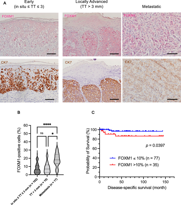
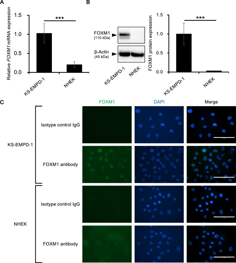
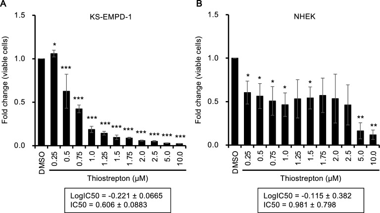
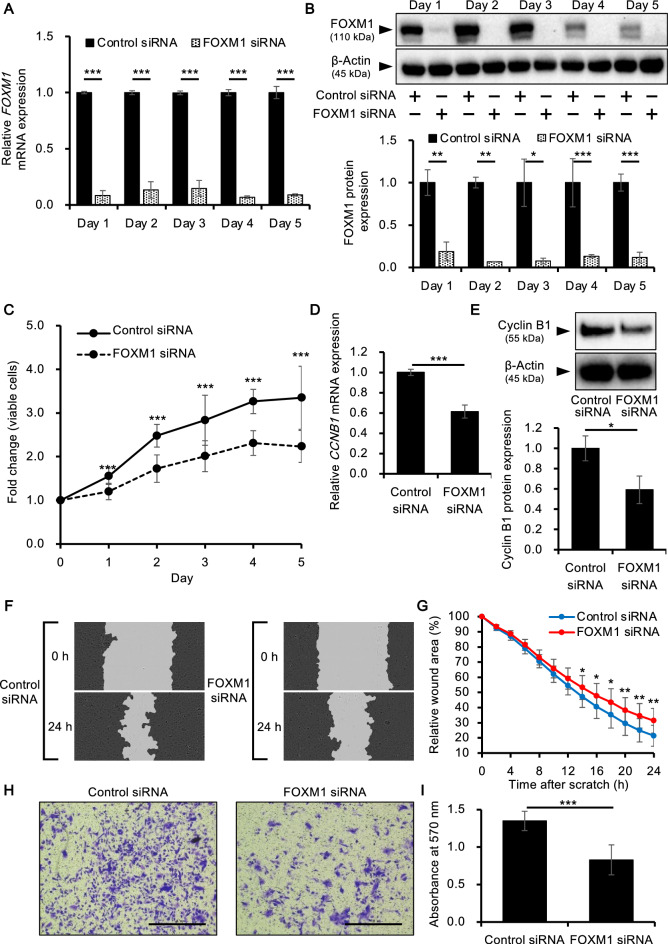
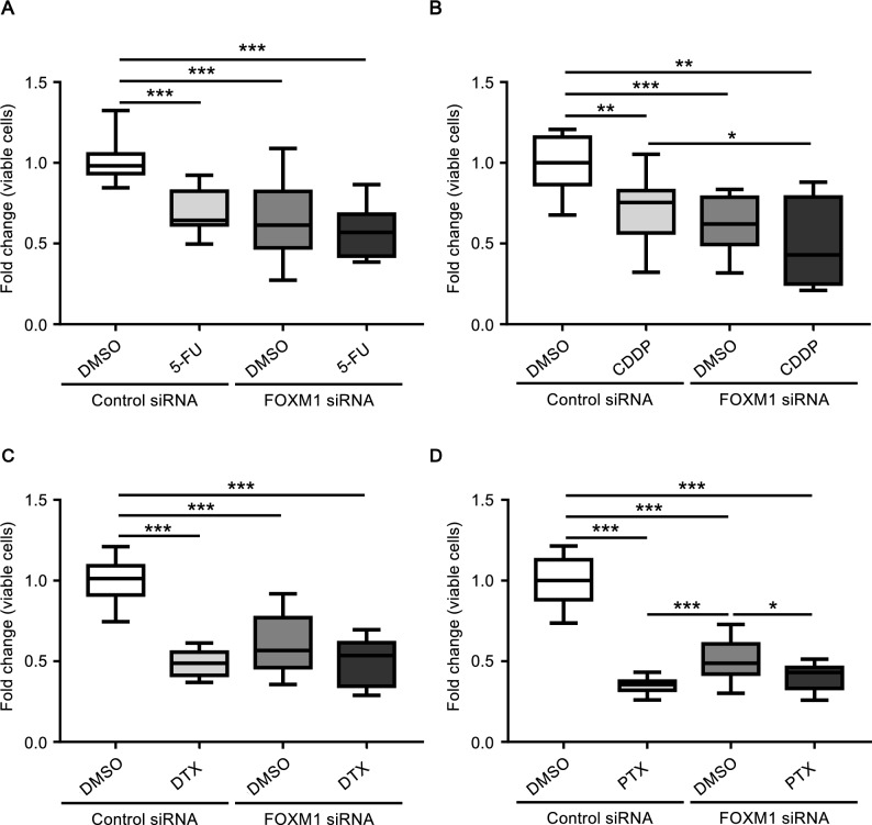
Extramammary Paget disease (EMPD) is a rare skin cancer that primarily affects older individuals predominantly in areas with apocrine sweat glands. Although most early EMPD lesions are indolent, patients with metastatic EMPD have a poor prognosis due to the lack of effective systemic treatment. In this study, we investigated the role of forkhead box M1 (FOXM1), a potent transcription factor, in EMPD and assessed the potential of FOXM1 as a therapeutic target. Immunohistochemistry of 112 primary and 17 metastatic EMPD samples revealed that FOXM1 expression increased with tumor progression. Patients in whom FOXM1 was expressed in more than 10% of tumor cells had significantly shorter disease-specific survival than the other patients (p = 0.0397). In in vitro studies using our newly established EMPD cell line, KS-EMPD-1, we found high expression of FOXM1. Knockdown of FOXM1 impaired tumor cell viability, migration, and invasion. Inhibition of FOXM1 using thiostrepton also reduced tumor cell viability in a dose-dependent manner. These findings suggest that FOXM1 is a promising therapeutic target for patients with EMPD.

Keywords: Cell line; FOXM1; Skin cancer; Targeted therapy; Thiostrepton.


Conflict of interest statement

The authors declare no competing interests.

Figures

Figure 1
Figure 1
FOXM1 expression in patients’ EMPD tissues. (A) Immunohistochemical images of FOXM1 in patients’ EMPD tissues. Representative images of early lesions [tumor thickness (TT) ≤ 3 mm, n = 102], locally advanced lesions (TT > 3 mm, n = 10), and metastatic lesions (n = 17) are shown. Scale bars = 100 μm. (B) Comparison of FOXM1-positive cells (%) among early, locally advanced, and metastatic lesions of patients’ tissues. *p < 0.05 and ***p < 0.001. (C) Survival curves of patients with ≤ 10% (n = 77) or > 10% (n = 35) FOXM1 staining positivity in immunohistochemistry. Patients with FOXM1 > 10% had significantly shorter disease-specific survival than those with FOXM1 ≤ 10% (p = 0.0397).
Figure 2
Figure 2
FOXM1 is highly expressed in KS-EMPD-1 cells. (A) FOXM1 mRNA expression in KS-EMPD-1 cells and NHEKs. Mean ± SD of FOXM1 obtained from three independent experiments is shown. ***p < 0.001. (B) FOXM1 protein expression in KS-EMPD-1 cells and NHEKs. Representative blot images (left) and mean ± SD of FOXM1 protein (right) are shown. Experiments were independently repeated three times. Original, uncropped images are shown in Supplementary Fig. S1. ***p < 0.001. (C) Immunocytochemical images of FOXM1 in KS-EMPD-1 cells and NHEKs. FOXM1 (green), DAPI (blue, nuclear), and merged images of FOXM1 and DAPI are shown. Scale bars = 100 μm.
Figure 3
Figure 3
KS-EMPD-1 is sensitive to a FOXM1 inhibitor, thiostrepton. KS-EMPD-1 cells and NHEKs were incubated with DMSO (0.1%) or various concentrations of thiostrepton (0.25–10.0 μM) for 72 h and evaluated for the number of viable cells using a formazan-based method. Mean ± SD of fold change (viable cells) calculated from three independent experiments is shown. The IC50 of thiostrepton is shown in the boxes below the graphs. *p < 0.05, **p < 0.01, and ***p < 0.001.
Figure 4
Figure 4
FOXM1 knockdown inhibits proliferation, migration, and invasion of KS-EMPD-1 cells. Cells were transfected with FOXM1 siRNA and evaluated for their proliferation, migration, and invasion. (A) Knockdown efficiency of FOXM1 mRNA. Mean ± SD of FOXM1 calculated from three independent experiments is shown. ***p < 0.001. (B) Knockdown efficiency of FOXM1 protein. Representative blot images (upper) and mean ± SD of FOXM1 protein (lower) are shown. Experiments were independently repeated three times. Original, uncropped images are shown in Supplementary Fig. S2A. *p < 0.05, **p < 0.01, and ***p < 0.001. (C) Viable cells in control and FOXM1 siRNA-transfected conditions were quantified using a formazan-based method. Experiments were independently repeated three times. ***p < 0.001. (D) CCNB1 mRNA expression in control or FOXM1 siRNA-transfected cells at 24 h post-transfection. Mean ± SD of FOXM1 expression calculated from three independent experiments is shown. ***p < 0.001. (E) Cyclin B1 protein expression in control or FOXM1 siRNA-transfected cells at 48 h post-transfection. Experiments were independently repeated three times and representative blot images (upper) and mean ± SD of cyclin B1 protein expression (lower) are shown. Original, uncropped images are shown in Supplementary Fig. S2B. *p < 0.05. Cells were transfected with siRNA for 48 h. The cell monolayers were then scratched and the wound area were captured for 24 h. Representative images (F) and wound area relative to that at 0 h (G) calculated from three independent experiments are shown. *p < 0.05 and **p < 0.01. Cells were transfected with siRNA for 48 h and evaluated for cell invasion. Representative images (H) and mean ± SD of absorbance of cell staining dye (I) are shown. Experiments were independently repeated three times. Scale bars = 1.0 mm. ***p < 0.001.
Figure 5
Figure 5
Knockdown of FOXM1 increases the chemosensitivity of KS-EMPD-1 cells. KS-EMPD-1 cells were transfected with control or FOXM1 siRNA for 24 h and further treated with vehicle control or anticancer drugs for 72 h. The viable cells were then quantitated using a formazan-based method. 5-Fluorouracil (5-FU, final concentration 100 nM), docetaxel (DTX, final concentration 5 μM), and paclitaxel (PTX, final concentration 1 μM) were dissolved in DMSO and cisplatin (CDDP, final concentration 10 μM) was dissolved in saline and further added to the culture medium. Concentration of anticancer drugs was determined based on the Cmax of each drug. Mean ± SD of fold change of viable cells calculated from three independent experiments is shown. *p < 0.05, **p < 0.01, and ***p < 0.001.

Similar articles

Cited by

References

    1. Kanitakis J. Mammary and extramammary Paget’s disease. J. Eur. Acad. Dermatol. Venereol. 2007;21(5):581–590. doi: 10.1111/j.1468-3083.2007.02154.x. - DOI - PubMed
    1. Ito T, Kaku-Ito Y, Furue M. The diagnosis and management of extramammary Paget’s disease. Expert Rev. Anticancer Ther. 2018;18(6):543–553. doi: 10.1080/14737140.2018.1457955. - DOI - PubMed
    1. Shepherd V, Davidson EJ, Davies-Humphreys J. Extramammary Paget’s disease. BJOG. 2005;112(3):273–279. doi: 10.1111/j.1471-0528.2004.00438.x. - DOI - PubMed
    1. Hashimoto H, Ito T. Current management and treatment of extramammary Paget’s disease. Curr. Treat. Options Oncol. 2022;23(6):818–830. doi: 10.1007/s11864-021-00923-3. - DOI - PubMed
    1. Ishizuki S, Nakamura Y. Extramammary Paget’s disease: Diagnosis, pathogenesis, and treatment with focus on recent developments. Curr. Oncol. 2021;28(4):2969–2986. doi: 10.3390/curroncol28040260. - DOI - PMC - PubMed

MeSH terms