. 2024 Jun;19(6):818-824.
doi: 10.1038/s41565-024-01620-6. Epub 2024 Feb 19.

Chimeric nanobody-decorated liposomes by self-assembly

Affiliations

Chimeric nanobody-decorated liposomes by self-assembly

Md Mofizur Rahman et al. Nat Nanotechnol. 2024 Jun.

Abstract

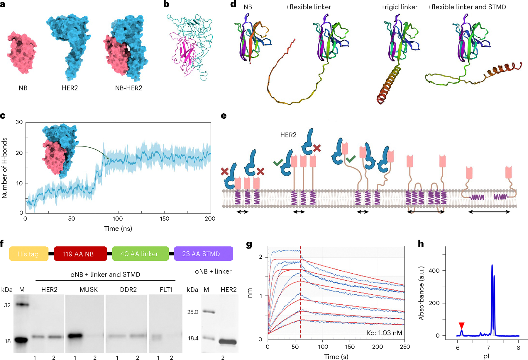
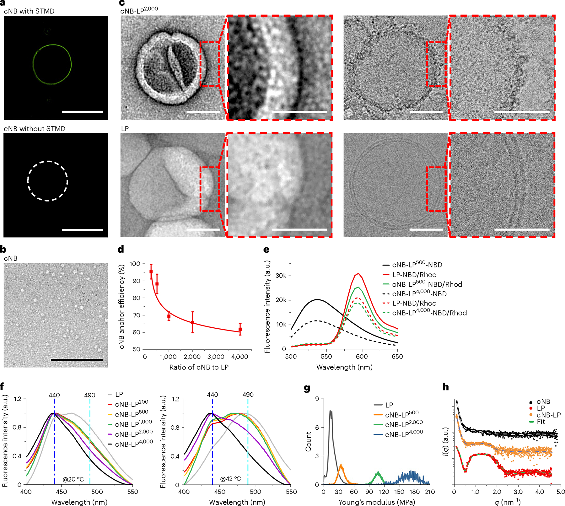
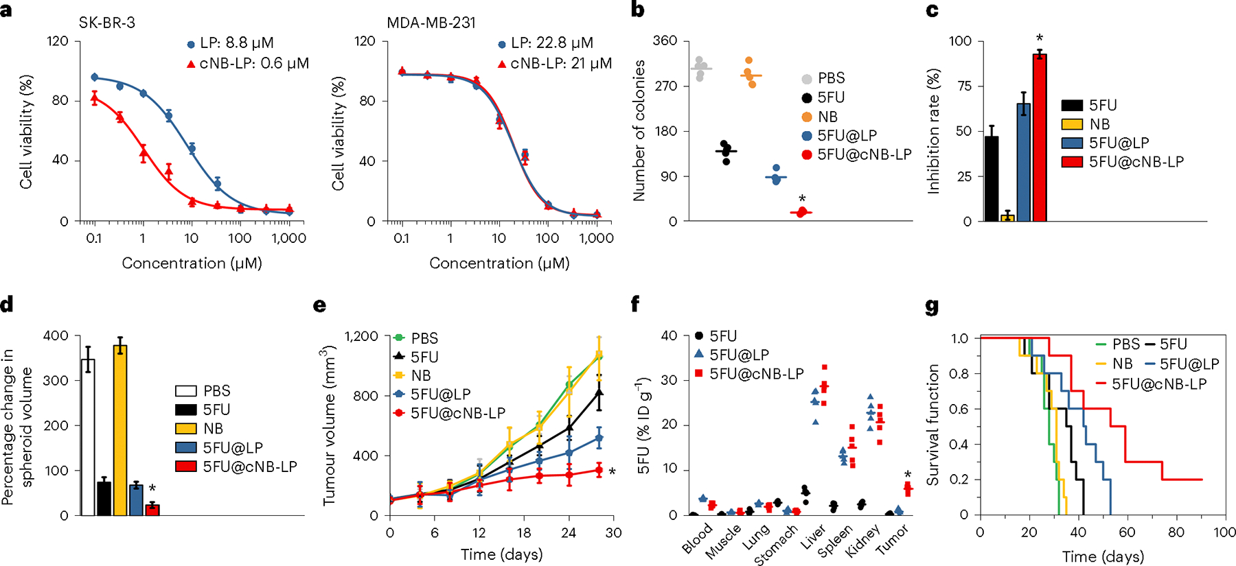
Liposomes as drug vehicles have advantages, such as payload protection, tunable carrying capacity and improved biodistribution. However, due to the dysfunction of targeting moieties and payload loss during preparation, immunoliposomes have yet to be favoured in commercial manufacturing. Here we report a chemical modification-free biophysical approach for producing immunoliposomes in one step through the self-assembly of a chimeric nanobody (cNB) into liposome bilayers. cNB consists of a nanobody against human epidermal growth factor receptor 2 (HER2), a flexible peptide linker and a hydrophobic single transmembrane domain. We determined that 64% of therapeutic compounds can be encapsulated into 100-nm liposomes, and up to 2,500 cNBs can be anchored on liposomal membranes without steric hindrance under facile conditions. Subsequently, we demonstrate that drug-loaded immunoliposomes increase cytotoxicity on HER2-overexpressing cancer cell lines by 10- to 20-fold, inhibit the growth of xenograft tumours by 3.4-fold and improve survival by more than twofold.


Conflict of interest statement

Competing interests

The authors declare no competing interests.

Figures

Fig. 1 |
Fig. 1 |. Schematic of immunoliposome preparation.
a, Flowchart illustrating the manufacturing processes of cNB. Alpacas were immunized with the HER2 extracellular domain, and subsequent isolation of peripheral blood mononuclear cells (PBMCs) was performed. Total RNA was extracted from the PBMCs followed by cDNA synthesis for amplifying variable heavy domain of heavy chain (VHH) gene regions. The PCR products were then ligated into the phagemid vector, and E. coli cells were transformed with the ligated products and cultured. Colonies were recovered for the biopanning of phage displayed VHH libraries. One specific VHH was selected and sequenced to determine its amino acid sequences. Subsequently, in the design process amino acids encoding a hydrophilic linker (shown in yellow) and a STMD (shown in purple) were added to the C-terminus of the NB. The corresponding cDNA was synthesized and integrated into plasmids for the expression of the cNB using E. coli cells. b, Overview of biophysical transformation due to size, surface charge, lipid fluidity, membrane stiffness and thermostability between liposome and cNB-LP. AA, amino acid.
Fig. 2 |
Fig. 2 |. Characterization of NB and engineered cNB.
a, Structural model of identified anti-HER2 NB, HER2 and the NB/HER2 complex. b, Crystal structure of NB/HER2 complex. c, Prediction of formed hydrogen bonds in NB/HER2 complex throughout 200 ns of molecular dynamics simulation (mean value ± s.d. shown). d, Crystal structure of NB, cNB that harbours only a flexible linker, cNB that harbours only a rigid linker, and cNB that harbours a flexible linker and STMD. e, Illustration of potential steric hindrance effect and cNB grafting density influenced different linkers or transmembrane domains. f, Designed amino acid (AA) sequence of cNB (top) and western blot of cNB with various STMD (bottom) prepared at 15 °C for 16 h (1) and at 37 °C for 4 h (2). Experiments were repeated five times. g, Kinetic analysis of cNB at pH 7.4. h, Isoelectric point analysis of cNB. Experiments were repeated five times.
Fig. 3 |
Fig. 3 |. Characterization of biophysical properties of cNB-LPs.
a, Immunofluorescence staining of a LP decorated with cNBs (top) and STMD-deficient cNBs (bottom). Scale bar, 5 μm. Experiments were repeated five times. b, TEM image shows the morphology of cNBs. Scale bar, 100 nm. Experiments were repeated three times. c, The cryo-TEM (left) and TEM (right) images show the membrane morphology of a typical cNB-LP2,000 and a typical plain LP. Scale bar, 50 nm. Experiments were repeated three times. d, Decoration efficiency of cNB as a function of cNB quantity (n = 12, mean value ± s.d.). e, Fluorescence signals of FRET-pair labelled cNB-LPs and LPs. f, The fluorescence signals of Laurdan emission from various cNB-LP at 20 °C and 42 °C. g, Stiffness distribution for free LPs and cNB-LPs (n = 100 in LP group; n = 87 in cNB-LP500 group; n = 51 in cNB-LP2,000 group; n = 34 in cNBLP4,000 group). h, SAXS scattering curves measured for plain LP, free cNB, and cNB-LP2,000 samples in solution.
Fig. 4 |
Fig. 4 |. Tumour treatment in vitro and in vivo.
a, Respective IC50 values of 5FU-loaded LPs and 5FU-loaded cNB-LPs in treatment of MDA-MB-231 cells and SK-BR-3 cells (n = 12, mean value ± s.d.). b, Growth inhibition on SK-BR-3 colonies in the respective group (n = 5, mean values shown; P < 0.001, one-way ANOVA). c, Inhibition of migration of SK-BR-3 cells in the respective group (n = 12, mean value ± s.d.; P < 0.001, one-way ANOVA). d, Volume changes in the SK-BR-3 spheroids in the respective group (n = 20, mean value ± s.d.; P < 0.001, one-way ANOVA). e, Tumour volume of SK-BR-3 orthotopic tumour xenograft in mice after drug or placebo administration. f, Biodistribution of 5FU in major organs at 24 h post-administration. Five biological replicates were measured (n = 5, mean values were shown; P < 0.001, one-way ANOVA). g, Survival curves for mice bearing SK-BR-3 orthotopic tumour after drug or placebo administration (ten mice in each group).

Similar articles

Cited by

References

    1. Sercombe L et al. Advances and challenges of liposome assisted drug delivery. Front. Pharmacol. 6, 286 (2015). - PMC - PubMed
    1. Liu Y, Castro Bravo KM & Liu J Targeted liposomal drug delivery: a nanoscience and biophysical perspective. Nanoscale Horiz. 6, 78–94 (2021). - PubMed
    1. Pattni BS, Chupin VV & Torchilin VP New developments in liposomal drug delivery. Chem. Rev. 115, 10938–10966 (2015). - PubMed
    1. Mitchell MJ et al. Engineering precision nanoparticles for drug delivery. Nat. Rev. Drug Discov. 20, 101–124 (2021). - PMC - PubMed
    1. Mamot C et al. Epidermal growth factor receptor-targeted immunoliposomes significantly enhance the efficacy of multiple anticancer drugs in vivo. Cancer Res. 65, 11631–11638 (2005). - PubMed

LinkOut - more resources