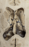
Stereotactic anatomy of the third ventricle
- PMID: 38374441
- PMCID: PMC10960742
- DOI: 10.1007/s00276-024-03312-1
Stereotactic anatomy of the third ventricle
Abstract
Purpose: Endoscopic third ventriculostomy (ETV) is a surgical procedure that can lead to complications and requires detailed preoperative planning. This study aimed to provide a more accurate understanding of the anatomy of the third ventricle and the location of important structures to improve the safety and success of ETV.
Methods: We measured the stereotactic coordinates of six points of interest relative to a predefined stereotactic reference point in 23 cadaver brain hemi-sections, 200 normal brain magnetic resonance imaging (MRI) scans, and 24 hydrocephalic brain MRI scans. The measurements were statistically analyzed, and comparisons were made.
Results: We found some statistically significant differences between genders in MRIs from healthy subjects. We also found statistically significant differences between MRIs from healthy subjects and both cadaver brains and MRIs with hydrocephalus, though their magnitude is very small and not clinically relevant. Some stereotactic points were more posteriorly and inferiorly located in cadaver brains, particularly the infundibular recess and the basilar artery. It was found that all stereotactic points studied were more posteriorly located in brains with hydrocephalus.
Conclusion: The study describes periventricular structures in cadaver brains and MRI scans from healthy and hydrocephalic subjects, which can guide neurosurgeons in planning surgical approaches to the third ventricle. Overall, the study contributes to understanding ETV and provides insights for improving its safety and efficacy. The findings also support that practicing on cadaveric brains can still provide valuable information and is valid for study and training of neurosurgeons unfamiliar with the ETV technique.
Keywords: Endoscopic third ventriculostomy; Hydrocephalus; Neuroanatomy; Neuronavigation; Stereotactic coordinates; Third ventricle.
© 2024. The Author(s).
Conflict of interest statement
The authors wish to declare no conflict of interests, and no commercial relationships.
Figures


References
-
- Decq P. Pediatric hydrocephalus. 2. Milano: Springer; 2005. Endoscopic anatomy of the ventricles; pp. 351–359.
MeSH terms
Grants and funding
LinkOut - more resources
Full Text Sources
Medical

