Review
. 2024 Mar 28;97(1156):695-704.
doi: 10.1093/bjr/tqae017.

State-of-the-art for contrast-enhanced mammography

Affiliations
Review

State-of-the-art for contrast-enhanced mammography

Matthew F Covington et al. Br J Radiol. .

Abstract

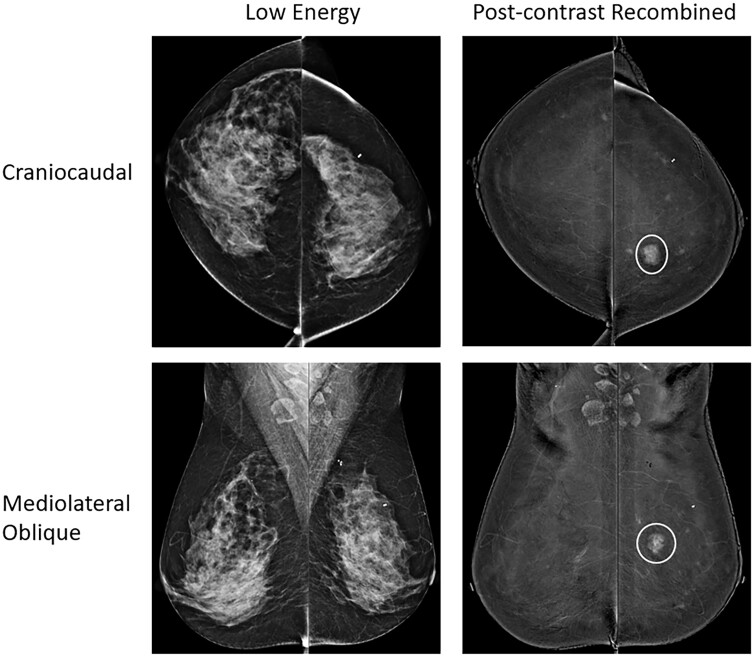
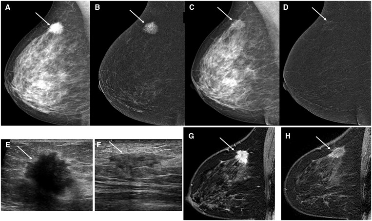
Contrast-enhanced mammography (CEM) is an emerging breast imaging technology with promise for breast cancer screening, diagnosis, and procedural guidance. However, best uses of CEM in comparison with other breast imaging modalities such as tomosynthesis, ultrasound, and MRI remain inconclusive in many clinical settings. This review article summarizes recent peer-reviewed literature, emphasizing retrospective reviews, prospective clinical trials, and meta-analyses published from 2020 to 2023. The intent of this article is to supplement prior comprehensive reviews and summarize the current state-of-the-art of CEM.

Keywords: CEDM; CEM; CESM; contrast-enhanced digital mammography; contrast-enhanced mammography; contrast-enhanced spectral mammography.


Conflict of interest statement

M.F.C.: Consultant for Invicro and GE Healthcare, and publishing royalties from Kindle Direct Publishing, outside of the scope of this topic. Institutional research support for contrast-enhanced mammography from Fujifilm Healthcare Americas and Bayer Healthcare. Research support from 5 For the Fight. S.S.: None. B.W.: None. L.L.F.: Consultant for WhiteRabbit.ai; Institutional research support for contrast-enhanced mammography from Fujifilm Healthcare Americas and Bayer Healthcare.

Figures

Figure 1.
Figure 1.
Standard contrast-enhanced mammography views are shown with craniocaudal (top row) and mediolateral oblique (bottom row) views of each breast depicted with low-energy images (right column) and post-contrast recombined images (left column). The white circles show an invasive ductal carcinoma in the medial left breast well-seen on the post-contrast recombined images, not seen on the low-energy images due to masking by dense breast tissue.
Figure 2.
Figure 2.
33-year-old woman with left breast calcifications found to represent ductal carcinoma in situ (DCIS) following core needle biopsy. Both CEM and MRI were performed for pre-surgical locoregional staging. (A) Left breast lateral magnification view demonstrates grouped mildly pleomorphic calcifications (white circle) and other more scattered round calcifications in the breast. (B) Low-energy CEM images in mediolateral oblique (MLO) projection show extremely dense breast tissue in both breasts with no additional significant mammographic finding. (C) Post-contrast recombined CEM images demonstrate widespread non-mass enhancement throughout the left breast on the MLO view suggesting multicentric DCIS (white arrows). An oval mass is also seen on the right MLO view which was not seen on standard mammography. (D) T1-fat saturated post-contrast sagittal breast MRI views demonstrate a similar degree of multicentric non-mass enhancement in the left breast (white arrows), and an oval enhancing mass in the right breast (white arrow). Final surgical pathology demonstrated 5.4 cm of multicentric DCIS in the left breast, corresponding with the extent of non-mass enhancement on CEM and MRI, and a right breast fibroadenoma accounting for the additional enhancing oval mass in the right breast on CEM and MRI.
Figure 3.
Figure 3.
53-year-old woman with histologically proven right breast invasive ductal carcinoma undergoing diagnostic imaging evaluation for response to neoadjuvant therapy. (A) Low-energy right breast mediolateral oblique (MLO) CEM image demonstrates a hyperdense spiculated mass in the superior posterior breast (white arrow) compatible with histologically proven right breast invasive ductal carcinoma. (B) Post-contrast recombined MLO view of the right breast shows the spiculated mass intensely enhances (white arrow) compatible with malignancy. (C) Low-energy MLO view of the right breast following neoadjuvant therapy demonstrates reduced size and density of known malignancy (white arrow) compatible with response to therapy. (D) Post-contrast recombined MLO view of the right breast following neoadjuvant therapy demonstrates mild residual enhancement at site of known malignancy (white arrow) suggesting treatment response with residual viable malignancy. (E) Right breast ultrasound image prior to neoadjuvant therapy demonstrates an irregular hypoechoic mass with posterior acoustic shadowing compatible with known malignancy. (F) Right breast ultrasound image following neoadjuvant therapy demonstrates reduced size of the known malignancy with some residual mass-like components (white arrow) suggesting partial treatment response. (G) T1-subtracted post-contrast MRI sagittal view of the right breast prior to neoadjuvant therapy demonstrates an intensely enhancing mass (white arrow) compatible with known malignancy. (H) T1-subtracted post-contrast MRI sagittal view of the right breast following neoadjuvant therapy demonstrates mild residual enhancement at site of known malignancy suggesting treatment response with residual viable malignancy. Final surgical pathology demonstrated treatment response of known invasive carcinoma with residual viable tumour compatible with the imaging findings on CEM, ultrasound, and MRI of partial response to therapy.

References

    1. Lewin JM, Patel BK, Tanna A.. Contrast-enhanced mammography: a scientific review. Journal of Breast Imaging. 2019;2(1):7–15. - PubMed
    1. Patel BK, Lobbes MBI, Lewin J.. Contrast enhanced spectral mammography: a review. Semin Ultrasound CT MR. 2018;39(1):70-79. - PubMed
    1. Hannsun G, Saponaro S, Sylvan P, Elmi A.. Contrast-enhanced mammography: technique, indications, and review of current literature. Curr Radiol Rep. 2021;9(11):911.
    1. Cozzi A, Magni V, Zanardo M, Schiaffino S, Sardanelli F.. Contrast-enhanced mammography: a systematic review and meta-analysis of diagnostic performance. Radiology. 2022;302(3):568-581. - PubMed
    1. Sogani J, Mango VL, Keating D, Sung JS, Jochelson MS.. Contrast-enhanced mammography: past, present, and future. Clin Imaging. 2021;69:269-279. https://www.sciencedirect.com/science/article/pii/S0899707120303454?via%... - PMC - PubMed