. 2024 Feb 5:12:1327772.
doi: 10.3389/fcell.2024.1327772. eCollection 2024.

Hypoxia at 3D organoid establishment selects essential subclones within heterogenous pancreatic cancer

Affiliations

Hypoxia at 3D organoid establishment selects essential subclones within heterogenous pancreatic cancer

Koichiro Kumano et al. Front Cell Dev Biol. .

Abstract

Pancreatic ductal adenocarcinoma (PDAC) is especially hypoxic and composed of heterogeneous cell populations containing hypoxia-adapted cells. Hypoxia as a microenvironment of PDAC is known to cause epithelial-mesenchymal transition (EMT) and resistance to therapy. Therefore, cells adapted to hypoxia possess malignant traits that should be targeted for therapy. However, current 3D organoid culture systems are usually cultured under normoxia, losing hypoxia-adapted cells due to selectivity bias at the time of organoid establishment. To overcome any potential selection bias, we focused on oxygen concentration during the establishment of 3D organoids. We subjected identical PDAC surgical samples to normoxia (O2 20%) or hypoxia (O2 1%), yielding glandular and solid organoid morphology, respectively. Pancreatic cancer organoids established under hypoxia displayed higher expression of EMT-related proteins, a Moffitt basal-like subtype transcriptome, and higher 5-FU resistance in contrast to organoids established under normoxia. We suggest that hypoxia during organoid establishment efficiently selects for hypoxia-adapted cells possibly responsible for PDAC malignant traits, facilitating a fundamental source for elucidating and developing new treatment strategies against PDAC.

Keywords: 3D organoid; hypoxia; pancreatic cancer; selection bias; tumor heterogeneity.


Conflict of interest statement

The authors declare that the research was conducted in the absence of any commercial or financial relationships that could be construed as a potential conflict of interest.

Figures

FIGURE 1
FIGURE 1
(A) Pancreatic cancer tissue used for organoids exhibits intratumoral heterogeneity. The histopathological diagnosis was pancreatic ductal adenocarcinoma (Pb, mod > por, pT2N2M0, pStageIII (UICC)). Immunohistochemistry staining of the primary tumor (H&E, E-cadherin, vimentin, and GATA6). Scale bars: 50 μm (B) Morphologic and immunohistochemical differences between NORMO-PCO and HYPO-PCO. Bright-field and immunohistochemistry staining of NORMO-PCO and HYPO-PCO (H&E, E-cadherin, vimentin, and GATA6). Scale bars: 50 μm (C) Proliferation and morphogenetic differences between NORMO-PCO and HYPO-PCO in pancreatic orthotopic mouse models. In both cases, tumors were identified in one of the two mice, with tumor diameters of 4 mm for NORMO-PCO and 8 mm for HYPO-PCO. No metastases were observed in both cases. Immunohistochemistry of xenograft sections from pancreatic orthotopic mouse models (H&E, E-cadherin and vimentin). Scale bars: 200 μm structured.
FIGURE 2
FIGURE 2
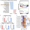
(A) Gene set enrichment analysis (GSEA) of NORMO-PCO relative to HYPO-PCO. EMT- and basal-like-related genes are highly expressed in HYPO-PCO. The red bar graph indicates gene sets enriched in NORMO-PCO and blue indicates gene sets enriched in HYPO-PCO. The horizontal axis shows NES value. On the right, enrichment scores for EMT (NES = 1.66, FDR = 0.073) and basal-like type (NES = 1.59, FDR = 0.065) by Moffitt classification are shown (B) Comparisons of expression of EMT-related genes (CDH1, VIM) and Moffit classification-related genes (GATA6, TP63, S100A2) in the data from RNA seq, respectively. *p < 0.05, ***p < 0.001 (unpaired Student’s t-test) (C) Normalized expression (log2 scale) of gene pairs assessed by purity independent subtyping of tumors (PurIST). Normalized expression (log2 scale).
FIGURE3
FIGURE3
Chemotherapy resistance experiments which conducted in biological triplicate revealed 5-FU resistance in HYPO-PCO. Cell viability was analyzed by using CellTiter-Glo 2.0. Dose response curves for gemcitabine range from 1.0 × 10⁻¹² to 1.0 × 10⁻⁴ mol/L and 5-FU range from 1.0 × 10⁻¹⁰ to 1.0 × 10⁻⁴. Red dots indicate NORMO-PCO; blue dots indicate HYPO-PCO. Drug concentration is in log10 [M].
FIGURE 4
FIGURE 4
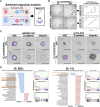
(A) Analysis after switching oxygen conditions reveals that NORMO-PCO and HYPO-PCO are irreversible. Experimental scheme. NORMO-PCO was cultured in hypoxia and HYPO-PCO in normoxia for three passages before morphology and transcriptome analysis (B) Bright field microscopy under each oxygen concentration. Scale bars: 50 μm. The growth curves of the organoids under each oxygen concentration. Proliferation normalized to 24 h (C) Immunohistochemistry of NORMO-PCO and HYPO-PCO (H&E, E-cadherin, vimentin). Scale bars: 50 μm (D) Comparison of GSEA analysis of NORMO-PCO and HYPO-PCO in matching oxygen niches. The red bar indicates gene sets enriched in NORMO-PCO, and blue indicates gene sets enriched in HYPO-PCO.

References

    1. abou Khouzam R., Rao S. P., Venkatesh G. H., Zeinelabdin N. A., Buart S., Meylan M., et al. (2021). An eight-gene hypoxia signature predicts survival in pancreatic cancer and is associated with an immunosuppressed tumor microenvironment. Front. Immunol. 12, 680435. 10.3389/fimmu.2021.680435 - DOI - PMC - PubMed
    1. Angst E., Sibold S., Tiffon C., Weimann R., Gloor B., Candinas D., et al. (2006). Cellular differentiation determines the expression of the hypoxia-inducible protein NDRG1 in pancreatic cancer. Br. J. Cancer 95, 307–313. 10.1038/sj.bjc.6603256 - DOI - PMC - PubMed
    1. Balamurugan K. (2016). HIF-1 at the crossroads of hypoxia, inflammation, and cancer. Int. J. Cancer 138, 1058–1066. 10.1002/ijc.29519 - DOI - PMC - PubMed
    1. Chang Q., Jurisica I., Do T., Hedley D. W. (2011). Hypoxia predicts aggressive growth and spontaneous metastasis formation from orthotopically grown primary xenografts of human pancreatic cancer. Cancer Res. 71, 3110–3120. 10.1158/0008-5472.CAN-10-4049 - DOI - PubMed
    1. Chen S., Chen J. Z., Zhang J. Q., Chen H. X., Yan M. L., Huang L., et al. (2016). Hypoxia induces TWIST-activated epithelial-mesenchymal transition and proliferation of pancreatic cancer cells in vitro and in nude mice. Cancer Lett. 383, 73–84. 10.1016/j.canlet.2016.09.027 - DOI - PubMed