. 2024 Feb 5:15:1332175.
doi: 10.3389/fmicb.2024.1332175. eCollection 2024.

Pseudorabies virus uses clathrin mediated endocytosis to enter PK15 swine cell line

Affiliations

Pseudorabies virus uses clathrin mediated endocytosis to enter PK15 swine cell line

Sabina Andreu et al. Front Microbiol. .

Abstract

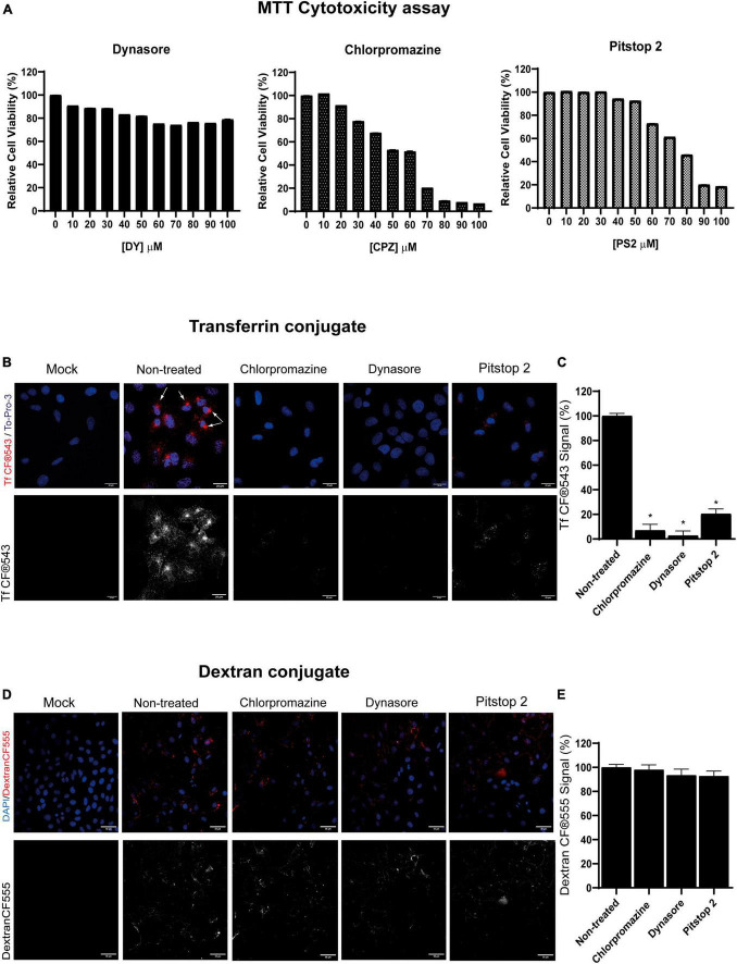
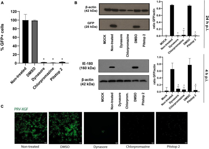
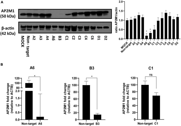
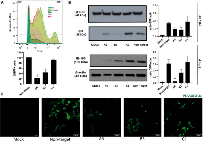
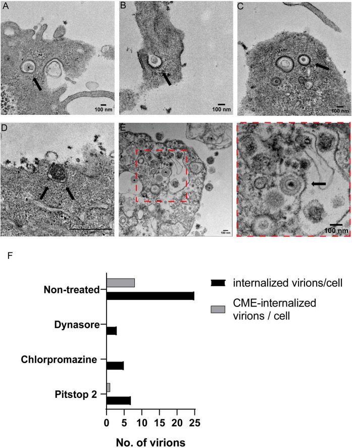
Pseudorabies virus (PRV), a herpesvirus responsible for Aujeszky's disease, causes high mortality in swine populations. To develop effective and novel antiviral strategies, it is essential to understand the mechanism of entry used by PRV to infect its host. Viruses have different ways of entering host cells. Among others, they can use endocytosis, a fundamental cellular process by which substances from the external environment are internalized into the cell. This process is classified into clathrin-mediated endocytosis (CME) and clathrin-independent endocytosis (CIE), depending on the role of clathrin. Although the involvement of cholesterol-rich lipid rafts in the entry of PRV has already been described, the importance of other endocytic pathways involving clathrin remains unexplored to date. Here, we characterize the role of CME in PRV entry into the PK15 swine cell line. By using CME inhibitory drugs, we report a decrease in PRV infection when the CME pathway is blocked. We also perform the shRNA knockdown of the μ-subunit of the adaptor protein AP-2 (AP2M1), which plays an important role in the maturation of clathrin-coated vesicles, and the infection is greatly reduced when this subunit is knocked down. Furthermore, transmission electron microscopy images report PRV virions inside clathrin-coated vesicles. Overall, this study suggests for the first time that CME is a mechanism used by PRV to enter PK15 cells and provides valuable insights into its possible routes of entry.

Keywords: PK15 cell line; clathrin; herpesvirus; pseudorabies virus; viral entry.


Conflict of interest statement

The authors declare that the research was conducted in the absence of any commercial or financial relationships that could be construed as a potential conflict of interest.

Figures

FIGURE 1
FIGURE 1
Clathrin-mediated endocytosis (CME) chemical inhibitors dynasore, chlorpromazine, and pitstop 2 disrupt transferrin but no dextran uptake at non-cytotoxic doses. (A) Cellular viability of PK15 cells exposed to dynasore, chlorpromazine, and pitstop 2 for 24 h. Cell viability was measured by MTT tetrazolium salt assay and calculated as the percentage of cell viability compared to untreated cells; columns represent the mean percentage of relative cellular viability ± S.D. (n = 4) after exposure to the drugs. (B) Transferrin uptake in PK15 cells is blocked by CME chemical inhibitors. PK15 cells were mock-treated or treated with either 20 μM chlorpromazine, 100 μM dynasore, or 50 μM pitstop 2 for 1 h and then incubated for 30 min on ice with Tf CF®543 (5 μg/ml). Finally, cells were fixed after 5 min of incubation at 37°C. Fluorescence microscopy images are shown, with Tf in red and cellular nuclei stained with To-Pro-3 in blue. Arrows point to the accumulation of Tf in the endosomal recycling compartments. (C) Quantification of Tf CF®543. Cells were acquired and analyzed as in (B,D); ROIs from groups of 30 cells and 3 areas of each image were measured. Mean percentage of fluorescence ± S.D. is shown. (D) Dextran-CF®555 uptake is not altered in PK15 cells treated with CME-inhibitory drugs. PK15 cells were incubated or mock-incubated as described in (B) but instead of Tf, they were incubated with CF®555 Labeled Dye Dextran 10,000 MW (5 μg/mL) for 15 min at 4°C. Cells were then transferred to 37°C for 10 min, washed, and then fixed with 4% PFA and stained as before. (E) Quantification of CF®555 Dextran. Cells were acquired and analyzed as previously described. Measurement of mean fluorescence intensity from 555 channel in a ROI was performed. Mean percentage of fluorescence ± S.D. is shown.
FIGURE 2
FIGURE 2
Pseudorabies virus (PRV)-XGF-N infection is blocked by CME chemical inhibitors. PK15 cells were pre-treated with either 0.1% (v/v) DMSO, 100 μM dynasore, 20 μM chlorpromazine, or 50 μM pitstop 2 for 1 h and then infected with PRV-XGF-N at an m.o.i of 0.5 (collected at 24 h p.i.) or moi 5 (collected at 4 h p.i.). Cells were maintained until they were collected in the presence of the drugs. (A) Flow cytometry data show the percentage of normalized infection 24 h p.i. (% GFP+ cells) ± S.D. for each condition. Triplicate experiments were performed for each data point (n = 3). *p < 0.05. (B) Western blot analysis of total cell lysates showing viral GFP and IE180 for each condition. β-actin was used as protein loading control. Values of Western blot quantification are reported as the mean ± S.D. (n = 3), *p < 0.05. (C) Fluorescence microscopy images show GFP+ signal corresponding to viral infection 24 h p.i. (n = 3).
FIGURE 3
FIGURE 3
Determination of AP2M1 shRNA knockdown efficiency in PK15 cells. PK15 cells were transfected with a pool of shRNA to KD the expression of AP2M1. KD efficiency was checked by both Western blot and RT-qPCR. (A) Western blot analysis of total cell lysates subjected to SDS-PAGE showing AP2M1 for each condition. β-actin was chosen as protein loading control. Values of immunoblot quantification are reported as the mean ± S.D. (n = 3); *p < 0.05. (B) qPCR amplification of AP2M1 mRNA from AP2M1 A6, B3, and C1 shRNA-treated PK15 cells in comparison to the non-targeted control. mRNA level is relative to β-actin (ACTB) control (n = 6, mean ± S.D.). *p < 0.05 was considered as statistically significant compared to the non-targeting shRNA-transfected cells.
FIGURE 4
FIGURE 4
The knockdown of AP2M1 in PK15 cells reduces PRV infection. KD PK15 cells A6, B3, and C1, and non-target cells were subjected to infection with PRV-XGF-N at an m.o.i of 0.5 (collected at 24 h p.i.) or moi 5 (collected at 4 h p.i.). After the corresponding time, samples were processed for the following experiments. (A) Flow cytometry analysis show the mean of the percentage of normalized infection 24 h p.i. (%GFP+ cells) ± S.D. (n = 4). Plots represent the histograms of GFP-positive (+) and GFP-negative (-) cells; *p < 0.05. (B) Western blot analysis of total cell lysates subjected to SDS-PAGE showing viral GFP or IE180 for each condition. β-actin was chosen as protein loading control. Values of immunoblot quantification are reported as the mean ± S.D. (n = 3); *p < 0.05. (C) Fluorescence microscopy images show GFP+ signal corresponding to viral infection 24 h p.i.
FIGURE 5
FIGURE 5
Transmission electron microscopy analysis of PRV-XGF-N virions inside clathrin vesicles in PK15 cells. Cells were infected with PRV-XGF-N at an m.o.i of 30 for 1 h at 4°C. Then they were maintained for 20 min at 37°C and processed for TEM. Arrows point to in (A–C) PRV-XGF-N virions inside clathrin vesicles, (D) virion entering a forming clathrin vesicle anchored in the cell membrane, and (E) overview of PRV-XGF-N surrounded by clathrin. (F) Quantification of the number of internalized virions per cell (n = 20 cells analyzed) in cells non-treated or treated with 100 μM dynasore, 20 μM chlorpromazine or 50 μM pitstop 2 for 1 h before infection, and during the 20 min of infection. The graph shows the number of virions inside the cell and from those, the virions entering via CME or internalized into CCVs.

Similar articles

Cited by

References

    1. Albecka A., Laine R. F., Janssen A. F. J., Kaminski C. F., Crump C. M. (2016). HSV-1 glycoproteins are delivered to virus assembly sites through dynamin-dependent endocytosis. Traffic 17 21–39. 10.1111/tra.12340 - DOI - PMC - PubMed
    1. Andreu S., Ripa I., Bello-Morales R., López-Guerrero J. A. (2020). Valproic acid and its amidic derivatives as new antivirals against alphaherpesviruses. Viruses 12:1356. 10.3390/v12121356 - DOI - PMC - PubMed
    1. Andreu S., Ripa I., Praena B., López-guerrero J. A., Bello-Morales R. (2021). The valproic acid derivative valpromide inhibits pseudorabies virus infection in swine epithelial and mouse neuroblastoma cell lines. Viruses 13:2522. 10.3390/v13122522 - DOI - PMC - PubMed
    1. Barrow E., Nicola A. V., Liu J. (2013). Multiscale perspectives of virus entry via endocytosis. Virol. J. 10:177. 10.1186/1743-422X-10-177 - DOI - PMC - PubMed
    1. Bayati A., Kumar R., Francis V., McPherson P. S. (2021). SARS-CoV-2 infects cells after viral entry via clathrin-mediated endocytosis. J. Biol. Chem. 296:100306. - PMC - PubMed