

Corneal Infantile Myofibromatosis Caused by Novel Activating Imatinib-Responsive Variants in PDGFRB
- PMID: 38374928
- PMCID: PMC10875226
- DOI: 10.1016/j.xops.2023.100444
Corneal Infantile Myofibromatosis Caused by Novel Activating Imatinib-Responsive Variants in PDGFRB
Abstract
Purpose: To investigate the genetic cause, clinical characteristics, and potential therapeutic targets of infantile corneal myofibromatosis.
Design: Case series with genetic and functional in vitro analyses.
Participants: Four individuals from 2 unrelated families with clinical signs of corneal myofibromatosis were investigated.
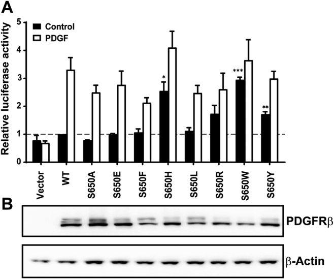
Methods: Exome-based panel sequencing for platelet-derived growth factor receptor beta gene (PDGFRB) and notch homolog protein 3 gene (NOTCH3) was performed in the respective index patients. One clinically affected member of each family was tested for the pathogenic variant detected in the respective index by Sanger sequencing. Immunohistochemical staining on excised corneal tissue was conducted. Functional analysis of the individual PDGFRB variants was performed in vitro by luciferase reporter assays on transfected porcine aortic endothelial cells using tyrosine kinase inhibitors. Protein expression analysis of mutated PDGFRB was analyzed by Western blot.
Main outcome measures: Sequencing data, immunohistochemical stainings, functional analysis of PDGFRB variants, and protein expression analysis.
Results: We identified 2 novel, heterozygous gain-of-function variants in PDGFRB in 4 individuals from 2 unrelated families with corneal myofibromatosis. Immunohistochemistry demonstrated positivity for alpha-smooth muscle actin and β-catenin, a low proliferation rate in Ki-67 (< 5%), marginal positivity for Desmin, and negative staining for Caldesmon and CD34. In all patients, recurrence of disease occurred after corneal surgery. When transfected in cultured cells, the PDGFRB variants conferred a constitutive activity to the receptor in the absence of its ligand and were sensitive to the tyrosine kinase inhibitor imatinib. The variants can both be classified as likely pathogenic regarding the American College of Medical Genetics and Genomics classification criteria.
Conclusions: We describe 4 cases of corneal myofibromatosis caused by novel PDGFRB variants with autosomal dominant transmission. Imatinib sensitivity in vitro suggests perspectives for targeted therapy preventing recurrences in the future.
Financial disclosures: Proprietary or commercial disclosure may be found in the Footnotes and Disclosures at the end of this article.
Keywords: Corneal myofibroma; Imatinib; Infantile myofibromatosis; PDGFRB; Receptor tyrosine kinase inhibitor.
© 2023 by the American Academy of Ophthalmology.
Figures


Similar articles
-
Association of PDGFRB Mutations With Pediatric Myofibroma and Myofibromatosis.JAMA Dermatol. 2019 Aug 1;155(8):946-950. doi: 10.1001/jamadermatol.2019.0114. JAMA Dermatol. 2019. PMID: 31017643 Free PMC article.
-
A germline PDGFRB splice site variant associated with infantile myofibromatosis and resistance to imatinib.Genet Med. 2025 Feb;27(2):101334. doi: 10.1016/j.gim.2024.101334. Epub 2024 Nov 21. Genet Med. 2025. PMID: 39580648
-
Novel PDGFRB rearrangement in multifocal infantile myofibromatosis is tumorigenic and sensitive to imatinib.Cold Spring Harb Mol Case Stud. 2019 Oct 23;5(5):a004440. doi: 10.1101/mcs.a004440. Print 2019 Oct. Cold Spring Harb Mol Case Stud. 2019. PMID: 31645346 Free PMC article.
-
Infantile Myofibromatosis With Intracranial Extradural Involvement and PDGFRB Mutation: A Case Report and Review of the Literature.Pediatr Dev Pathol. 2019 May-Jun;22(3):258-264. doi: 10.1177/1093526618787736. Epub 2018 Aug 13. Pediatr Dev Pathol. 2019. PMID: 30103666 Review.
-
PDGF receptor mutations in human diseases.Cell Mol Life Sci. 2021 Apr;78(8):3867-3881. doi: 10.1007/s00018-020-03753-y. Epub 2021 Jan 15. Cell Mol Life Sci. 2021. PMID: 33449152 Free PMC article. Review.
Cited by
-
Current Advances in Corneal Stromal Stem Cell Biology and Therapeutic Applications.Cells. 2024 Jan 16;13(2):163. doi: 10.3390/cells13020163. Cells. 2024. PMID: 38247854 Free PMC article. Review.
References
-
- Gommans L.N.M., Spring in 't Veld L.G., van der Putten M.E., Wijnen M.H. [Infantile myofibroma: a neonate with a swelling on the arm] Ned Tijdschr Geneeskd. 2015;159 - PubMed
-
- Wiswell T.E., Davis J., Cunningham B.E., et al. Infantile myofibromatosis: the most common fibrous tumor of infancy. J Pediatr Surg. 1988;23:314–318. - PubMed
-
- Choopong P., Nielsen P.G., Perlman E.M., et al. Solitary myofibroma of the sclera. Cornea. 2007;26:114–116. - PubMed
-
- Chung E., Enzinger F.M. Infantile myofibromatosis. Cancer. 1981;48:1807–1818. - PubMed
-
- Chung E.M., Smirniotopoulos J.G., Specht C.S., et al. Pediatric orbit tumors and tumorlike lesions: nonosseous lesions of the extraocular orbit. Radiographics. 2007;27:1777–1799. - PubMed
LinkOut - more resources
Full Text Sources
Miscellaneous
