

Neural correlates of fine motor grasping skills: Longitudinal insights into motor cortex activation using fNIRS
- PMID: 38376039
- PMCID: PMC10784192
- DOI: 10.1002/brb3.3383
Neural correlates of fine motor grasping skills: Longitudinal insights into motor cortex activation using fNIRS
Abstract
Background: Motor learning is essential for performing specific tasks and progresses through distinct stages, including the rapid learning phase (initial skill acquisition), the consolidation phase (skill refinement), and the stable performance phase (skill mastery and maintenance). Understanding the cortical activation dynamics during these stages can guide targeted rehabilitation interventions.
Methods: In this longitudinal randomized controlled trial, functional near-infrared spectroscopy was used to explore the temporal dynamics of cortical activation in hand-related motor learning. Thirty-one healthy right-handed individuals were randomly assigned to perform either easy or intricate motor tasks with their non-dominant hand over 10 days. We conducted 10 monitoring sessions to track cortical activation in the right hemisphere (according to lateralization principles, the primary hemisphere for motor control) and evaluated motor proficiency concurrently.
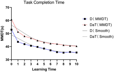
Results: The study delineated three stages of nondominant hand motor learning: rapid learning (days 1 and 2), consolidation (days 3-7), and stable performance (days 8-10). There was a power-law enhancement of motor skills correlated with learning progression. Sustained activation was observed in the supplementary motor area (SMA) and parietal lobe (PL), whereas activation in the right primary motor cortex (M1R) and dorsolateral prefrontal cortex (PFCR) decreased. These cortical activation patterns exhibited a high correlation with the augmentation of motor proficiency.
Conclusions: The findings suggest that early rehabilitation interventions, such as transcranial magnetic stimulation and transcranial direct current stimulation (tDCS), could be optimally directed at M1 and PFC in the initial stages. In contrast, SMA and PL can be targeted throughout the motor learning process. This research illuminates the path for developing tailored motor rehabilitation interventions based on specific stages of motor learning.
New and noteworthy: In an innovative approach, our study uniquely combines a longitudinal design with the robustness of generalized estimating equations (GEEs). With the synergy of functional near-infrared spectroscopy (fNIRS) and the Minnesota Manual Dexterity Test (MMDT) paradigm, we precisely trace the evolution of neural resources during complex, real-world fine-motor task learning. Centering on right-handed participants using their nondominant hand magnifies the intricacies of right hemisphere spatial motor processing. We unravel the brain's dynamic response throughout motor learning stages and its potent link to motor skill enhancement. Significantly, our data point toward the early-phase rehabilitation potential of TMS and transcranial direct current stimulation on the M1 and PFC regions. Concurrently, SMA and PL appear poised to benefit from ongoing interventions during the entire learning curve. Our findings carve a path for refined motor rehabilitation strategies, underscoring the importance of timely noninvasive brain stimulation treatments.
Keywords: cortical activation; functional near-infrared spectroscopy (fNIRS); longitudinal study; motor learning; motor rehabilitation.
© 2024 The Authors. Brain and Behavior published by Wiley Periodicals LLC.
Conflict of interest statement
The authors declare no conflicts of interest.
Figures


Similar articles
-
Mapping Motor Learning Stages: A Longitudinal fNIRS-Based Assessment of Cortical Activation.Curr Protoc. 2025 Jun;5(6):e70147. doi: 10.1002/cpz1.70147. Curr Protoc. 2025. PMID: 40536095
-
Age-specific neural responses to SMA and M1 stimulation during implicit motor sequence learning: Insights from a concurrent tDCS-fNIRS approach.Neuroscience. 2025 Jun 21;577:240-251. doi: 10.1016/j.neuroscience.2025.05.002. Epub 2025 May 8. Neuroscience. 2025. PMID: 40345479
-
Transcranial direct current stimulation to the left dorsolateral prefrontal cortex enhances early dexterity skills with the left non-dominant hand: a randomized controlled trial.J Transl Med. 2023 Feb 24;21(1):143. doi: 10.1186/s12967-023-03989-9. J Transl Med. 2023. PMID: 36823635 Free PMC article. Clinical Trial.
-
Transcranial electrical stimulation for procedural learning and rehabilitation.Neurobiol Learn Mem. 2024 Sep;213:107958. doi: 10.1016/j.nlm.2024.107958. Epub 2024 Jul 5. Neurobiol Learn Mem. 2024. PMID: 38971460 Review.
-
Motor learning in man: a review of functional and clinical studies.J Physiol Paris. 2006 Jun;99(4-6):414-24. doi: 10.1016/j.jphysparis.2006.03.007. Epub 2006 May 26. J Physiol Paris. 2006. PMID: 16730432 Review.
Cited by
-
Degradation-aware neural imputation: Advancing decoding stability in brain machine interfaces.APL Bioeng. 2025 Apr 16;9(2):026106. doi: 10.1063/5.0250296. eCollection 2025 Jun. APL Bioeng. 2025. PMID: 40247859 Free PMC article.
References
Publication types
MeSH terms
LinkOut - more resources
Full Text Sources
Miscellaneous
