

Functional neural networks stratify Parkinson's disease patients across the spectrum of cognitive impairment
- PMID: 38376051
- PMCID: PMC10808882
- DOI: 10.1002/brb3.3395
Functional neural networks stratify Parkinson's disease patients across the spectrum of cognitive impairment
Abstract
Introduction: Cognitive impairment (CI) is a significant non-motor symptoms in Parkinson's disease (PD) that often precedes the emergence of motor symptoms by several years. Patients with PD hypothetically progress from stages without CI (PD-normal cognition [NC]) to stages with Mild CI (PD-MCI) and PD dementia (PDD). CI symptoms in PD are linked to different brain regions and neural pathways, in addition to being the result of dysfunctional subcortical regions. However, it is still unknown how functional dysregulation correlates to progression during the CI. Neuroimaging techniques hold promise in discriminating CI stages of PD and further contribute to the biomarker formation of CI in PD. In this study, we explore disparities in the clinical assessments and resting-state functional connectivity (FC) among three CI stages of PD.
Methods: We enrolled 88 patients with PD and 26 healthy controls (HC) for a cross sectional clinical study and performed intra- and inter-network FC analysis in conjunction with comprehensive clinical cognitive assessment.
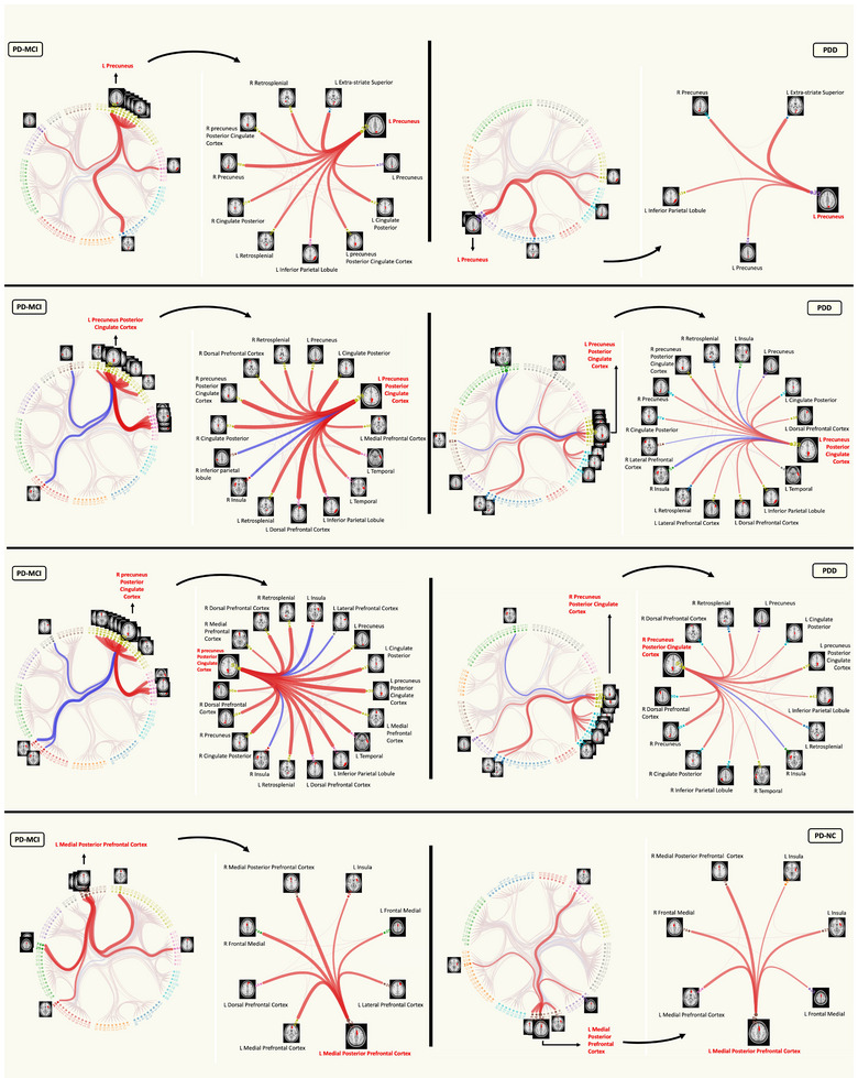
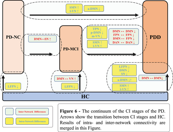
Results: Our findings underscore the significance of several neural networks, namely, the default mode network (DMN), frontoparietal network (FPN), dorsal attention network, and visual network (VN) and their inter-intra-network FC in differentiating between PD-MCI and PDD. Additionally, our results showed the importance of sensory motor network, VN, DMN, and salience network (SN) in the discriminating PD-NC from PDD. Finally, in comparison to HC, we found DMN, FPN, VN, and SN as pivotal networks for further differential diagnosis of CI stages of PD.
Conclusion: We propose that resting-state networks (RSN) can be a discriminating factor in distinguishing the CI stages of PD and progressing from PD-NC to MCI or PDD. The integration of clinical and neuroimaging data may enhance the early detection of PD in clinical settings and potentially prevent the disease from advancing to more severe stages.
Keywords: MCI; Parkinson's disease; cognitive impairment; dementia; fMRI; resting-state.
© 2024 The Authors. Brain and Behavior published by Wiley Periodicals LLC.
Conflict of interest statement
The authors declare no conflicts of interest.
Figures


Similar articles
-
A Cross Sectional and Longitudinal Assessment of Neuropsychiatric Symptoms and Brain Functional Connectivity in Patients With Mild Cognitive Impairment, Cerebrovascular Disease and Parkinson Disease.Int J Geriatr Psychiatry. 2025 Apr;40(4):e70075. doi: 10.1002/gps.70075. Int J Geriatr Psychiatry. 2025. PMID: 40246706 Free PMC article.
-
Dynamic functional connectivity in Parkinson's disease patients with mild cognitive impairment and normal cognition.Neuroimage Clin. 2017 Dec 9;17:847-855. doi: 10.1016/j.nicl.2017.12.013. eCollection 2018. Neuroimage Clin. 2017. PMID: 29527489 Free PMC article.
-
Detection of visual and frontoparietal network perfusion deficits in Parkinson's disease dementia.Eur J Radiol. 2021 Nov;144:109985. doi: 10.1016/j.ejrad.2021.109985. Epub 2021 Sep 28. Eur J Radiol. 2021. PMID: 34619619
-
Meta-Analysis of Cognition in Parkinson's Disease Mild Cognitive Impairment and Dementia Progression.Neuropsychol Rev. 2022 Mar;32(1):149-160. doi: 10.1007/s11065-021-09502-7. Epub 2021 Apr 16. Neuropsychol Rev. 2022. PMID: 33860906 Review.
-
Cognitive training interventions for dementia and mild cognitive impairment in Parkinson's disease.Cochrane Database Syst Rev. 2020 Feb 26;2(2):CD011961. doi: 10.1002/14651858.CD011961.pub2. Cochrane Database Syst Rev. 2020. PMID: 32101639 Free PMC article.
References
-
- Amboni, M. , Tessitore, A. , Esposito, F. , Santangelo, G. , Picillo, M. , Vitale, C. , Giordano, A. , Erro, R. , de Micco, R. , Corbo, D. , Tedeschi, G. , & Barone, P. (2015). Resting‐state functional connectivity associated with mild cognitive impairment in Parkinson's disease. Journal of Neurology, 262(2), 425–434. 10.1007/s00415-014-7591-5 - DOI - PubMed
-
- Aracil‐Bolaños, I. , Sampedro, F. , Marin, J. , Horta‐Barba, A. , Martinez‐Horta, S. , Gónzalez‐de‐Echávarri, J. , Pérez‐Pérez, J. , Bejr‐Kasem, H. , Md, B. , Botí, M. , Campolongo, A. , Izquierdo, C. , Gironell, A. , Gómez‐Ansón, B. , Kulisevsky, J. , & Md, J. (2022). Tipping the scales: How clinical assessment shapes the neural correlates of Parkinson's disease mild cognitive impairment. Brain Imaging and Behavior, 16, 761–772. 10.1007/s11682-021-00543-3 - DOI - PubMed
Publication types
MeSH terms
Grants and funding
LinkOut - more resources
Full Text Sources
Medical
