. 2024 Mar 5;63(5):625-631.
doi: 10.1021/acs.biochem.3c00647. Epub 2024 Feb 20.

Lipid-Dependent Activation of the Orphan G Protein-Coupled Receptor, GPR3

Affiliations

Lipid-Dependent Activation of the Orphan G Protein-Coupled Receptor, GPR3

Isabella C Russell et al. Biochemistry. .

Abstract

The class A orphan G protein-coupled receptor (GPCR), GPR3, has been implicated in a variety of conditions, including Alzheimer's and premature ovarian failure. GPR3 constitutively couples with Gαs, resulting in the production of cAMP in cells. While tool compounds and several putative endogenous ligands have emerged for the receptor, its endogenous ligand, if it exists, remains a mystery. As novel potential drug targets, the structures of orphan GPCRs have been of increasing interest, revealing distinct modes of activation, including autoactivation, presence of constitutively activating mutations, or via cryptic ligands. Here, we present a cryo-electron microscopy (cryo-EM) structure of the orphan GPCR, GPR3 in complex with DNGαs and Gβ1γ2. The structure revealed clear density for a lipid-like ligand that bound within an extended hydrophobic groove, suggesting that the observed "constitutive activity" was likely due to activation via a lipid that may be ubiquitously present. Analysis of conformational variance within the cryo-EM data set revealed twisting motions of the GPR3 transmembrane helices that appeared coordinated with changes in the lipid-like density. We propose a mechanism for the binding of a lipid to its putative orthosteric binding pocket linked to the GPR3 dynamics.


Conflict of interest statement

The authors declare the following competing financial interest(s): P.M.S. is a co-founder and shareholder of Septerna Inc. and DACRA Tx. D.W. is a shareholder in Septerna Inc. and co-founder and shareholder of DACRA Tx. M.M.F. is an employee of AstraZeneca.

Figures

Figure 1
Figure 1
(A) Normalized bioluminescence resonance energy transfer (BRET) between Rluc8-tagged GPR3 and split-Venus tagged Gβ1γ2. Upon addition of GDP-βs (100 μM; black line) to apyrase-treated permeabilized cells, the constitutively active receptor stimulates turnover of the precomplexed Gαs heterotrimer. Gαs heterotrimer dissociation from the receptor is indicated by the reduction in the BRET ratio due to separation of the Venus and Rluc8 tags. BRET ratios (ΔBRET) were baseline corrected twice. First, the average of the first two data points was subtracted from the data points of each individual experiment. Second, the values from experiments where no GDP-βs was added were subtracted from the corresponding values of experiments where GDP-βs was added after the second baseline read. The data shown in panel A represent the average (± SEM) difference from the baseline of four independent experiments (n = 4), each performed in technical duplicate. (B) Basal cAMP accumulation over 60 min expressed as pM/well. HEK293a cells were transiently transfected in suspension with a tagged GPR3 or pcDNA3.1 vector control. Data shown as the means ± SEM where n = 3 independent experiments with individual mean experimental data points displayed. (C) Sequence comparison of key conserved class A GPCR motifs. The related orphan receptors, GPR3 and GPR12, are compared to the prototypical class A GPCR, β1-adrenoreceptor (β1AR), and the related lipid receptor, sphingosine-1-phosphate-receptor 1 (S1P1R). Residues are color coded as follows: red, acidic/negatively charged; orange, aliphatic; yellow, aromatic; green, polar/neutral; blue, basic/positively charged. Sequence comparison was performed using GPCRdb (www.gpcrdb.org), and aligned residues are numbered using the Ballesteros–Weinstein numbering system.
Figure 2
Figure 2
(A) Cryo-EM structure of GPR3:DNGαs:Gβ1γ2. GPR3: cyan, lipid: red, DNGαs: black, Gβ1: dark gray, Gγ2: light gray. (B) The placement of ECL2, as well as the extracellular portion of the TMD, largely occludes access to the intracellular domain, although a small entrance can still be observed. (C) Model of potential lipid(s) in the TM cavity. The lipid was modeled as a simple 16 carbon saturated lipid, with two individual lipids modeled (red and yellow) to account for the extended density in the cryo-EM map. Receptor residues that are in the proximity of the lipid are represented as sticks. These residues create a hydrophobic “channel” where the lipid is proposed to bind. These residues include key class A activation motifs: the PIF motif and the W6.48 toggle switch. (D) Hydrophobic surface representation of the GPR3, including the placement of the lipid in its “upper” position.
Figure 3
Figure 3
(A) Cartoon schematic illustrating the rearrangement of the ECL2 and TMD observed in the 3DVA (further details supplied in the legend for Supplementary Video 1). (B) Placement of the lipid in its “upper” position (left) and its “lower” position (right).
Figure 4
Figure 4
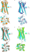
Side and top views of GPR3 superimposed with similar lipid binding receptors. (A) S1P1R with its endogenous lipid S1P (PDB: 7VIE). (B) GPR119 with its endogenous lipid (PDB: 7XZ5). (C) CB1R was bound to an endocannabinoid-like analogue (PDB: 8GHV). (D) GPR174 was bound to the endogenous lipid LPC (7XV3). The placement of the TMs is equivalent in each panel. In all comparisons, the modeled lipid in GPR3 is colored red, with the backbone of the ligands present in the comparator structures color matched to the receptor.

References

    1. Sriram K.; Insel P. A. G protein-coupled receptors as targets for approved drugs: how many targets and how many drugs?. Molecular pharmacology 2018, 93, 251–258. 10.1124/mol.117.111062. - DOI - PMC - PubMed
    1. Sveidahl Johansen O.; Ma T.; Hansen J. B.; Markussen L. K.; Schreiber R.; Reverte-Salisa L.; Dong H.; Christensen D. P.; Sun W.; Gnad T.; Karavaeva I.; Nielsen T. S.; Kooijman S.; Cero C.; Dmytriyeva O.; Shen Y.; Razzoli M.; O’Brien S. L.; Kuipers E. N.; Nielsen C. H.; Orchard W.; Willemsen N.; Jespersen N. Z.; Lundh M.; Sustarsic E. G.; Hallgren C. M.; Frost M.; McGonigle S.; Isidor M. S.; Broholm C.; Pedersen O.; Hansen J. B.; Grarup N.; Hansen T.; Kjær A.; Granneman J. G.; Babu M. M.; Calebiro D.; Nielsen S.; Rydén M.; Soccio R.; Rensen P. C. N.; Treebak J. T.; Schwartz T. W.; Emanuelli B.; Bartolomucci A.; Pfeifer A.; Zechner R.; Scheele C.; Mandrup S.; Gerhart-Hines Z. Lipolysis drives expression of the constitutively active receptor GPR3 to induce adipose thermogenesis. Cell 2021, 184, 3502–3518. 10.1016/j.cell.2021.04.037. - DOI - PMC - PubMed
    1. Zhou Q.; Yang D.; Wu M.; Guo Y.; Guo W.; Zhong L.; Cai X.; Dai A.; Jang W.; Shakhnovich E. I.; Liu Z.-J.; Stevens R. C.; Lambert N. A.; Babu M. M.; Wang M.-W.; Zhao S. Common activation mechanism of class A GPCRs. eLife 2019, 8, e5027910.7554/eLife.50279. - DOI - PMC - PubMed
    1. Huang Y.; Rafael Guimarães T.; Todd N.; Ferguson C.; Weiss K. M.; Stauffer F. R.; McDermott B.; Hurtle B. T.; Saito T.; Saido T. C.; MacDonald M. L.; Homanics G. E.; Thathiah A. G protein–biased GPR3 signaling ameliorates amyloid pathology in a preclinical Alzheimer’s disease mouse model. Proc. Natl. Acad. Sci. U. S. A. 2022, 119, e220482811910.1073/pnas.2204828119. - DOI - PMC - PubMed
    1. Vaccari S.; Horner K.; Mehlmann L. M.; Conti M. Generation of mouse oocytes defective in cAMP synthesis and degradation: Endogenous cyclic AMP is essential for meiotic arrest. Dev. Biol. 2008, 316, 124–134. 10.1016/j.ydbio.2008.01.018. - DOI - PMC - PubMed

Publication types

LinkOut - more resources