. 2024 Mar 19;98(3):e0201023.
doi: 10.1128/jvi.02010-23. Epub 2024 Feb 20.

Absence of CD80 reduces HSV-1 replication in the eye and delays reactivation but not latency levels

Affiliations

Absence of CD80 reduces HSV-1 replication in the eye and delays reactivation but not latency levels

Ujjaldeep Jaggi et al. J Virol. .

Abstract

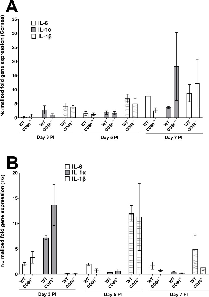
Herpes simplex virus-1 (HSV-1) infections are among the most frequent serious viral eye infections in the U.S. and are a major cause of viral-induced blindness. HSV-1 infection is known to induce T cell activation, proliferation, and differentiation that play crucial roles in the development of virus-induced inflammatory lesions, leading to eye disease and causing chronic corneal damage. CD80 is a co-stimulatory molecule and plays a leading role in T cell differentiation. Previous efforts to limit lesion severity by controlling inflammation at the cellular level led us to ask whether mice knocked out for CD80 would show attenuated virus replication following reactivation. By evaluating the effects of CD80 activity on primary and latent infection, we found that in the absence of CD80, virus replication in the eyes and virus reactivation in latent trigeminal ganglia were both significantly reduced. However, latency in latently infected CD80-/- mice did not differ significantly from that in wild-type (WT) control mice. Reduced virus replication in the eyes of CD80-/- mice correlated with significantly expanded CD11c gene expression as compared to WT mice. Taken together, our results indicate that suppression of CD80 could offer significant beneficial therapeutic effects in the treatment of Herpes Stromal Keratitis (HSK).IMPORTANCEOf the many problems associated with recurrent ocular infection, reducing virus reactivation should be a major goal of controlling ocular herpes simplex virus-1 (HSV-1) infection. In this study, we have shown that the absence of CD80 reduces HSV-1 reactivation, which marks the establishment of a previously undescribed mechanism underlying viral immune evasion that could be exploited to better manage HSV infection.

Keywords: CD8; PD-1; corneal scarring; latency; reactivation; virus replication.


Conflict of interest statement

The authors declare no conflict of interest.

Figures

Fig 1
Fig 1
Viral titers and gB copy number in WT and CD80−/− mouse eyes following ocular infection with HSV-1 McKrae strain. (A) Virus replication in infected eyes. WT and CD80−/− mice were infected with 2 × 10⁵ PFU/eye of McKrae virus. The presence of infectious virus in the eyes of mice was monitored daily for 7 days by collecting tear films and quantifying the virus using standard plaque assays as described (see Materials and Methods). Each point represents the mean ± SEM from 50 eyes for both infected mouse groups; and (B) gB copy number during primary infection. Eyes of three WT and three CD80−/− mice were ocularly infected as described above. On days 3, 5, and 7 PI, six corneas and six TG were harvested and the gB copy number was determined by qPCR. No differences in gB copy number were seen between the two groups in either tissue except for significantly higher expression of gB copy number on day 3 PI in TG of CD80−/− mice. The experiment was repeated twice. (A) Virus replication in infected eyes; and (B) gB copy number in infected eyes and TG.
Fig 2
Fig 2
Quantification of CD4 and CD8α RNA transcripts in the corneas and TG of infected WT and CD80−/− mice. (A) Expression in infected corneas. Corneas from WT and CD80−/−-infected mice (three mice/group) were harvested on days 3, 5, and 7 PI from mice infected with 2 × 10⁵ PFU/eye HSV-1 McKrae. Total RNA was isolated from each cornea, and GAPDH expression was used to normalize the expression of CD4 and CD8α transcripts in the corneas of ocularly infected mice. No differences in corneal expression were observed between the two infected mice groups. Each bar represents the mean expression ± SEM in six corneas from both infected mouse groups. (B) Expression in infected TG. TG were harvested on days 3, 5, and 7 PI from the above infected mice. Total RNA was isolated from each TG, and GAPDH expression was used to normalize the expression of each transcript in the TG of ocularly infected mice. CD8α expression was lower in CD80−/− mice on day 7 PI (P < 0.0001). Each bar represents the mean expression ± SEM in six TG from both infected mouse groups. Only differences that are statistically significant are shown for each gene.
Fig 3
Fig 3
Quantification of F4/80, CD11c, and NK1.1 RNA transcripts in the corneas and TG of infected WT and CD80−/− mice. (A) Expression in infected corneas. WT and CD80−/− mice were infected as described in Fig. 2. Total RNA was isolated from each cornea, and GAPDH expression was used to normalize the expression of F4/80, CD11c, and NK1.1 transcripts in the corneas of ocularly infected mice. CD11c expression was higher in CD80−/− mice on day 7 PI (P = 0.0008). Each bar represents the mean expression ± SEM in six corneas for both infected mice groups. (B) Expression in infected TG. TG were harvested on days 3, 5, and 7 PI from infected mice. Total RNA was isolated from each TG, and GAPDH expression was used to normalize the expression of each transcript in the TG of ocularly infected mice. No significant differences in expression were detected among genes in either infected mouse group (P > 0.05). Each bar represents the mean expression ± SEM in six TG from both infected mouse groups. Only statistically significant differences are shown for each gene.
Fig 4
Fig 4
Quantification of IL-6, IL-1α, and IL-1β RNA transcripts in the corneas and TG of infected WT and CD80−/− mice. (A) Expression in infected corneas. WT and CD80−/− mice were infected as described in Fig. 2. Total RNA was isolated from each cornea, and GAPDH expression was used to normalize the expression of IL-6, IL-1α, and IL-1β transcripts in the corneas of ocularly infected mice. No significant expression differences were detected among the genes in the two infected mouse groups. Each bar represents the mean expression ± SEM in six corneas for both infected mouse groups. (B) Expression in infected TG. TG were harvested on days 3, 5, and 7 PI from infected mice. Total RNA was isolated from each TG, and GAPDH expression was used to normalize the expression of each transcript in the TG of ocularly infected mice. Each bar represents the mean expression ± SEM in six TG from both infected mouse groups. Only differences that are statistically significant are shown for each gene.
Fig 5
Fig 5
Quantification of GzmA, GzmB, and perforin RNA transcripts in the corneas and TG of infected WT and CD80−/−- mice. (A) Expression in infected corneas. WT and CD80−/− mice were infected as described in Fig. 2. Total RNA was isolated from each cornea, and GAPDH expression was used to normalize the expression of GzmA, GzmB, and perforin transcripts in the corneas of ocularly infected mice. GzmB expression was significantly lower in CD80−/− mice than in WT mice on day 5 PI (P = 0.012). Each bar represents the mean expression ± SEM in six corneas from both infected mouse groups. (B) Expression in infected TG. TG were harvested on days 3, 5, and 7 PI from infected mice. Total RNA was isolated from each TG, and GAPDH expression was used to normalize the expression of each transcript in the TG of ocularly infected mice. No significant differences among the genes were observed in infected mouse groups. Each bar represents the mean expression ± SEM in six TG from both infected mouse groups. Only differences that are statistically significant are shown for each gene.
Fig 6
Fig 6
PD-L1, CD45, and TNF-α expression in the corneas and TG of infected WT and CD80−/− mice. (A) Expression of PD-L1 in infected corneas and TG. Corneas and TG from WT and CD80−/−-infected mice were harvested on days 3, 5, and 7 PI from mice infected with 2 × 10⁵ PFU/eye of HSV-1 McKrae. Total RNA was isolated from each cornea, and GAPDH expression was used to normalize the expression of PD-L1 RNA transcripts in the corneas and TG of ocularly infected mice. Each bar represents the mean expression ± SEM in six corneas and mean expression ± SEM in six TG. (B) Expression of CD45 in infected corneas and TG. Corneas and TG from WT- and CD80−/−-infected mice were harvested on days 3, 5, and 7 PI from mice infected with 2 × 10⁵ PFU/eye HSV-1 McKrae. Total RNA was isolated from each cornea, and GAPDH expression was used to normalize the expression of CD45 RNA transcripts in the corneas and TG of ocularly infected mice. Each bar represents the mean expression ± SEM from six corneas and mean expression ± SEM from six TG. (C) Expression of TNF-α in infected corneas and TG. Corneas and TG from WT and CD80−/−-infected mice were harvested on days 3, 5, and 7 PI from mice infected with 2 × 10⁵ PFU/eye HSV-1 McKrae. Total RNA was isolated from each cornea, and GAPDH expression was used to normalize the expression of CD45 RNA transcripts in the corneas and TG of ocularly infected mice. No significant differences were observed among the genes measured in either infected mouse group (P > 0.05). Each bar represents the mean expression ± SEM from six corneas and mean expression ± SEM from six TG. Only differences that are statistically significant are shown for each gene.
Fig 7
Fig 7
Absence of CD80 does not affect survival and eye disease. (A) Survival. Mice were ocularly infected with 2 × 10⁵ PFU/eye of HSV-1 McKrae as described above. Survival of WT and CD80−/− mice was monitored over a 28-day period after infection. An average of five independent experiments is graphed. (B) Eye disease. A total of 68 eyes from WT and 70 eyes from CD80−/−-infected mice used for survival were used to measure CS. Severity of CS in mouse corneas was examined in all groups by slit lamp biomicroscopy. Severity was scored on day 28 PI.
Fig 8
Fig 8
Levels of latency, latent gB expression, and duration of explant reactivation following ocular infection of WT and CD80−/− mice. (A) LAT RNA transcript in latent TG. Eyes from WT and CD80−/− mice were infected with 2 × 10⁵ PFU/eye of McKrae virus. On day 28 PI, TG from infected mice were harvested and LAT expression was analyzed by RT-PCR. qRT-PCR was performed on each individual TG. The estimated relative copy number of HSV-1 LAT was calculated in each experiment using standard curves generated from pGem5317. The plasmid template was serially diluted 10-fold such that 10 µL contained from 103 to 1011 copies of LAT. Serial dilutions were then analyzed by TaqMan Real-time PCR with the same probe set. The copy number for each reaction was determined by comparing the normalized threshold cycle of each sample to the standard threshold cycle. GAPDH expression was used to normalize the relative viral LAT RNA expression in the TG. Each bar represents mean copy number ± SEM from 31 TG for infected WT mice and 31 TG for CD80−/− infected mice. (B) gB DNA copy number in latent TG. A total of 11 TG from WT- and CD80−/−-infected mice were isolated on day 28 PI. Expression of gB DNA was determined using qPCR, and gB copy number was measured as described in Materials and Methods. (C) Explant reactivation in latent TG. On day 28 PI, TG from infected WT and CD80−/− mice were isolated and incubated in 1.5 mL of tissue culture media at 37°C, and the presence of infectious virus was monitored as described in Materials and Methods. The results are shown as the number of TG that reactivated daily. Each point represents mean reactivated TG ± SEM of 20 TG for WT mice and 19 TG for CD80−/− from two independent experiments. (A) LAT RNA; (B) gB DNA; and (C) explant reactivation.
Fig 9
Fig 9
Levels of CD80, CD86, CD28, CD4, CD8, PD-1, PD-L1, CTLA4, IFN-γ, IFN-α2A, and IFN-β. (A) Expression of CD80, CD86, and CD28. Mice were infected with 2 × 10⁵ PFU/eye of HSV-1 McKrae as described above. TG from infected mice were extracted on day 28 PI. Total RNA was isolated from each TG, and GAPDH expression was used to normalize the expression of each transcript in the TG of ocularly infected mice. Each bar represents the mean expression ± SEM from 10 TG for each mouse strain. (B) Expression of CD4, CD8, PD-1, PD-L1, and CTLA4. RNA extracted from the TG of latently infected mice described above was used to measure the expression of CD4, CD8, PD-1, PD-L1, and CTLA4. Each bar represents the mean expression ± SEM from 10 TG. (C) Expression of IFN-γ, IFN-α2A, and IFN-β. RNA extracted from the TG of latently infected mice as described above was used to measure IFN-γ, IFN-α2A, and IFN-β TG expression on day 28 PI. Each bar represents the mean expression ± SEM from 10 TG per mouse strain.

Similar articles

Cited by

References

    1. Iwasaki A, Pillai PS. 2014. Innate immunity to influenza virus infection. Nat Rev Immunol 14:315–328. doi:10.1038/nri3665 - DOI - PMC - PubMed
    1. Zhu S, Viejo-Borbolla A. 2021. Pathogenesis and virulence of herpes simplex virus. Virulence 12:2670–2702. doi:10.1080/21505594.2021.1982373 - DOI - PMC - PubMed
    1. Roizman B, Whitley RJ. 2013. An inquiry into the molecular basis of HSV latency and reactivation. Annu Rev Microbiol 67:355–374. doi:10.1146/annurev-micro-092412-155654 - DOI - PubMed
    1. Webre JM, Hill JM, Nolan NM, Clement C, McFerrin HE, Bhattacharjee PS, Hsia V, Neumann DM, Foster TP, Lukiw WJ, Thompson HW. 2012. Rabbit and mouse models of HSV-1 latency, reactivation, and recurrent eye diseases. J Biomed Biotechnol 2012:612316. doi:10.1155/2012/612316 - DOI - PMC - PubMed
    1. Marcocci ME, Napoletani G, Protto V, Kolesova O, Piacentini R, Li Puma DD, Lomonte P, Grassi C, Palamara AT, De Chiara G. 2020. Herpes simplex virus-1 in the brain: the dark side of a sneaky infection. Trends Microbiol 28:808–820. doi:10.1016/j.tim.2020.03.003 - DOI - PubMed