. 2024 Feb 27;121(9):e2315985121.
doi: 10.1073/pnas.2315985121. Epub 2024 Feb 20.

Diverse cytomegalovirus US11 antagonism and MHC-A evasion strategies reveal a tit-for-tat coevolutionary arms race in hominids

Affiliations

Diverse cytomegalovirus US11 antagonism and MHC-A evasion strategies reveal a tit-for-tat coevolutionary arms race in hominids

Cosima Zimmermann et al. Proc Natl Acad Sci U S A. .

Abstract

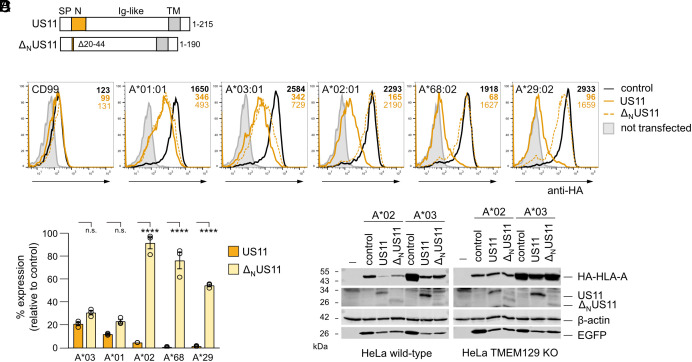
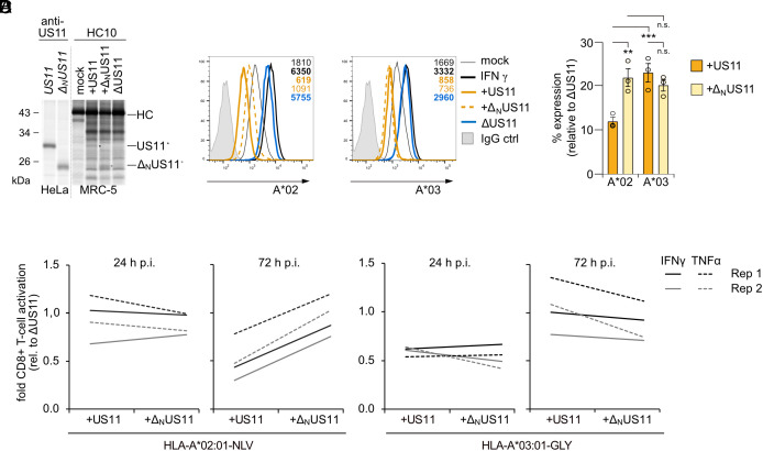
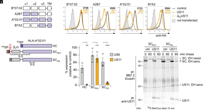
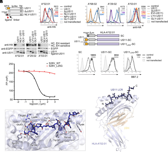
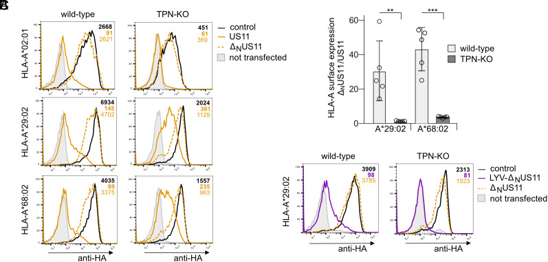
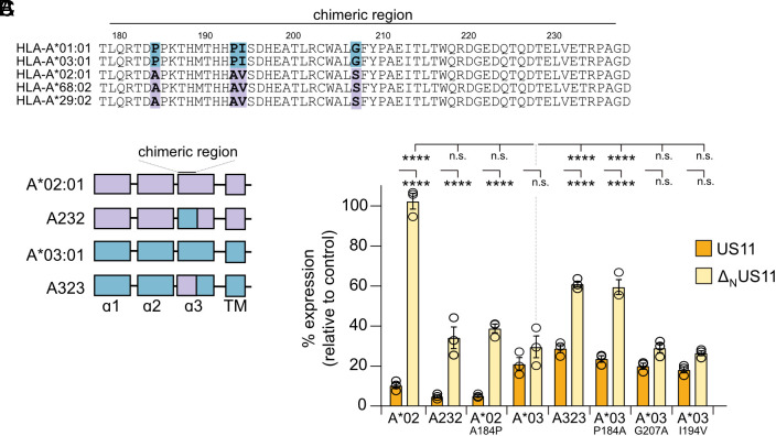
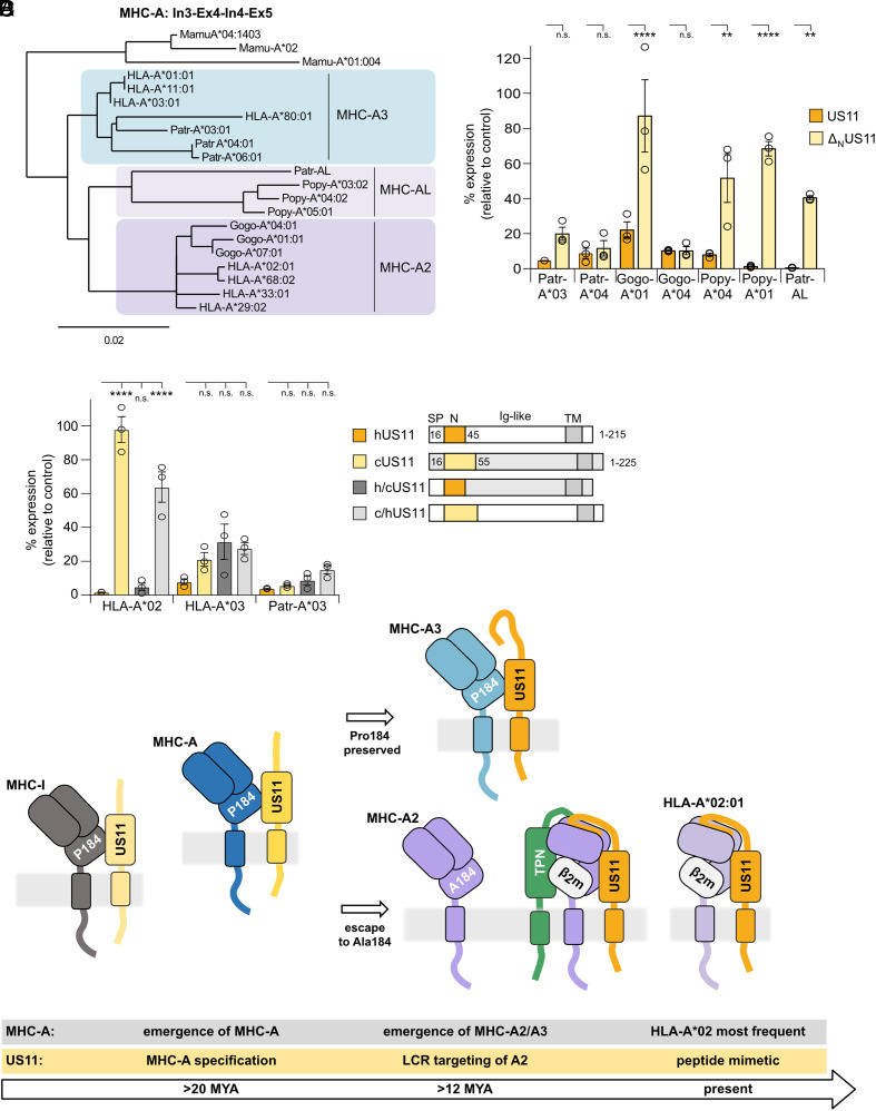
Recurrent, ancient arms races between viruses and hosts have shaped both host immunological defense strategies as well as viral countermeasures. One such battle is waged by the glycoprotein US11 encoded by the persisting human cytomegalovirus. US11 mediates degradation of major histocompatibility class I (MHC-I) molecules to prevent CD8+ T-cell activation. Here, we studied the consequences of the arms race between US11 and primate MHC-A proteins, leading us to uncover a tit-for-tat coevolution and its impact on MHC-A diversification. We found that US11 spurred MHC-A adaptation to evade viral antagonism: In an ancestor of great apes, the MHC-A A2 lineage acquired a Pro184Ala mutation, which confers resistance against the ancestral US11 targeting strategy. In response, US11 deployed a unique low-complexity region (LCR), which exploits the MHC-I peptide loading complex to target the MHC-A2 peptide-binding groove. In addition, the global spread of the human HLA-A*02 allelic family prompted US11 to employ a superior LCR strategy with an optimally fitting peptide mimetic that specifically antagonizes HLA-A*02. Thus, despite cytomegaloviruses low pathogenic potential, the increasing commitment of US11 to MHC-A has significantly promoted diversification of MHC-A in hominids.

Keywords: HLA-A; MHC class I; coevolution; cytomegalovirus; tapasin.


Conflict of interest statement

Competing interests statement:The authors declare no competing interest.

Figures

Fig. 1.
Fig. 1.
The HCMV-encoded immunoevasin US11 differentially regulates the surface expression of HLA-A allotypes. (A) Schematic representation of full-length US11 and ΔNUS11. SP, signal peptide, Ig-like, immunoglobulin-like domain; TM, transmembrane domain. (B) HeLa cells were transiently co-transfected with plasmids encoding HA-tagged HLA-I and CD99 and pIRES-EGFP encoding either US11, ΔNUS11, or a control protein. At 20 h post-transfection, surface expression was determined by flow cytometry (anti-HA) on EGFP-positive cells. Representative histograms are shown. Example of gating is shown in SI Appendix, Fig. S1B. (C) The percentage of MFI (mean fluorescence intensity) as compared to control transfected cells was determined from analysis in (B). Dots represent individual values and bars mean values ± SEM from three independent experiments. Significance was analyzed by two-way ANOVA. (D) HeLa wild-type cells and TMEM129 KO cells were transfected as in (B). At 20 h post-transfection, cells were lysed and western blot analysis was performed with antibodies detecting indicated proteins (anti-HA for detection of HA-tagged HLA-A).
Fig. 2.
Fig. 2.
The US11 LCR protects HCMV from HLA-A*02:01-restricted CD8+ T-cells. (A) US11 or ΔNUS11 in pIRES-EGFP were transiently transfected into HeLa cells. MRC-5 fibroblast were mock treated or infected with an MOI of 7 with indicated HCMV mutants. At 48 h p.i., cells were metabolically labeled for 2 h. The anti-US11 antiserum or the mAb HC10 were applied for immunoprecipitation. Retrieved proteins were separated on sodium dodecyl sulfate – polyacrylamid gel electrophoresis (SDS-PAGE) and detected by autoradiography. (B) HF-α fibroblasts were treated as indicated and infected with an MOI of 8. At 48 h p.i., HLA-A*02:01 and A*03:01 cell surface expression was determined by flow cytometry. (C) The percentage of MFI as compared to ΔUS11-infected cells was determined from the analysis in (A). Dots represent individual values and bars mean values ± SEM from three independent experiments. Significance was determined using one-way ANOVA. (D) The fold change in the percentage of activated CD8+ T cells as compared to CD8+ T cells co-cultured with ΔUS11-infected cells is depicted. HF-α fibroblasts were mock treated or infected as indicated with an MOI of 8. At 24 and 72 h p.i., the cells were co-cultured for 5 h with HLA-A*02:01NLV or HLA-A*03:01GLY specific polyclonal ex vivo expanded CD8+ T cells. Activation of CD8 and tetramer-positive T cells was determined by staining of intracellular IFNγ and TNFα.
Fig. 3.
Fig. 3.
Peptide-loaded HLA-A*02:01 confers resistance to US11. (A) Schematic representation of HA-HLA-A*02:01 and B*07:02 chimeras. (B) HA-HLA-I surface expression was determined as in Fig. 1B. Representative histograms are shown. Statistical analysis is depicted in SI Appendix, Fig. S3A. (C) Schematic representation of HLA-A*02:01 SC constructs. HASP, HA SP; A2SP, HLA-A*02 SP; S-S, disulfide stabilization. (D) HLA-A*02:01 HC or SC constructs encoded in pcDNA3.1 were transiently co-transfected with pIRES-EGFP encoding a control protein, US6 or US11 into HeLa cells. At 20 h post-transfection HLA-A*02:01 cell surface expression was analyzed by flow cytometry using the mAb BB7.2. The percentage of MFI as compared to control transfected cells was determined. Dots represent individual values and bars mean values ± SEM from three independent experiments. Significance was analyzed by two-way ANOVA. (E) Cells were transfected as in (D) with indicated constructs. At 20 h post-transfection, cells were metabolically labeled for 15 min and a pulse-chase experiment was performed. The mAb BB7.2 and anti-US11 were applied for immunoprecipitation. Retrieved proteins were EndoH (EH) digested as indicated prior to separation on SDS-PAGE and detection by autoradiography.
Fig. 4.
Fig. 4.
The US11 LCR mimics peptide binding to HLA-A*02:01. (A) Schematic representation of chimeric HLA-A ligand-US11 constructs. (B and C) Panels show flow cytometry histograms of HA-tagged HLA-A expressed by transient co-transfection with pIRES-EGFP encoding ligand-US11 variants or a control. At 20 h post-transfection, surface expression was determined by flow cytometry (anti-HA) on EGFP-positive cells. Representative histograms are shown. (D) Cells were transfected as in (B and C). At 20 h post-transfection, cells were lysed and lysates were treated with EndoH. Proteins were detected by western blot analysis with indicated antibodies. (E) Schematic representation of HLA-A*02:01 SC constructs. (F) HLA-A*02:01 SC constructs encoded in pcDNA3.1 were transiently co-transfected with pIRES-EGFP encoding a control protein or US6 into HeLa cells. At 20 h post-transfection, SC cell surface expression was analyzed by flow cytometry using the mAb BB7.2. Representative histograms are shown. Statistical analysis is depicted in SI Appendix, Fig. S4B. (G) Effect of the US11 peptide S28V_WT (amino acids 17 to 44) and the mutated peptide S28V_L25G on binding of the fluorescein isothiocyanate (FITC)-labeled GV9 peptide to HLA-A*02:01 in an MHC-I fluorescence polarization assay. A representative experiment out of three independent experiments to determine the IC50 value is shown. Primary data are shown in SI Appendix, Fig. S4C. (H) X-ray crystal structure of a US1117-43-HLA-A*02:01 SC. Cartoon representation of HLA-A*02:01 and β2m, colored in lavender and wheat, respectively. The US11 LCR (blue) is shown in stick format with the N and C termini marked (PDB ID: 8FRT). (I) Close-up of the peptide-binding groove of H with US11 LCR residues indicated in single-letter code, and key HLA-A*02:01 residues in stick format and in three-letter code. Hydrogen bonds are displayed as dashed lines.
Fig. 5.
Fig. 5.
Optimal US11 LCR targeting of HLA-A*29:02 and A*68:02 is tapasin dependent. (A) Wild-type and TPN-KO HeLa cells were transiently co-transfected with plasmids encoding HA-HLA-I and pIRES-EGFP encoding US11, ΔNUS11, or a control protein. At 20 h post-transfection, HA-HLA-I surface expression was determined by flow cytometry (anti-HA) on EGFP-positive cells. Representative histograms are shown. (B) The ratio of MFI determined in ΔNUS11 and US11-expressing cells in A. Dots represent individual values and bars mean values ± SEM from five independent experiments. Significance was calculated using two-way ANOVA. (C) Flow cytometry analysis as described in A with US11 variants as indicated. Representative histograms are shown.
Fig. 6.
Fig. 6.
A single residue in the α3 domain dictates N-terminus–independent regulation of HLA-A by US11. (A) HLA-A protein sequence alignment. (B) Schematic representation of HA-HLA-A*02:01 (lavender) and A*03:01 (blue) chimeras with a swapped α3 segment (residues 179 to 236). (C) HeLa cells were transiently co-transfected with plasmids encoding HA-HLA-A and pIRES-EGFP encoding US11 versions as indicated or a control protein. At 20 h post-transfection, HA-HLA-A surface expression was determined by flow cytometry (anti-HA) on EGFP-positive cells. The percentage of MFI as compared to control transfected cells was determined. Dots represent individual values and bars mean values ± SEM from three independent experiments. Significance was calculated using two-way ANOVA.
Fig. 7.
Fig. 7.
US11 evolved an LCR-dependent strategy specifically to counteract A2 lineage escape from N-terminus–independent antagonism. (A) Alignment of MHC-A genomic sequences from intron 3, exon 4, intron 4, and exon 5. Phylogeny.fr was used to build the phylogenetic tree [84]). (B and C) HA-MHC-A surface expression was determined as in Fig. 1B. (C) In the Right panel, a schematic representation of hUS11, cUS11, h/cUS11, and c/hUS11 is depicted. (D) Model of US11 and MHC-A coevolution. After the split of New World monkeys and Old World monkeys, the MHC-A locus emerged, which might have resulted in better control of CMV, spurring dedication of US11 to regulate MHC-A. The lineages A2 and A3 evolved in an ancestor of great apes. The A2 lineage is characterized by Ala at position 184, resisting control by the concurrent US11. US11 gained a new function at the N-terminus; in a tapasin-dependent manner, this sequence is able to interact with the peptide-binding domain of assembled A2 molecules and redirects the target molecule for degradation. In the human population, the HLA-A*02 allelic family became highly prevalent prompting US11 to target this allotype specifically by a peptide mimetic in the LCR.

Similar articles

Cited by

References

    1. Dos Santos Francisco R., et al. , HLA supertype variation across populations: New insights into the role of natural selection in the evolution of HLA-A and HLA-B polymorphisms. Immunogenetics 67, 651–663 (2015). - PMC - PubMed
    1. Hertz T., et al. , Mapping the landscape of host-pathogen coevolution: HLA class I binding and its relationship with evolutionary conservation in human and viral proteins. J. Virol. 85, 1310–1321 (2011). - PMC - PubMed
    1. Stern-Ginossar N., et al. , Decoding human cytomegalovirus. Science 338, 1088–1093 (2012). - PMC - PubMed
    1. Berry R., Watson G. M., Jonjic S., Degli-Esposti M. A., Rossjohn J., Modulation of innate and adaptive immunity by cytomegaloviruses. Nat. Rev. Immunol. 20, 113–127 (2020). - PubMed
    1. Guma M., et al. , Imprint of human cytomegalovirus infection on the NK cell receptor repertoire. Blood 104, 3664–3671 (2004). - PubMed