. 2024 Feb 27;121(9):e2317322121.
doi: 10.1073/pnas.2317322121. Epub 2024 Feb 20.

The global RNA-RNA interactome of Klebsiella pneumoniae unveils a small RNA regulator of cell division

Affiliations

The global RNA-RNA interactome of Klebsiella pneumoniae unveils a small RNA regulator of cell division

Eric Ruhland et al. Proc Natl Acad Sci U S A. .

Abstract

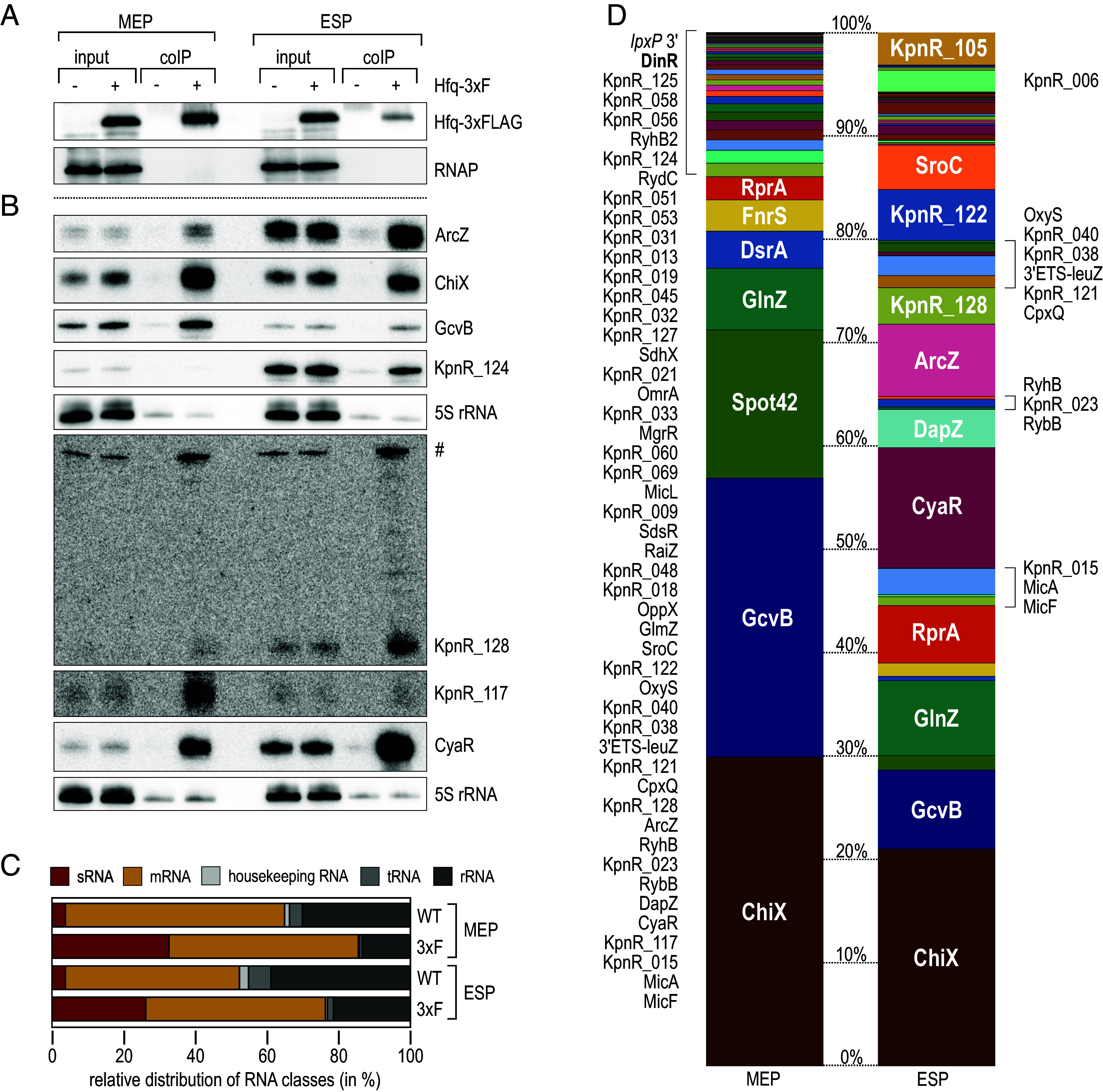
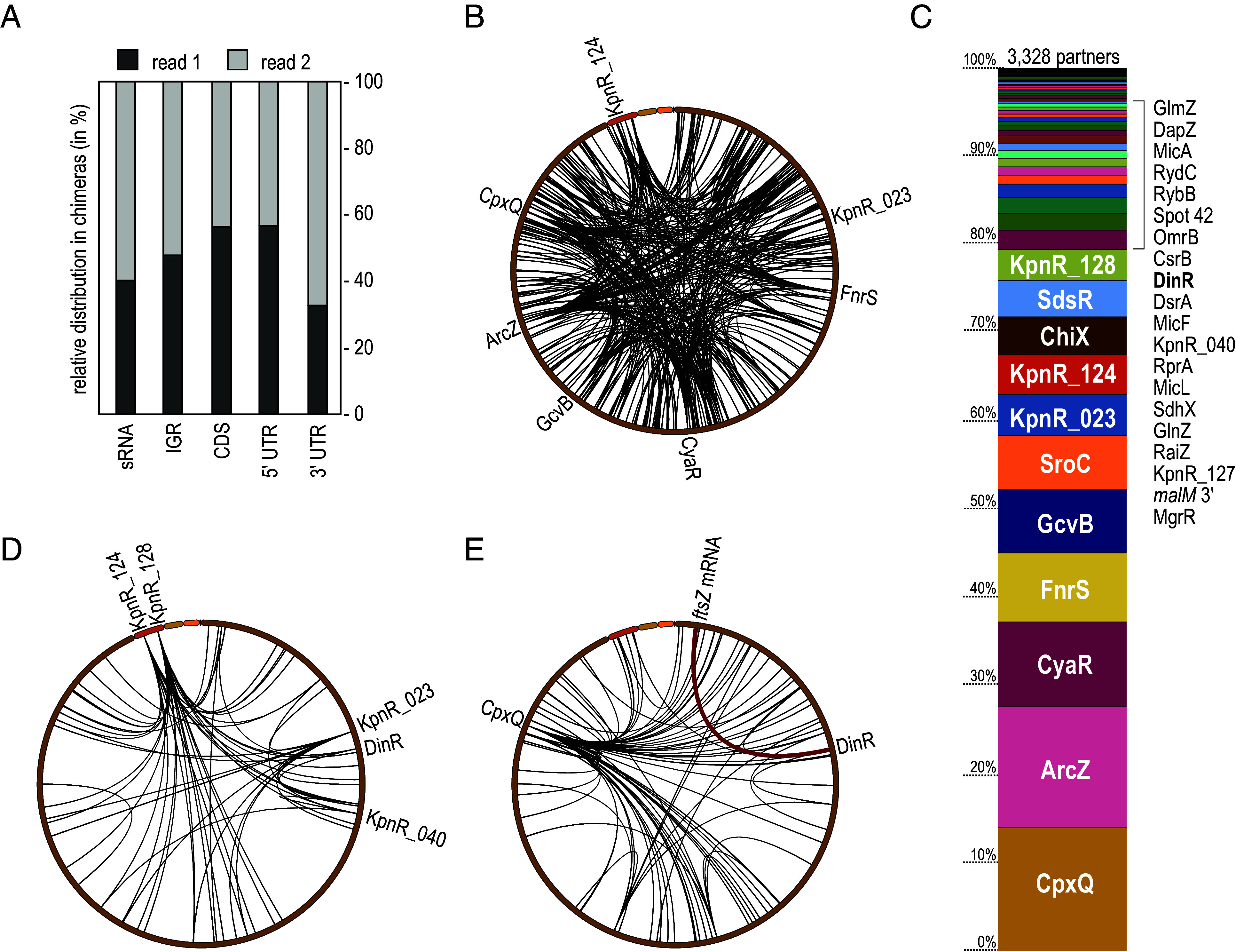
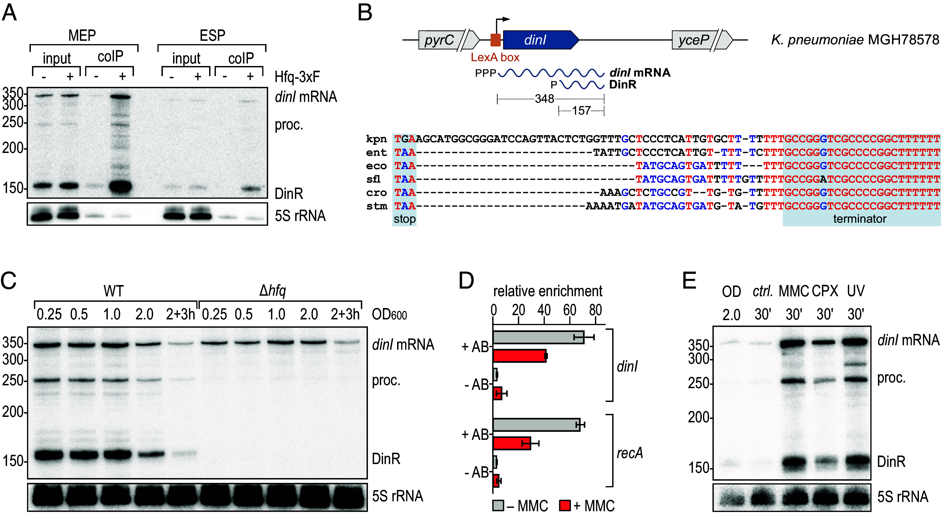
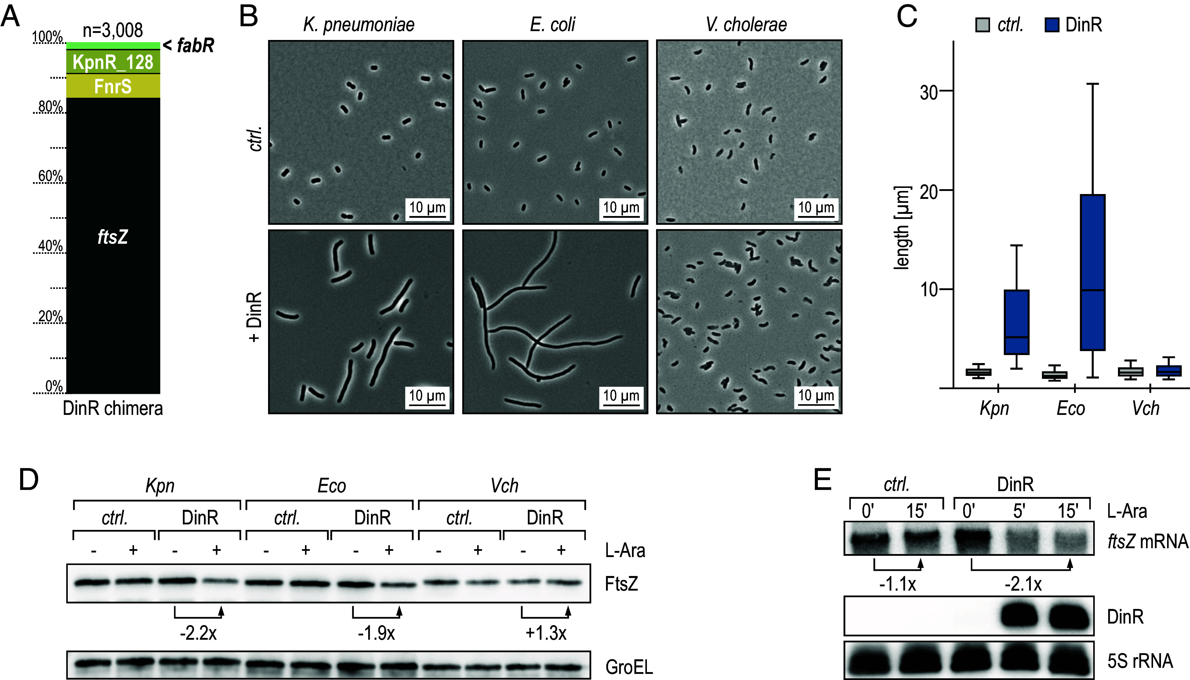
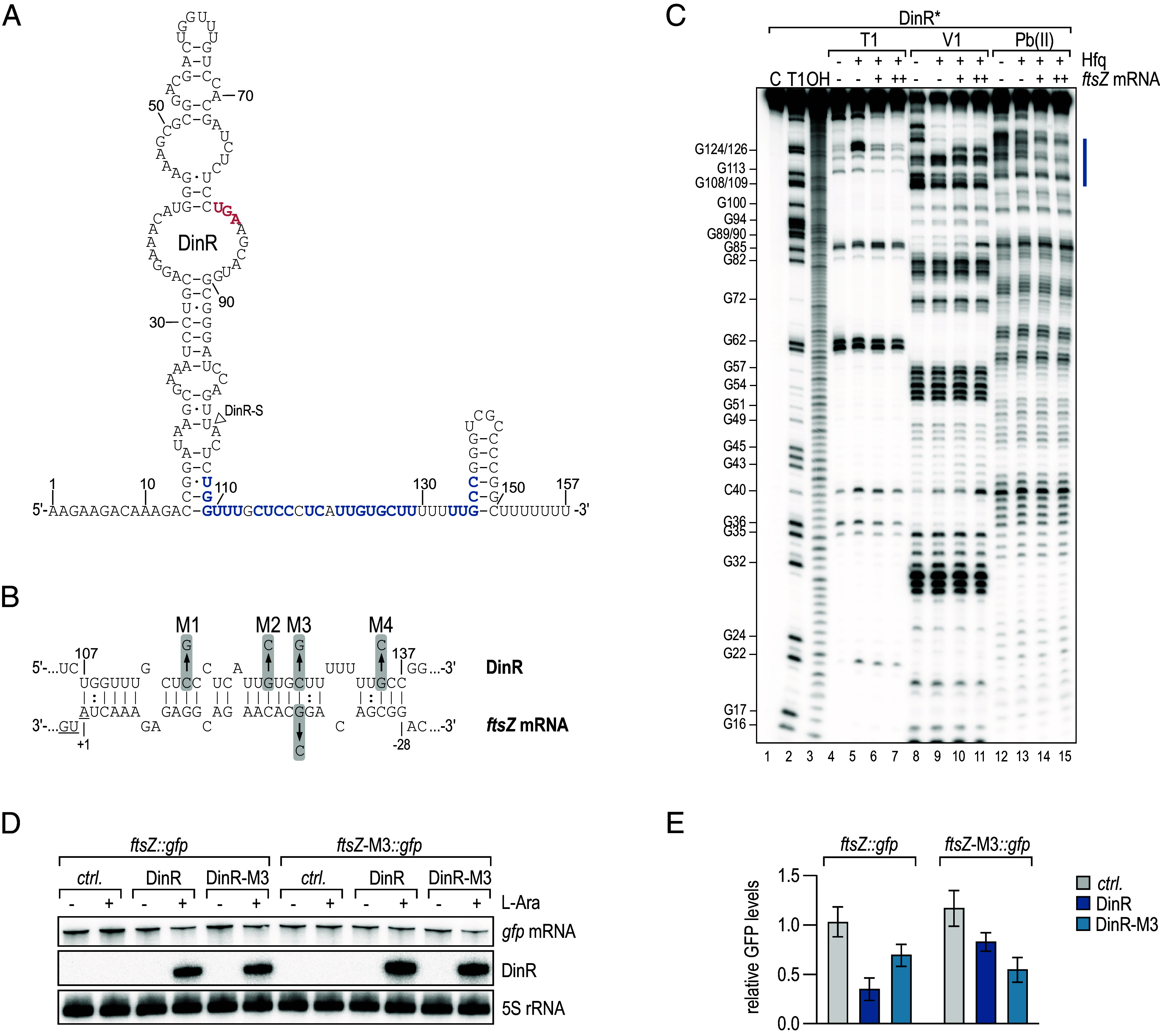
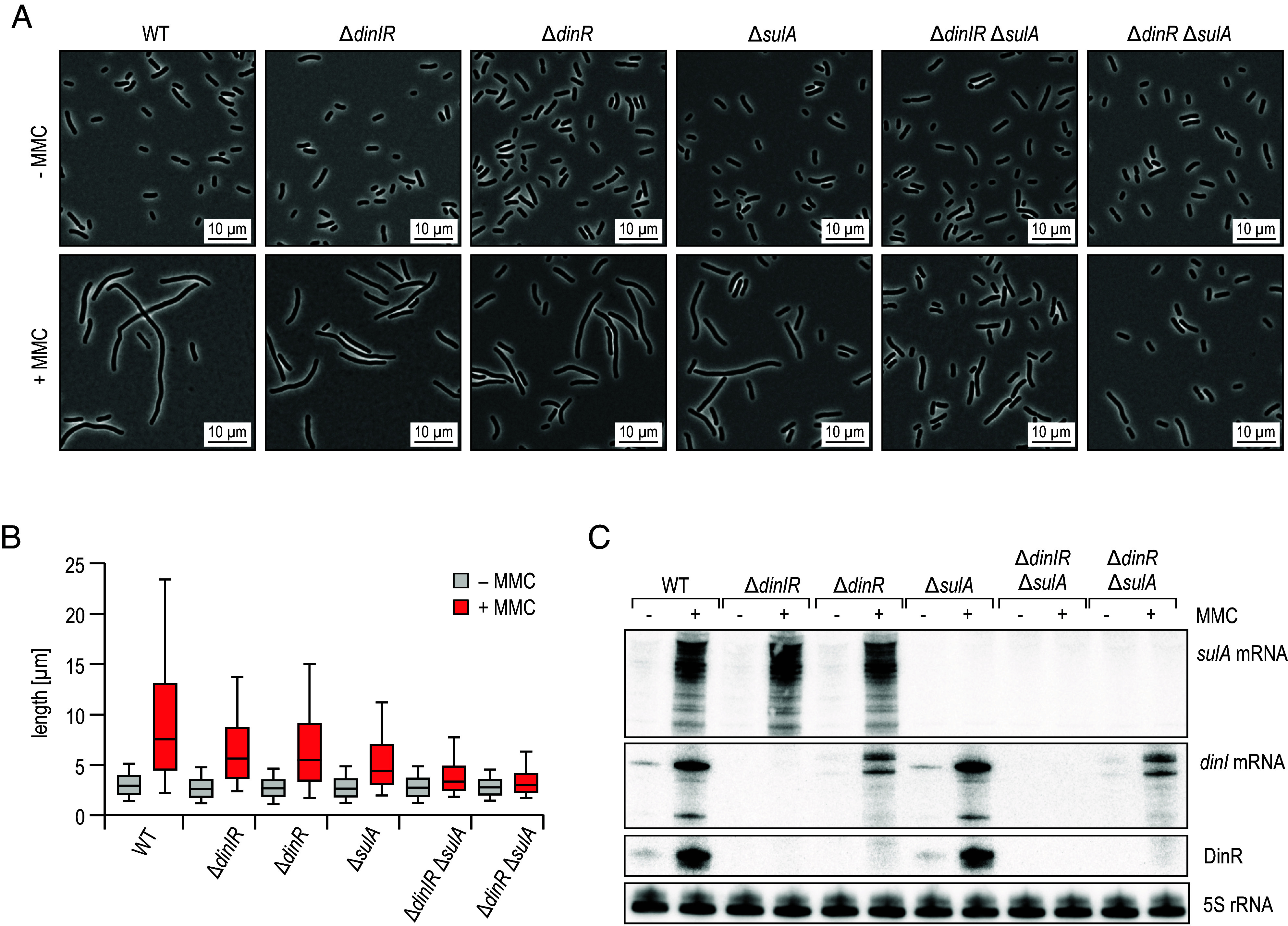
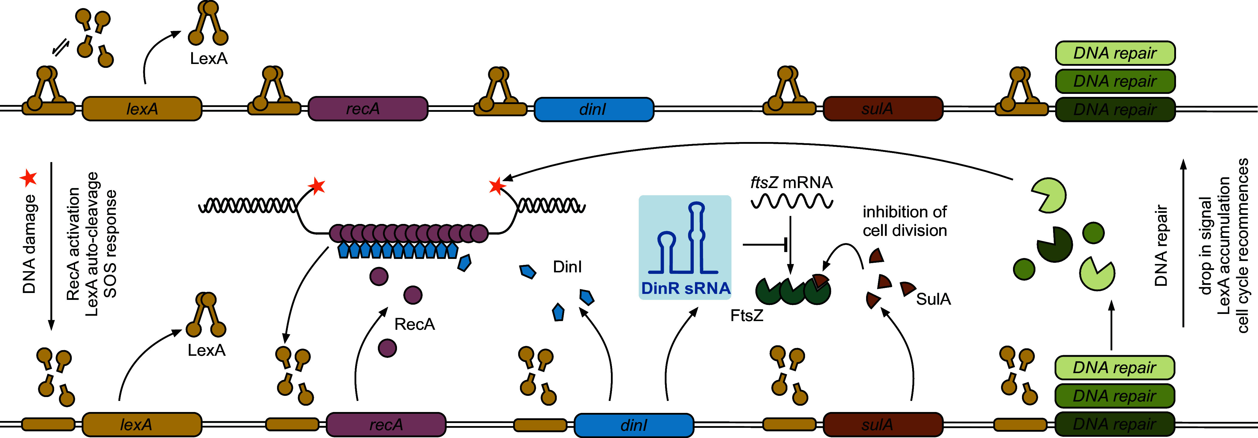
The ubiquitous RNA chaperone Hfq is involved in the regulation of key biological processes in many species across the bacterial kingdom. In the opportunistic human pathogen Klebsiella pneumoniae, deletion of the hfq gene affects the global transcriptome, virulence, and stress resistance; however, the ligands of the major RNA-binding protein in this species have remained elusive. In this study, we have combined transcriptomic, co-immunoprecipitation, and global RNA interactome analyses to compile an inventory of conserved and species-specific RNAs bound by Hfq and to monitor Hfq-mediated RNA-RNA interactions. In addition to dozens of RNA-RNA pairs, our study revealed an Hfq-dependent small regulatory RNA (sRNA), DinR, which is processed from the 3' terminal portion of dinI mRNA. Transcription of dinI is controlled by the master regulator of the SOS response, LexA. As DinR accumulates in K. pneumoniae in response to DNA damage, the sRNA represses translation of the ftsZ transcript by occupation of the ribosome binding site. Ectopic overexpression of DinR causes depletion of ftsZ mRNA and inhibition of cell division, while deletion of dinR antagonizes cell elongation in the presence of DNA damage. Collectively, our work highlights the important role of RNA-based gene regulation in K. pneumoniae and uncovers the central role of DinR in LexA-controlled division inhibition during the SOS response.

Keywords: Hfq; Klebsiella pneumoniae; RIL-seq; SOS response; small RNA.


Conflict of interest statement

Competing interests statement:The authors declare no competing interest.

Figures

Fig. 1.
Fig. 1.
RIP-seq analysis uncovers Hfq-associated sRNAs in K. pneumoniae. (A and B) K. pneumoniae MGH 78578 expressing FLAG-tagged Hfq (3xF; +) from the native chromosomal locus were cultivated in LB to MEP (OD600 of 0.25) or ESP (OD600 of 2.0) and subjected to immunoprecipitation with an anti-FLAG antibody. An untagged WT strain (−) served as control. Protein and RNA samples were collected prior to (input) and after purification (coIP). (A) Protein samples were analyzed on western blots to confirm expression and enrichment of the Hfq-3xFLAG protein. RNAP was probed as loading control. (B) RNA samples were analyzed on Northern blots to determine expression and enrichment of indicated sRNAs using gene-specific probes. KpnR_128 is expressed from its own promoter internal to an upstream gene. Both transcripts share the same termination site and the ~300 nt upstream transcript (#) is thus detected by the KpnR_128 probe. 5S rRNA served as loading control. (C) Relative abundance of different RNA classes recovered from RNA coIPs from WT and cells expressing FLAG-tagged Hfq (3xF) in MEP and ESP. (D) Read distribution of sRNAs enriched ≥threefold after coIP with FLAG-tagged Hfq in MEP (Left) and ESP (Right).
Fig. 2.
Fig. 2.
RIL-seq reveals the K. pneumoniae Hfq-dependent RNA interactome. (A) Relative distribution of RNA classes and their organization in chimeric reads. (B) Circos plot of all RIL-seq interactions recovered in both replicates involving K. pneumoniae sRNAs. Interaction hubs of selected sRNAs are indicated. (C) Relative distribution of numbers of interaction partners in Hfq RIL-seq for all sRNAs. (D) Circos plot of all RIL-seq interactions recovered in both replicates involving previously unknown candidate sRNAs. Selected sRNAs with multiple partners are indicated. (E) Circos plot of all RIL-seq interactions recovered in both replicates involving sRNA derived from mRNA 3′ ends. CpxQ as an interaction hub is marked. The interaction between DinR and ftsZ mRNA is highlighted in red.
Fig. 3.
Fig. 3.
DinR is an Hfq-associated sRNA produced in response to DNA damage. (A) RNA samples recovered from the Hfq RIP-seq analysis (Fig. 1) were analyzed on Northern blots and probed with a DinR-specific riboprobe to determine expression and enrichment of dinI mRNA and its processing products including DinR. 5S rRNA served as loading control. (B) Upper: Organization of the dinI locus on the main chromosome of K. pneumoniae MGH 78578. A LexA-box overlapping the dinI TSS is indicated by an orange box. The dinI primary transcript (348 nt) is processed, releasing DinR (157 nt). Lower: Nonredundant alignment of the dinI 3′UTR in diverse enterobacteria (kpn: Klebsiella pneumoniae MGH 78578; ent: Enterobacter sp. 638; eco: E. coli MG1655; sfl: Shigella flexneri 301; cro: Citrobacter rodentium ICC168; stm: Salmonella Typhimurium LT2); the dinI stop codon and the Rho-independent transcriptional termination site are shaded in gray. Nucleotides are colored regarding their degree of conservation (red: high conservation; blue: partial conservation; black: little or no conservation). (C) Expression of dinI mRNA and DinR sRNA in WT and Δhfq K. pneumoniae. RNA samples were collected at different time points over growth (OD600 from 0.25 to 2.0, and 3 h after cells had reached OD600 of 2.0) and analyzed by Northern blotting. 5S rRNA served as loading control. (D) In vivo binding of LexA to the K. pneumoniae dinI promoter. Association of LexA before and after addition of MMC was determined by ChIP (+AB: anti-LexA antibody; −AB: no antibody control) followed by quantitative PCR. Relative enrichment of DNA fragments was calculated as a ratio between the tested promoter region and a control region located within the sgrR CDS. (E) Expression of DinR in response to DNA damage. WT cells were cultivated in LB to OD600 of 2.0. RNA samples were collected prior to and 30 min after induction of DNA damage with MMC, CPX or UV, or from an untreated control (ctrl.). Expression of dinI mRNA and DinR was assessed by Northern blot analysis; 5S rRNA served as loading control. See SI Appendix, Fig. S7 for a relative comparison of dinI mRNA and DinR expression levels.
Fig. 4.
Fig. 4.
RIL-seq determines ftsZ mRNA as the main target of DinR. (A) Relative distribution of RIL-seq chimeras of DinR represented in both replicates. (B) K. pneumoniae, E. coli, and V. cholerae carrying either an empty control vector (pBADKP-ctrl., pBADEC-ctrl., or pBADVC-ctrl., respectively) or pBAD variants for the expression of DinR (pBADKP-DinR, pBADEC-DinR, or pBADVC-DinR, respectively) were diluted from overnight cultures into fresh medium, and sRNA expression was induced by the addition of arabinose. Cell morphology was assessed by phase contrast microscopy after 5 h. (C) Analysis of cell lengths in samples described in (B). The center line indicates the median, boxes represent the 25th and 75th percentiles, and lower and upper whiskers represent the 10th and 90th percentiles, respectively. (D) FtsZ protein levels were determined by western blot analysis using a FtsZ-specific antiserum in total protein samples collected from strains described in (B), which were diluted from overnight cultures into fresh medium, and cultivated for 5 h in the absence (−) or presence of arabinose (+). GroEL served as loading control. (E) K. pneumoniae carrying either pBADKP-ctrl. or pBADKP-DinR were grown to OD600 of 2.0 when expression from the araBAD promoter was induced by the addition of arabinose. Expression of DinR and ftsZ mRNA was determined by Northern blot analysis of RNA samples collected at indicated time points. 5S rRNA served as loading control.
Fig. 5.
Fig. 5.
DinR represses ftsZ mRNA through a direct RNA–RNA interaction. (A) Secondary structure of DinR as determined by bioinformatics predictions and chemical probing shown in (C). The dinI stop codon is marked in red; the nucleotides predicted to interact with ftsZ mRNA are highlighted in blue. The 5′ end of DinR-S is indicated by a triangle. (B) Predicted base-pairing interaction forming between DinR and ftsZ mRNA. For DinR, the positions are numbered relative to the sRNA start site. For ftsZ mRNA, the positions are numbered relative to the start codon (underlined). Positions of single-nucleotide exchanges generating mutants M1, M2, M4, and the compensatory mutant M3 are indicated. (C) In vitro structure probing of 5′-end-labeled DinR (0.4 pmol) sRNA with RNase T1 (lanes 4 to 7), RNase V1 (lanes 8 to 11), and lead(II) acetate (lanes 12 to 15) in the absence (−) or presence (+) of 5x Hfq protein and 10x (+) or 25x (++) ftsZ mRNA. RNase T1 and alkaline ladders of DinR were used to map the positions of individual nucleotides. The putative ftsZ mRNA binding site is marked in blue. (D) E. coli carrying either an empty control vector pBADEC-ctrl., pBADEC-DinR or pBADEC-DinR-M3 in combination with the posttranscriptional reporters ftsZ::gfp or ftsZ-M3::gfp, respectively, were cultivated to OD600 of 0.5, and then, expression from the araBAD promoter was induced. RNA samples collected prior to and 5 min after addition of arabinose were analyzed by Northern blotting to determine the expression of DinR and ftsZ::gfp mRNA and their respective variants; 5S rRNA served as loading control. (E) Quantification of ftsZ::gfp or ftsZ-M3::gfp mRNA levels 5 min after addition of arabinose determined as described in (D). mRNA levels were determined relative to the expression levels prior to addition of the inducer; error bars represent the SD calculated from three independent biological replicates.
Fig. 6.
Fig. 6.
DinR and SulA contribute to cell filamentation in response to DNA damage. (A) K. pneumoniae WT, ΔdinIR, ΔdinR, ΔsulA, ΔdinIR ΔsulA, and ΔdinR ΔsulA cells were diluted from overnight cultures into fresh medium, and grown for 30 min. Cultivation was continued for 5 h in the presence (+) or absence (−) of MMC to induce DNA damage. Cell morphology was assessed by phase contrast microscopy. Details on mutant design at the dinIR locus are provided in SI Appendix, Fig. S12A. (B) Analysis of cell lengths in samples described in (A). The center line indicates the median, boxes represent the 25th and 75th percentiles, and lower and upper whiskers represent the 10th and 90th percentiles, respectively. (C) RNA samples were collected from cells cultivated as described in (A) for 30 min in the presence (+) or absence (−) of MMC.
Fig. 7.
Fig. 7.
DinR sRNA functions in the SOS response to DNA damage. In the absence of DNA damage (Upper part), LexA repressor dimers are bound to operator sequences (SOS boxes) within the promoters of SOS genes. Under SOS-inducing conditions (Lower part), RecA assembles on ssDNA to form a nucleoprotein filament which catalyzes LexA autoproteolysis, resulting in SOS gene derepression. Repair pathways of different fidelity are induced to ensure removal of DNA lesions. SulA protein constrains FtsZ activity and Z-ring formation to allow the bacterium to repair its DNA prior to cell division. In this study, we identify the Hfq-dependent sRNA DinR as an additional inhibitor of cytokinesis in K. pneumoniae, repressing ftsZ mRNA at the posttranscriptional level through a direct base-pairing interaction. DinR is processed from the 3′ end of dinI mRNA, encoding for a small protein modulating RecA filament stability. With lexA being a SOS gene itself, a negative-feedback loop ensures re-establishment of LexA-mediated repression once the stress signal is ceased.

Similar articles

Cited by

References

    1. Podschun R., Ullmann U., Klebsiella spp. as nosocomial pathogens: Epidemiology, taxonomy, typing methods, and pathogenicity factors. Clin. Microbiol. Rev. 11, 589–603 (1998). - PMC - PubMed
    1. Russo T. A., Marr C. M., Hypervirulent Klebsiella pneumoniae. Clin. Microbiol. Rev. 32, e00001-19 (2019). - PMC - PubMed
    1. Martin R. M., Bachman M. A., Colonization, infection, and the accessory genome of Klebsiella pneumoniae. Front. Cell Infect. Microbiol. 8, 4 (2018). - PMC - PubMed
    1. Tacconelli E., et al. , Discovery, research, and development of new antibiotics: The WHO priority list of antibiotic-resistant bacteria and tuberculosis. Lancet Infect. Dis. 18, 318–327 (2018). - PubMed
    1. Sommer M. O. A., Munck C., Toft-Kehler R. V., Andersson D. I., Prediction of antibiotic resistance: Time for a new preclinical paradigm? Nat. Rev. Microbiol. 15, 689–696 (2017). - PubMed

MeSH terms