Review
. 2024 May-Jun;90(3):101374.
doi: 10.1016/j.bjorl.2023.101374. Epub 2023 Dec 8.

Task force of the Brazilian Society of Otology - evaluation and management of peripheral facial palsy

Affiliations
Review

Task force of the Brazilian Society of Otology - evaluation and management of peripheral facial palsy

Henrique Furlan Pauna et al. Braz J Otorhinolaryngol. 2024 May-Jun.

Abstract

Objective: To review key evidence-based recommendations for the diagnosis and treatment of peripheral facial palsy in children and adults.

Methods: Task force members were educated on knowledge synthesis methods, including electronic database search, review and selection of relevant citations, and critical appraisal of selected studies. Articles written in English or Portuguese on peripheral facial palsy were eligible for inclusion. The American College of Physicians' guideline grading system and the American Thyroid Association's guideline criteria were used for critical appraisal of evidence and recommendations for therapeutic interventions.

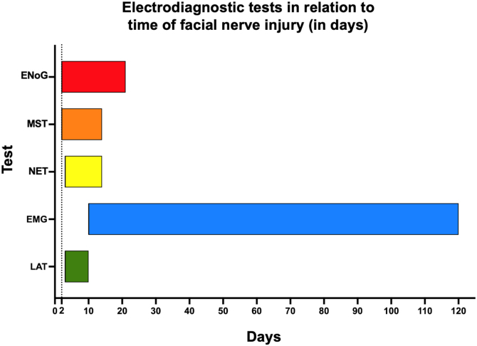
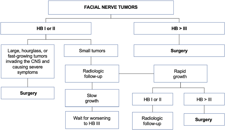
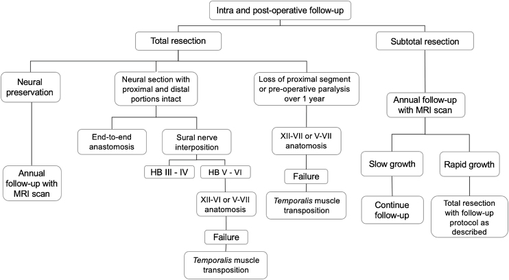
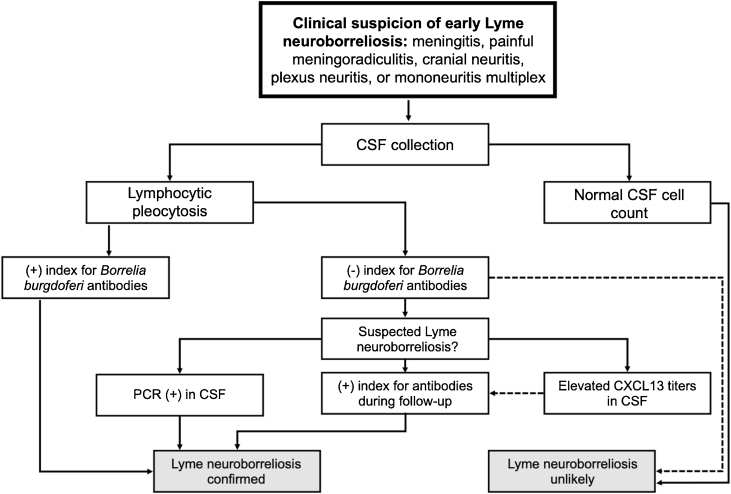
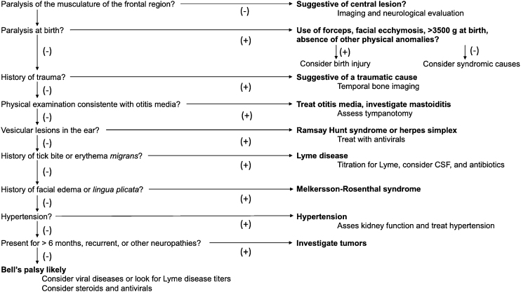
Results: The topics were divided into 2 main parts: (1) Evaluation and diagnosis of facial palsy: electrophysiologic tests, idiopathic facial palsy, Ramsay Hunt syndrome, traumatic peripheral facial palsy, recurrent peripheral facial palsy, facial nerve tumors, and peripheral facial palsy in children; and (2) Rehabilitation procedures: surgical decompression of the facial nerve, facial nerve grafting, surgical treatment of long-term peripheral facial palsy, and non-surgical rehabilitation of the facial nerve.

Conclusions: Peripheral facial palsy is a condition of diverse etiology. Treatment should be individualized according to the cause of facial nerve dysfunction, but the literature presents better evidence-based recommendations for systemic corticosteroid therapy.

Keywords: Bell palsy; Facial nerve disease; Facial nerve trauma; Facial palsy; Facial paralysis; Guidelines; Herpes Zoster Oticus; Microvascular decompression surgery.


Figures

Figure 1
Figure 1
Schematic representation of nerve structure.
Figure 2
Figure 2
Anatomical schematic representation of the facial nerve divided into segments and intracranial and extracranial innervated structures.
Figure 3
Figure 3
Histological sections of temporal bone at 20× magnification, right ear, stained with hematoxylin & Eosin. 1: Facial nerve, labyrinthine segment; 2: Geniculate ganglion; 3: Facial nerve, tympanic segment; 4: Mastoid segment of the facial nerve; 5: Facial nerve, mastoid segment; 6: Internal auditory canal; 7: Cochlea; 8: Superior semicircular canal; 9: Lateral semicircular canal; 10: Posterior semicircular canal; 11: Utricle; 12: Malleus; 13: Incus; 14: Stapes; 15: Tympanic sinus; 16: Facial recess; 17: Tympanic membrane; 18: Pyramidal eminence; 19: Oval window niche; 20: Oval window; 21: Carotid artery.
Figure 4
Figure 4
Histological images of the evolution of an axonotmesis-like lesion of the facial nerve. Axonotmesis: facial nerve injury by compression and evolution at 6 weeks. (A) Normal facial nerve. (B) Facial nerve 1 week after the injury showing significant degeneration, with loss of the myelin sheath. (C) Facial nerve 2 weeks after injury. (D) Facial nerve 4 weeks after injury. (E) Facial nerve 6 weeks after the injury with a structure already quite similar to the normal facial nerve.
Figure 5
Figure 5
Indication of electrodiagnostic tests according to the degree of evolution of peripheral facial palsy. EMG, Electromyography; ENoG, Electroneurography; LAT, Latency test; MST, Maximal Stimulation Test; NET, Nerve Excitability Test.
Figure 6
Figure 6
Characteristics of Melkersson–Rosenthal Syndrome. Facial paralysis in (A) and lingua plicata in (B).
Figure 7
Figure 7
Proposed algorithm for follow-up and treatment of intrinsic facial nerve tumors. Adapted from Prasad et al.
Figure 8
Figure 8
Recommendations regarding intraoperative and postoperative management in the treatment of facial nerve injuries.
Figure 9
Figure 9
Flowchart for assessment of Lyme neuroborreliosis.
Figure 10
Figure 10
Algorithm for etiological diagnosis of peripheral facial paralysis in children.

References

    1. Kim S.J., Lee H.Y. Acute peripheral facial palsy: a systematic review of the literature. J Korean Med Sci. 2020;35:e245. - PMC - PubMed
    1. Fonseca K.M.O., Mourão A.M., Motta A.R., Vicente L.C.C. Scales of degree of facial paralysis: b Limits of agreement. Braz J Otorhinolaryngol. 2015;81:288–293. - PMC - PubMed
    1. Malik M., Cubitt J.J. Paediatric facial paralysis: an overview and insights into management. J Paediatr Child Health. 2021;57:786–790. - PubMed
    1. Wohrer D., Moulding T., Titomanlio L., Lenglart L. Acute facial nerve palsy in children: gold standard management. Children (Basel). 2022;9:273. - PMC - PubMed
    1. Lorch M., Teach S.J. Facial nerve palsy: etiology and approach to diagnosis and treatment. Pediatr Emerg Care. 2010;26:763–769. - PubMed

LinkOut - more resources