. 2024 May:71:103087.
doi: 10.1016/j.redox.2024.103087. Epub 2024 Feb 13.

METTL17 coordinates ferroptosis and tumorigenesis by regulating mitochondrial translation in colorectal cancer

Affiliations

METTL17 coordinates ferroptosis and tumorigenesis by regulating mitochondrial translation in colorectal cancer

Hao Li et al. Redox Biol. 2024 May.

Abstract

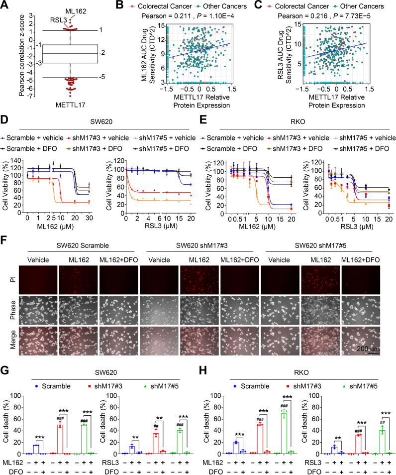
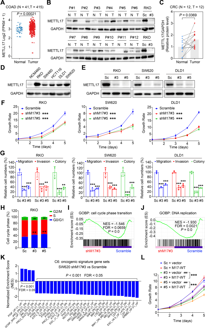
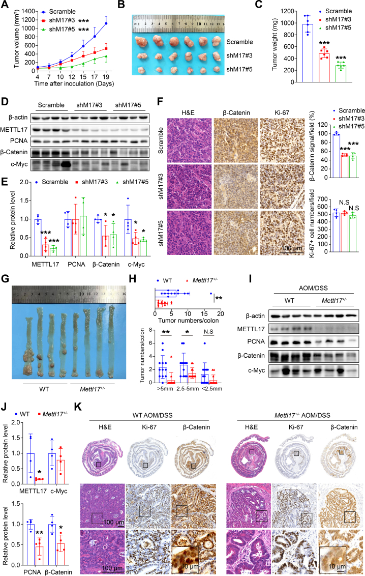
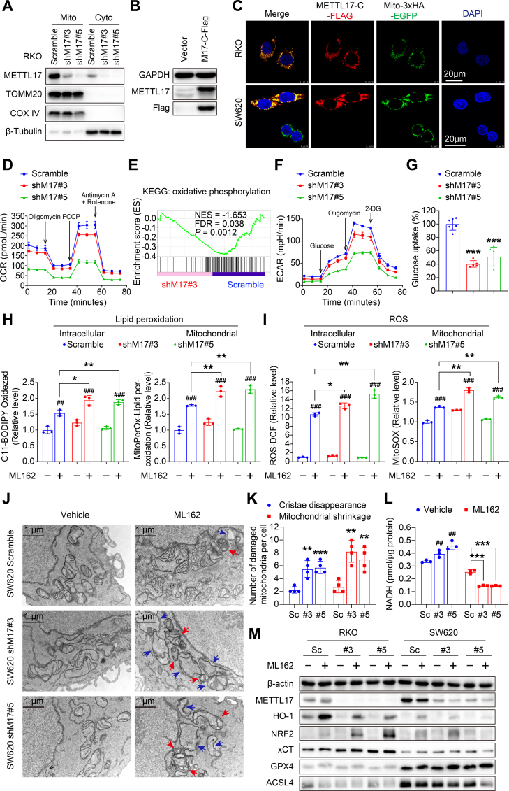
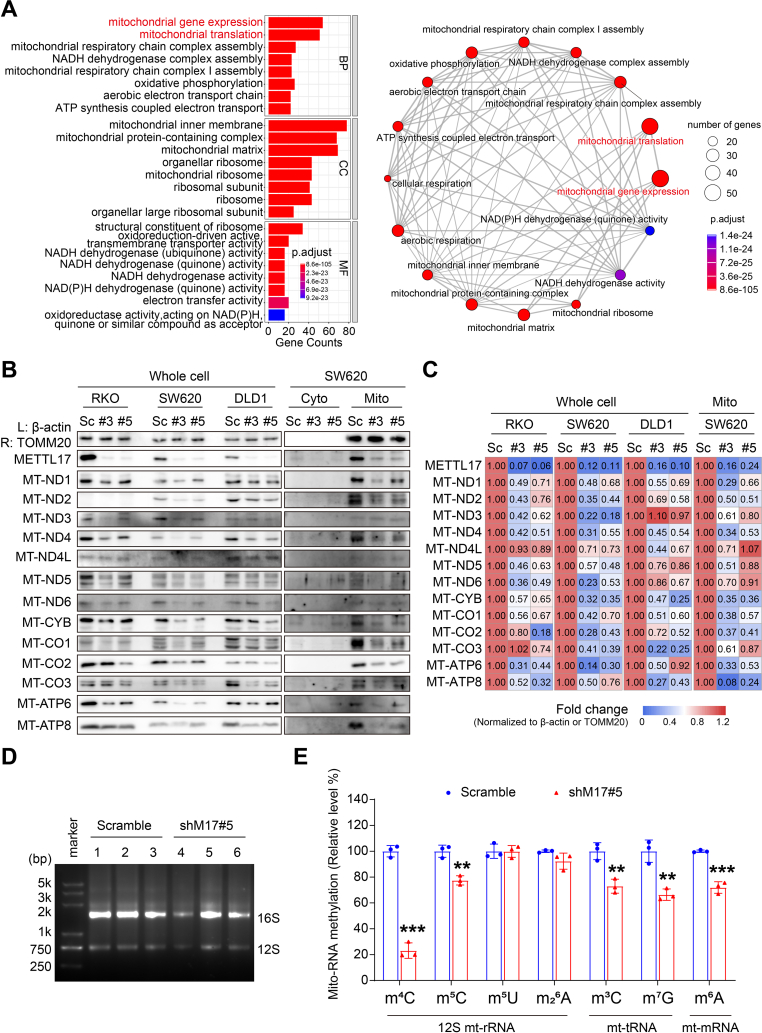
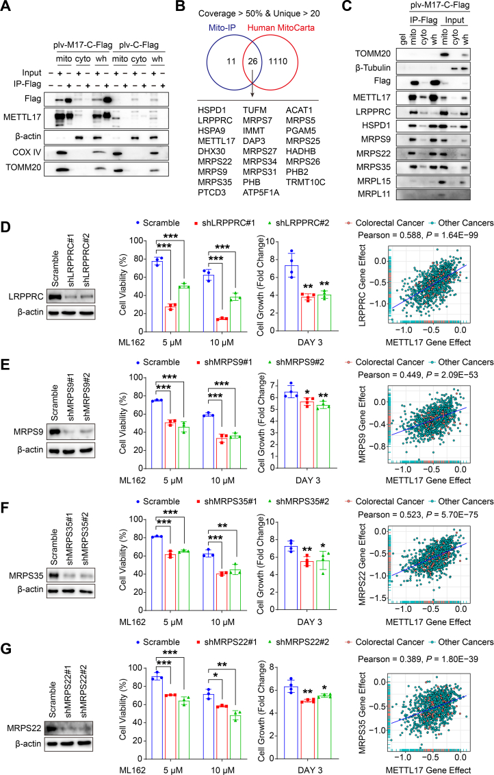
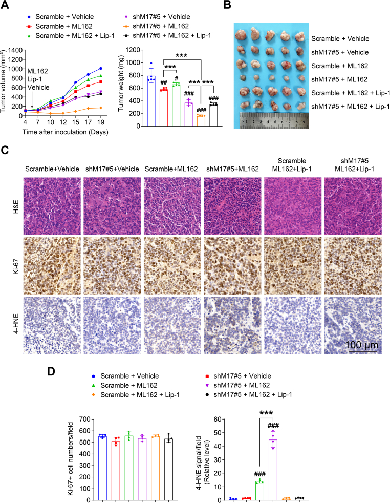
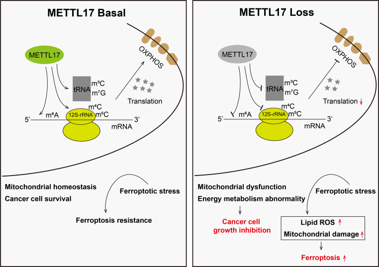
Ferroptosis, an iron-dependent lipid peroxidation-induced form of regulated cell death, shows great promise as a cancer therapy strategy. Despite the critical role of mitochondria in ferroptosis regulation, the underlying mechanisms remain elusive. This study reveals that the mitochondrial protein METTL17 governs mitochondrial function in colorectal cancer (CRC) cells through epigenetic modulation. Bioinformatic analysis establishes that METTL17 expression positively correlates with ferroptosis resistance in cancer cells and is up-regulated in CRC. Depletion of METTL17 sensitizes CRC cells to ferroptosis, impairs cell proliferation, migration, invasion, xenograft tumor growth, and AOM/DSS-induced CRC tumorigenesis. Furthermore, suppression of METTL17 disrupts mitochondrial function, energy metabolism, and enhances intracellular and mitochondrial lipid peroxidation and ROS levels during ferroptotic stress. Mechanistically, METTL17 inhibition significantly reduces mitochondrial RNA methylation, including m4C, m5C, m3C, m7G, and m6A, leading to impaired translation of mitochondrial protein-coding genes. Additionally, the interacting proteins associated with METTL17 are essential for mitochondrial gene expression, and their knockdown sensitizes CRC cells to ferroptosis and inhibits cell proliferation. Notably, combined targeting of METTL17 and ferroptosis in a therapeutic approach effectively suppresses CRC xenograft growth in vivo. This study uncovers the METTL17-mediated defense mechanism for cell survival and ferroptosis in mitochondria, highlighting METTL17 as a potential therapeutic target for CRC.

Keywords: Colorectal cancer (CRC); Ferroptosis; METTL17; Mitochondrial RNA methylation.


Conflict of interest statement

Declaration of competing interest The authors declare no competing interests.

Figures

Fig. 1
Fig. 1
METTL17 expression is positively correlated with ferroptosis-resistance of cancer cells and knockdown of METTL17 sensitizes CRC cells to ferroptosis. A. METTL17 expression exhibited positive correlation with resistance to GPX4 inhibitors (ML162 and RSL3) in cancer cells. Data in plots were mined from the CTRP database and each plot represented a certain anti-tumor compound. B and C. Scatter plots showing the correlation between ML162 (B), RSL3 (C) AUC drug sensitivity and METTL17 protein level in colorectal cancer or other cancer cell lines. Data were mined from the DepMap database and each plot represented a certain cancer cell line. D and E. Cell viability of control (Scramble) and METTL17 knockdown (shM17#3 and shM17#5) SW620 cells (D) or RKO cells (E) treated with ML162 or RSL3 for 4 h after pretreatment with or without DFO (100 μM) for 1 h n = 3–4 per group. F. Microscopy showing cell death. SW620 cells were treated with ML162 (10 μM) for 4 h following pretreatment with or without DFO (100 μM) for 1 h. Upper panel: propidium iodide (PI) staining for dead cells; middle panel, phase-contrast; lower panel: merge image. Scale bar = 200 μm. G and H. Quantification of ferroptotic cells determined by PI-positive staining in SW620 cells (G) or RKO cells (H) treated with ML162 (10 μM) or RSL3 (10 μM) for 4 h following pretreatment with or without DFO (100 μM) for 1 h. The number of cell deaths was counted from Fig. 1F and Supplementary Fig. 1 n = 3 per group. Data are shown as mean ± SD. ##p < 0.01, ###p < 0.001 compared to ML162 or RSL3-treated Scramble group, and **p < 0.01, ***p < 0.001 compared with the indicated two groups, based on two-sided Student's t-test or Pearson r test.
Fig. 2
Fig. 2
METTL17 is highly expressed in CRC and knockdown of METTL17 inhibits CRC cell proliferation, migration, invasion, and oncogenic signatures in vitro. A. The mRNA levels of METTL17 were up-regulated in colon adenocarcinoma (COAD) compared to adjacent normal tissues based on TCGA analysis. Normal tissue (N), n = 41. Tumor tissues (T), n = 415. B and C. The protein levels of METTL17 were measured in 12 pairs of human CRC tumor tissues (T) and the corresponding surrounding non-tumorous tissues (N) from 12 patients (abbreviated to P#1–12) (B). Relative protein level of METTL17 normalized to GAPDH was quantified by Image J (C). n = 12 per group. The METTL17 knockdown (#3) and Scramble (Sc) RKO cells were used as control. D. METTL17 was highly expressed in human CRC cell lines compared to human normal colonic epithelial cell line (NCM460) detected by western blotting. E. The knockdown efficiency of METTL17 in RKO, SW620, and DLD1 cells was confirmed by Western blot assays. Sc, Scramble. #3, shMETTL17#3. #5, shMETTL17#5. F. Crystal violet staining assays showed that knockdown of METTL17 reduced cell proliferation of RKO, SW620, and DLD1. n = 5–6 per group. shM17#3, shMETTL17#3. shM17#5, shMETTL17#5. G. Knockdown of METTL17 inhibited cell migration, invasion, and colony formation in RKO, SW620, and DLD1 cells measured through trans-well migration and invasion assays followed by crystal violet staining. The relative levels respectively were counted from Supplementary Fig. 3A, n = 4 per group. H, I and J. Knockdown of METTL17 caused cell cycle arrest at G0/G1 phase in RKO cells (H) (n = 3 per group), and GSEA analysis revealed disruptions in cell cycle phase transition (I) and DNA replication (J) in METTL17-deficient RKO cells. GOBP, Gene Ontology Biological Process. FDR, false discovery rate. K. GSEA analysis of RNA-Seq data identified that METTL17 knockdown in SW620 cells suppressed multiple oncogenic pathways. L. Exogenous overexpression of METTL17 via lentivirus rescued the inhibited cell proliferation in METTL17-deficient SW620 cells. n = 5–6 per group. M17-WT, human wide type METTL17. Data are shown as mean ± SD. ##p < 0.01, ###p < 0.001 compared to Scramble group, and **p < 0.01, ***p < 0.001 compared to Scramble group or the indicated two groups, based on two-sided Student's t-test or one-way ANOVA.
Fig. 3
Fig. 3
METTL17 deficiency reduces xenograft tumor growth and AOM/DSS-induced CRC tumorigenesis in vivo. A, B and C. Knockdown of METTL17 inhibited SW620 xenograft tumor growth (A) and reduced tumor size (B) and weights (C) in mice. n = 6 per group. D and E. Knockdown of METTL17 led to a reduction in the protein expression of β-Catenin and c-Myc, while the PCNA protein level remained unchanged, as analyzed by Western blot (D). The relative expression was quantified by normalizing to β-actin (E). n = 4 per group. F. Knockdown of METTL17 caused reduced β-Catenin expression, but unchanged Ki-67 positive cells in SW620 xenograft tumors. H&E, β-Catenin, and Ki-67 staining were performed on tissue sections from Scramble and METTL17 knockdown SW620 tumors (Scale bar = 100 μm). The β-Catenin signal and Ki-67-positive tumor cells were quantified in the 400 × field, n = 4 per group. G and H.Mettl17 heterozygote mice (Mettl17+/−) showed a decreased incidence of AOM/DSS-induced CRC formation compared to WT mice. Representative images of intact tumors were shown (G). Mettl17+/− mice exhibited fewer and smaller colon tumors after AOM/DSS treatment, as indicated by the number of total tumors per colon (up) and different sizes' tumors per colon (down) (H). n = 12 per group. I and J. Colon tumors in Mettl17+/− mice exhibited reduced levels of PCNA and β-Catenin proteins, with no significant change observed in c-Myc, as analyzed by Western blot (I). The relative expression was respectively quantified by normalizing to β-actin (J). n = 4 per group. K. H&E, Ki-67, and β-Catenin staining were performed on colon sections from WT and Mettl17+/− mice after AOM/DSS treatment. Data are shown as mean ± SD. *p < 0.05, **p < 0.01, ***p < 0.001, based on two-sided Student's t-test.
Fig. 4
Fig. 4
METTL17 loss causes significant mitochondrial dysfunction and promotes mitochondrial lipid peroxidation in ferroptosis. A. Western blot analysis revealed a higher expression of METTL17 in the mitochondrial fraction compared to the cytoplasm in RKO cells. Mito, mitochondria. Cyto, cytoplasm. B. Lentivirus mediated-overexpression of METTL17-C-Flag (M17-C-Flag) in RKO WT cells was confirmed by Western blot analysis. C. Immunofluorescence revealed co-localization between overexpressed METTL17 and mitochondria in RKO and SW620 cells, where mitochondria were stably tagged with 3xHA-EGFP (Scale bar = 20 μm). D. Seahorse analysis showed that METTL17 knockdown reduced oxygen consumption rate (OCR) in SW620 cells. n = 5–6 per group. E. GSEA analysis exhibited poor oxidative phosphorylation in METTL17 loss SW620 cells. The representative down-regulated genes were presented in Supplementary Fig. 13A. F. Seahorse analysis showed that METTL17 knockdown reduced extracellular acidification rate (ECAR) in SW620 cells. n = 5–6 per group. G. Knockdown of METTL17 significantly suppressed the glucose uptake ability of RKO cells. n = 5–6 per group. H and I. Knockdown of METTL17 increased intracellular and mitochondrial lipid peroxidation (H) and ROS (I) in ML162 (10 μM, 3 h)-treated SW620 cells, which were stained by indicated fluorescent dyes followed by flow cytometry detection. Relative levels were respectively quantified. n = 3 per group. J and K. Knockdown of METTL17 caused worse mitochondrial damage in ML162 (10 μM, 3 h)-treated SW620 cells detected by transmission electron microscope. Representative images were shown (Scale bar = 1 μm), with red arrows indicating mitochondrial shrinkage and blue arrows indicating the disappearance of mitochondrial cristae (J). The number of damaged mitochondria per cell was counted in ML162-treated SW620 cells (K), n = 4 per group. L. Knockdown of METTL17 in SW620 cells dramatically decreased cellular NADH content compared to Scramble cells after ML162 treatment (10 μM, 4 h), while METTL17 knockdown alone increased the level of NADH. n = 3 per group. M. Western blot analysis revealed alterations in ferroptosis markers mediated by METTL17 knockdown under ML162 (10 μM, 4 h) challenge in RKO and SW620 cells. The relative protein levels were performed semi-quantitative analysis, showing as a heatmap in Supplementary Fig. 6C. Data are shown as mean ± SD. ##p < 0.01, ###p < 0.001 compared to Scramble group, and *p < 0.05, **p < 0.01, ***p < 0.001 compared to Scramble group or the indicated two groups, based on two-sided Student's t-test.
Fig. 5
Fig. 5
METTL17 is required for global mitochondrial translation in mitochondrial RNA methylation manner in CRC. A. GO enrichment analysis revealed that the top 100 METTL17 DepMap co-dependent genes predominantly associated with mitochondria, influencing the regulation of mitochondrial gene expression and mitochondrial translation. The bar plot illustrated enrichments in multiple mitochondria-related gene sets (left), while the visualized network highlighted the close connections among these gene sets (right). BP, biological process. CC, cellular component. MF, molecular function. B and C. Knockdown of METTL17 down-regulated the protein expression of mitochondrial coding genes in CRC cells detected by Western blot (B). Semi-quantitative analysis of the relative protein levels was performed and visualized in the heatmap of fold change, normalized to Scramble (C). D and E. The quality of mitochondrial RNA from RKO cells was identified by agarose gel assay (D). HPLC-MS analysis of mitochondrial RNA showed that knockdown of METTL17 significantly reduced m4C, m5C, m3C, m7G, and m6A levels within mitochondrial RNA methylations in RKO cells (E). Relative levels were quantified by comparing to scramble, n = 3 per group. Data are shown as mean ± SD. **p < 0.01, ***p < 0.001 compared to Scramble group, based on a two-sided Student's t-test.
Fig. 6
Fig. 6
Targeting METTL17 interacting proteins sensitizes cancer cells to ferroptosis. A. RKO cells overexpressing METTL17-C-Flag through plv lentivirus (plv-M17-C-Flag) were isolated into mitochondrial (mito), cytoplasmic (cyto), and whole cell (wh) fractions. Immunoprecipitation with anti-Flag affinity gel (IP-Flag) was performed, and the samples were subsequently identified by Western blot. B. Mitochondrial precipitants (Mito-IP) were subjected to mass spectrometry analysis to identify the protein complexes binding to METTL17-C-Flag. Venn diagram analysis, based on mapping Mito-IP with Human MitoCarta, identified 26 METTL17 interactors with high affinity to METTL17, filtered by coverage >50% and unique peptide >20. C. RKO cells overexpressing METTL17-C-Flag (plv-M17-C-Flag) were isolated into mitochondrial (mito), cytoplasmic (cyto), and whole cell (wh) fractions, and immunoprecipitated by anti-Flag affinity gel (IP-Flag). Western blot analysis of METTL17 precipitants revealed strong interactions between METTL17 and LRPPRC, HSPD1, MRPS9, MRPS22, and MRPS35. D, E, F and H. Knockdown of METTL17-interacting proteins, LRPPRC (D), MRPS9 (E), MRPS35 (F), and MRPS22 (G) using lentivirus pLKO-shRNA, sensitized RKO cells to ML162-induced ferroptosis (n = 3 per group). Moreover, their knockdown inhibited cell proliferation in RKO cells (n = 4 per group), and the gene effect of METTL17 highly correlated with those of LRPPRC, MRPS9, MRPS35 and MRPS22. Data on gene knockout effect on cancer cells were mined from the DepMap database, with each plot representing a certain cancer cell line. Data are shown as mean ± SD. *p < 0.05, **p < 0.01 ***p < 0.001, based on two-sided Student's t-test or Pearson r-test.
Fig. 7
Fig. 7
Combination therapy by targeting METTL17 and ferroptosis suppress CRC tumorigenesis. A and B. The tumor growth volume, weight, and size of SW620 xenograft tumors were significantly attenuated in the combination group receiving shRNA targeting METTL17 and ML162 treatment compared to monotherapy with either METTL17 knockdown or ML162 treatment. However, the efficacy of the combination treatment was compromised by the administration of Lip-1, a potent ferroptosis inhibitor by blocking lipid peroxidation. Vehicle control, ML162 (10 mg/kg per mouse) and Lip-1 (10 mg/kg per mouse) were intraperitoneally administrated every day starting from day 5 post SW620 inoculation. n = 5 per group. C and D. H&E, Ki-67, and 4-HNE staining of tissue sections from SW620 xenograft tumors (C). Scale bar = 100 μm. Ki-67-positive tumor cells and 4-HNE signal were quantified at the 400 × field (D), n = 4 per group. Data are shown as mean ± SD. #p < 0.05, ##p < 0.01, ###p < 0.001 compared to Scramble vehicle group, and ***p < 0.001 compared to indicated two groups, based on two-sided Student's t-test.
Fig. 8
Fig. 8
Schematic model for the mechanism by which METTL17 coordinates ferroptosis and tumorigenesis by regulating mitochondrial translation in CRC. METTL17 methylates m4C/m5C on 12S-rRNA, m3C/m7G on mt-RNA, and m6A on mt-mRNA within mitochondria, which affect mitochondrial translation efficiency and respiratory chain activity. METTL17 loss results in the suppression of these modifications and mitochondrial translation, leading to mitochondrial dysfunction and energy metabolism abnormality, thus inhibiting CRC cell growth and sensitizing CRC cells to ferroptosis.

Similar articles

Cited by

References

    1. Dixon S.J., Lemberg K.M., Lamprecht M.R., Skouta R., Zaitsev E.M., Gleason C.E., et al. Ferroptosis: an iron-dependent form of nonapoptotic cell death. Cell. 2012;149:1060–1072. - PMC - PubMed
    1. Chen X., Kang R., Kroemer G., Tang D.L. Broadening horizons: the role of ferroptosis in cancer. Nat. Rev. Clin. Oncol. 2021;18:280–296. - PubMed
    1. Lei G., Zhuang L., Gan B.Y. Targeting ferroptosis as a vulnerability in cancer. Nat. Rev. Cancer. 2022;22:381–396. - PMC - PubMed
    1. Hassannia B., Vandenabeele P., Vanden Berghe T. Targeting ferroptosis to iron out cancer. Cancer Cell. 2019;35:830–849. - PubMed
    1. Liang C., Zhang X.L., Yang M.S., Dong X.C. Recent progress in ferroptosis inducers for cancer therapy. Adv. Mater. 2019;31 - PubMed