. 2024 Dec;15(1):2316459.
doi: 10.1080/21505594.2024.2316459. Epub 2024 Feb 20.

Intranasal B5 promotes mucosal defence against Actinobacillus pleuropneumoniae via ameliorating early immunosuppression

Affiliations

Intranasal B5 promotes mucosal defence against Actinobacillus pleuropneumoniae via ameliorating early immunosuppression

Jingsheng Huang et al. Virulence. 2024 Dec.

Abstract

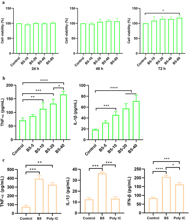
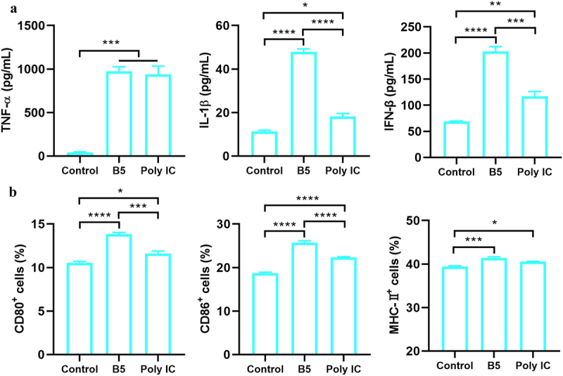
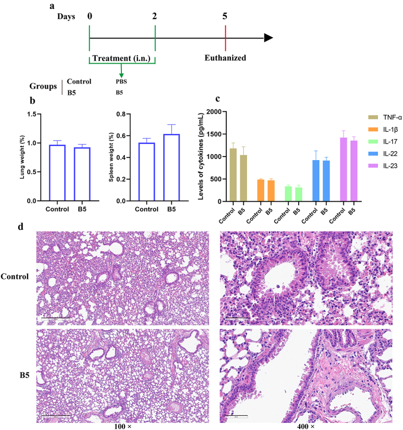
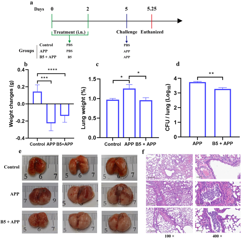
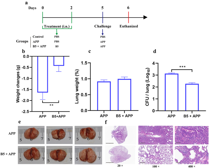
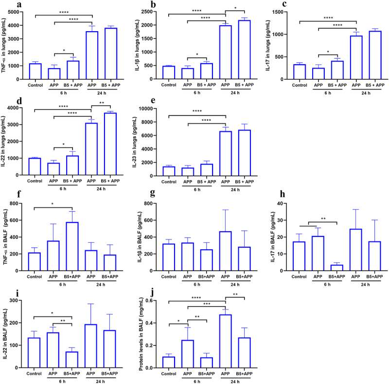
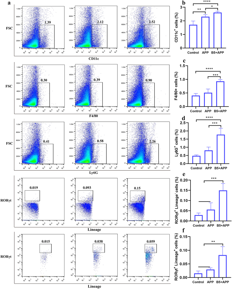
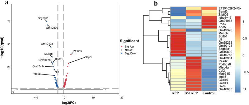
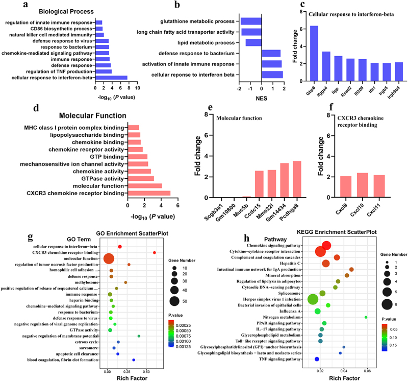
Actinobacillus pleuropneumoniae (APP) is an important pathogen of the porcine respiratory disease complex, which leads to huge economic losses worldwide. We previously demonstrated that Pichia pastoris-producing bovine neutrophil β-defensin-5 (B5) could resist the infection by the bovine intracellular pathogen Mycobacterium bovis. In this study, the roles of synthetic B5 in regulating mucosal innate immune response and protecting against extracellular APP infection were further investigated using a mouse model. Results showed that B5 promoted the production of tumour necrosis factor (TNF)-α, interleukin (IL)-1β, and interferon (IFN)-β in macrophages as well as dendritic cells (DC) and enhanced DC maturation in vitro. Importantly, intranasal B5 was safe and conferred effective protection against APP via reducing the bacterial load in lungs and alleviating pulmonary inflammatory damage. Furthermore, in the early stage of APP infection, we found that intranasal B5 up-regulated the secretion of TNF-α, IL-1β, IL-17, and IL-22; enhanced the rapid recruitment of macrophages, neutrophils, and DC; and facilitated the generation of group 3 innate lymphoid cells in lungs. In addition, B5 activated signalling pathways associated with cellular response to IFN-β and activation of innate immune response in APP-challenged lungs. Collectively, B5 via the intranasal route can effectively ameliorate the immune suppression caused by early APP infection and provide protection against APP. The immunization strategy may be applied to animals or human respiratory bacterial infectious diseases. Our findings highlight the potential importance of B5, enhancing mucosal defence against intracellular bacteria like APP which causes early-phase immune suppression.

Keywords: Actinobacillus pleuropneumoniae; Group 3 innate lymphoid cells; bovine neutrophil β-defensin-5; immune suppression; mucosal defence.


Conflict of interest statement

No potential conflict of interest was reported by the author(s).

Figures

Figure 1.
Figure 1.
B5 activates macrophages. a, the effects of B5 on the viability in RAW264.7 cells. Cells were incubated with 10–80 µg/ml of B5 for 24 h, 48 h, or 72 h, and cell viability was detected using CCK8 solution. b, levels of TNF-α and IL-1β in B5-treated RAW264.7 cells. Cells were incubated with 5–40 µg/ml of B5 for 24 h. c, levels of TNF-α, IL-1β and IFN-β in bone marrow-derived macrophages (BMDMs) treated with B5 or poly IC. Data shown are means ± SD. Data are representative of three independent experiments (n = 3). The significance of differences between the groups was determined by ANOVA with post-hoc Tukey’s multiple comparison test (*p < 0.033, **p < 0.0021, ***p < 0.0002, ****p < 0.0001).
Figure 2.
Figure 2.
B5 promotes the maturation of functional DC. a, levels of TNF-α, IL-1β, and IFN-β in bone marrow-derived dendritic cells (BMDCs) stimulated with B5 or poly IC. b, expression levels of CD80, CD86, and MHC-II in BMDC stimulated with B5 or poly IC. Data shown are means ± SD. Data are representative of three independent experiments (n = 3). The significance of differences between the groups was determined by ANOVA with post-hoc Tukey’s multiple comparison test (*p < 0.033, **p < 0.0021, ***p < 0.0002, ****p < 0.0001).
Figure 3.
Figure 3.
Intranasal B5 safety evaluation. a, schematic representation of treatment model. Mice were treated through intranasal route with 20 μg of B5 (twice, 2-d interval). Three days after the last treatment, mice were euthanized for safety evaluation. b, weight of lung and spleen tissues after treatment with B5. c, levels of TNF-α, IL-1β, IL-17, IL-22 and IL-23 in lungs. d, histopathological images of lungs performed with HE staining (left images scale bar: 300 μm; right images scale bar: 60 μm). Data shown are means ± SD. Data are representative of two independent experiments (n = 3 mice per group). The significance of differences between two groups was determined by Student’s t-test.
Figure 4.
Figure 4.
Intranasal B5 provides early protection against APP. a, schematic representation of pretreatment/challenge model. Mice were pretreated intranasally with 20 μg of B5 (twice, 2-d interval) and challenged with APP 3 d after the last pretreatment and euthanized for protective evaluation 6 h after challenge. b, body weight changes. c, weight of lung tissues. d, bacterial load in lungs. e, gross pathology of lungs. f, histopathological images of lungs performed with HE staining (left images scale bar: 300 μm; right images scale bar: 60 μm). Data shown are means ± SD. Data are representative of two independent experiments (n = 6 mice per group). The significance of differences between the groups was determined by ANOVA with post-hoc Tukey’s multiple comparison test (*p < 0.033, **p < 0.0021, ***p < 0.0002, ****p < 0.0001).
Figure 5.
Figure 5.
Intranasal B5 maintains protection in the later stage of APP infection. a, schematic representation of pretreatment/challenge model. Mice were pretreated intranasally with 20 μg of B5, challenged with APP 3 d after the last pretreatment, and euthanized for protective evaluation 24 h after challenge. b, body weight changes. c, weight of lung tissues. d, bacterial load in lungs. e, gross pathology of lungs. f, histopathological images of lungs performed with HE staining (left images scale bar: 2 mm; middle images scale bar: 300 μm; right images scale bar: 60 μm). Data shown are means ± SD. Data are representative of two independent experiments (n = 6 mice per group). The significance of differences between the groups was determined by ANOVA with post-hoc Tukey’s multiple comparison test (**p < 0.0021, ***p < 0.0002).
Figure 6.
Figure 6.
Intranasal B5 promotes cytokine secretion in lungs during APP infection. Mice were pretreated intranasally with 20 μg of B5, challenged with APP 3 d after the last pretreatment, and euthanized for cytokines detection 6 h and 24 h after challenge. a-e, levels of TNF-α (a), IL-1β (b), IL-17 (c), IL-22 (d), and IL-23 (e) in lungs. f-i, levels of TNF-α (f), IL-1β (g), IL-17 (h), and IL-22 (i) in BALF. j, protein levels in BALF. Data shown are means ± SD. Data are representative of two independent experiments (n = 3–4 mice per group). The significance of differences between the groups was determined by ANOVA with post-hoc Tukey’s multiple comparison test (*p < 0.033, **p < 0.0021, ***p < 0.0002, ****p < 0.0001).
Figure 7.
Figure 7.
Intranasal B5 induces the generation of innate immune cells in lungs. Mice were pretreated intranasally with 20 μg of B5, challenged with APP 3 d after the last pretreatment, and euthanized for innate immune cells detection 6 h after challenge. a, Representative FACS blots of DC (CD11c+), macrophages (F4/80+), neutrophils (Ly6G+), ILC3s (Lineage Rorγt+), and Lineage+ Rorγt+ cells in lungs. b-f, percentage of DC (b), macrophages (c), neutrophils (d), ILC3s (e), and Lineage+ Rorγt+ cells (f). Data shown are means ± SD. Data are representative of two independent experiments (n = 4–6 mice per group). The significance of differences between the groups was determined by ANOVA with post-hoc Tukey’s multiple comparison test (*p < 0.033, **p < 0.0021, ***p < 0.0002, ****p < 0.0001).
Figure 8.
Figure 8.
Analysis of differentially expressed genes. a, differentially expressed genes were shown in volcano plots (B5 + APP group vs APP group). b, representative differentially expressed genes were shown in heat map.
Figure 9.
Figure 9.
Gene ontology (GO) term analysis of differentially expressed genes. B5 + APP group vs APP group: a, gene enrichment in biological process. b, gene set enrichment analysis (GSEA). c, fold changes of genes involving cellular response to interferon-beta d, gene enrichment in molecular function. e-f, fold changes of genes involving molecular function (e) and CXCR3 chemokine receptor binding (f). g-h, GO enrichment scatterplot (g) and KEGG enrichment scatterplot (h).

Similar articles

Cited by

References

    1. Gottschalk M, Broes A.. Actinobacillosis. In Zimmerman JJ, Karriker LA, Ramirez A, et al., editors. Diseases of swine. Chichester: Wiley; 2019. p. 749–17.
    1. Sassu EL, Bossé JT, Tobias TJ, et al. Update on Actinobacillus pleuropneumoniae-knowledge, gaps and challenges. Transbound Emerg Dis. 2018;1:72–90. doi: 10.1111/tbed.12739 - DOI - PubMed
    1. Qi W, Zhu R, Bao C, et al. Porcine circovirus type 2 promotes Actinobacillus pleuropneumoniae survival during coinfection of porcine alveolar macrophages by inhibiting ROS production. Vet Microbiol. 2019;233:93–101. doi: 10.1016/j.vetmic.2019.04.028 - DOI - PubMed
    1. Stringer OW, Bossé JT, Lacouture S, et al. Proposal of Actinobacillus pleuropneumoniae serovar 19, and reformulation of previous multiplex PCRs for capsule-specific typing of all known serovars. Veterinary Microbiology. 2021;255:109021. doi: 10.1016/j.vetmic.2021.109021 - DOI - PubMed
    1. Kardos G, Sárközi R, Laczkó L, et al. Genetic Diversity of Actinobacillus pleuropneumoniae Serovars in Hungary[J]. Vet Sci. 2022;9(10):511. doi: 10.3390/vetsci9100511 - DOI - PMC - PubMed

Substances