

Macular vessel density in the superficial plexus is not a proxy of cerebrovascular damage in non-demented individuals: data from the NORFACE cohort
- PMID: 38378643
- PMCID: PMC10877901
- DOI: 10.1186/s13195-024-01408-9
Macular vessel density in the superficial plexus is not a proxy of cerebrovascular damage in non-demented individuals: data from the NORFACE cohort
Abstract
Introduction: Optical coherence tomography angiography (OCT-A) is a novel tool that allows the detection of retinal vascular changes. We investigated the association of macular vessel density (VD) in the superficial plexus assessed by OCT-A with measures of cerebrovascular pathology and atrophy quantified by brain magnetic resonance imaging (MRI) in non-demented individuals.
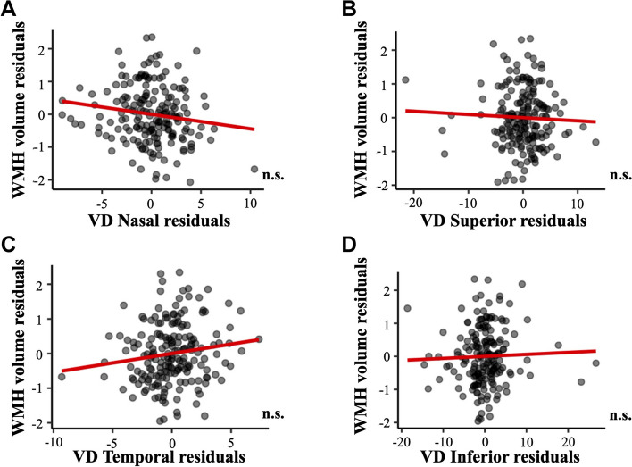
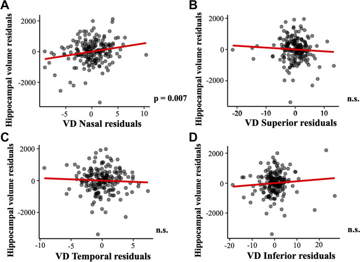
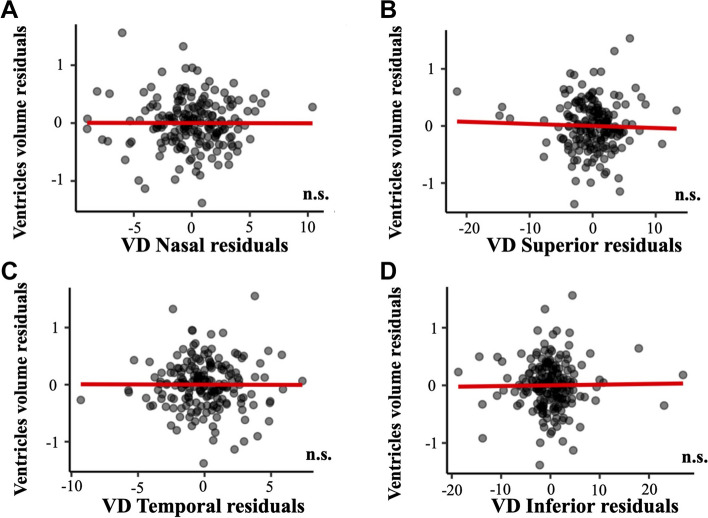
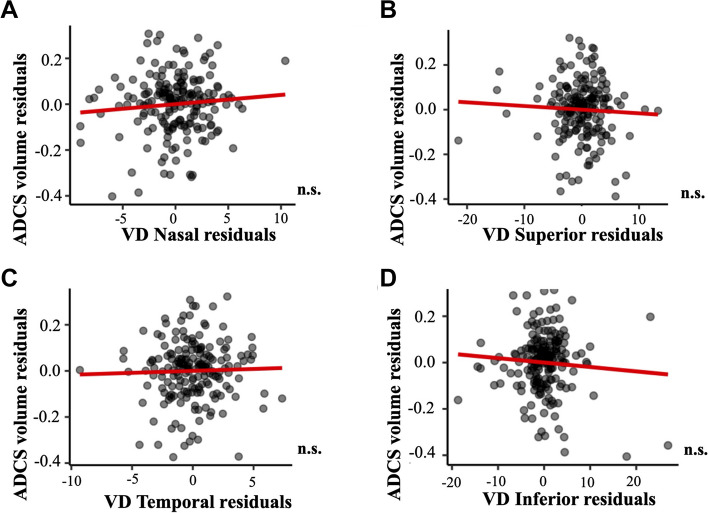
Methods: Clinical, demographical, OCT-A, and brain MRI data from non-demented research participants were included. We analyzed the association of regional macular VD with brain vascular burden using the Fazekas scale assessed in a logistic regression analysis, and the volume of white matter hyperintensities (WMH) assessed in a multiple linear regression analysis. We also explored the associations of macular VD with hippocampal volume, ventricle volume and Alzheimer disease cortical signature (ADCS) thickness assessed in multiple linear regression analyses. All analyses were adjusted for age, sex, syndromic diagnosis and cardiovascular variables.
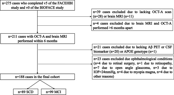
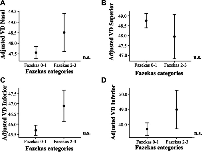
Results: The study cohort comprised 188 participants: 89 with subjective cognitive decline and 99 with mild cognitive impairment. No significant association of regional macular VD with the Fazekas categories (all, p > 0.111) and WMH volume (all, p > 0.051) were detected. VD in the nasal quadrant was associated to hippocampal volume (p = 0.007), but no other associations of macular VD with brain atrophy measures were detected (all, p > 0.05).
Discussion: Retinal vascular measures were not a proxy of cerebrovascular damage in non-demented individuals, while VD in the nasal quadrant was associated with hippocampal atrophy independently of the amyloid status.
Keywords: BIOFACE; Brain atrophy; Cerebrovascular damage; FACEHBI; NORFACE; Optical coherence tomography-angiography; Vessel density.
© 2024. The Author(s).
Conflict of interest statement
MB has consulted for Araclon, Avid, Grifols, Lilly, Nutricia, Roche, Eisai and Servier. She received fees from lectures and funds for research from Araclon, Biogen, Grifols, Nutricia, Roche and Servier. She reports grants/research funding from Abbvie, Araclon, Biogen Research Limited, Bioiberica, Grifols, Lilly, S.A, Merck Sharp & Dohme, Kyowa Hakko Kirin, Laboratorios Servier, Nutricia SRL, Oryzon Genomics, Piramal Imaging Limited, Roche Pharma SA, and Schwabe Farma Iberica SLU, all outside the submitted work. She has not received personal compensations from these organizations. AR is member of scientific advisory board of Landsteiner Genmed and Grifols SA. AR has stocks of Landsteiner Genmed. MM has consulted for F. Hoffmann-La Roche Ltd. The rest of authors declare that they have no competing interests.
Figures
References
-
- Gorelick PB, Scuteri A, Black SE, Decarli C, Greenberg SM, Iadecola C, et al. Vascular contributions to cognitive impairment and dementia: A statement for healthcare professionals from the American Heart Association/American Stroke Association. Stroke; 2011. p. 2672–713. Available from: https://pubmed.ncbi.nlm.nih.gov/21778438/ - PMC - PubMed
-
- Attems J, Jellinger KA. The overlap between vascular disease and Alzheimer’s disease--lessons from pathology. BMC Med. 2014;12. Available from: https://pubmed.ncbi.nlm.nih.gov/25385447/ - PMC - PubMed
Publication types
MeSH terms
Grants and funding
LinkOut - more resources
Full Text Sources
