. 2024 Mar;43(6):993-1014.
doi: 10.1038/s44318-024-00054-z. Epub 2024 Feb 20.

Increases in cyclin A/Cdk activity and in PP2A-B55 inhibition by FAM122A are key mitosis-inducing events

Affiliations

Increases in cyclin A/Cdk activity and in PP2A-B55 inhibition by FAM122A are key mitosis-inducing events

Benjamin Lacroix et al. EMBO J. 2024 Mar.

Abstract

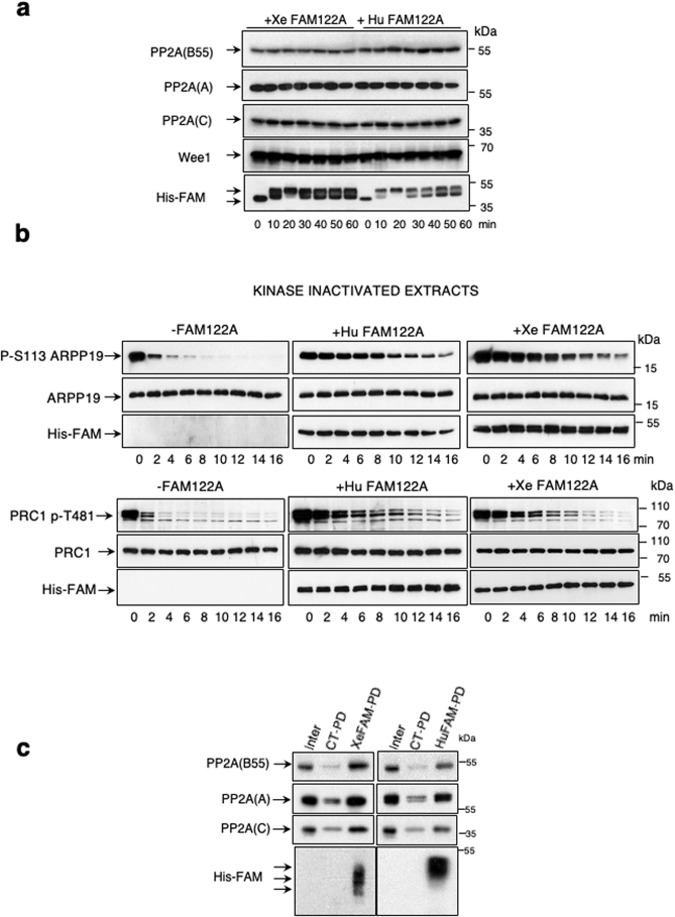
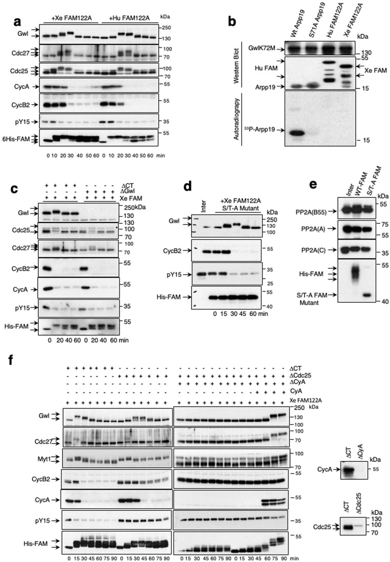
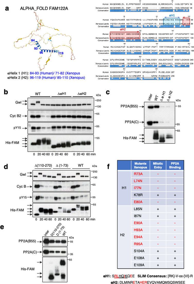
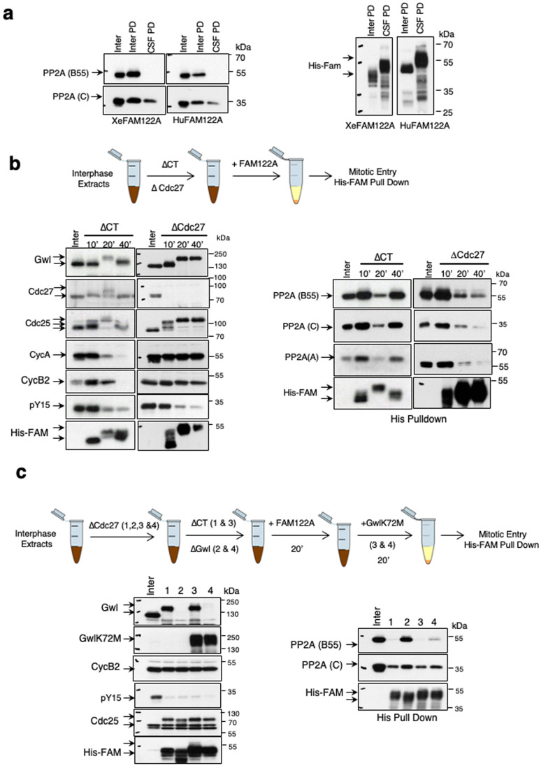
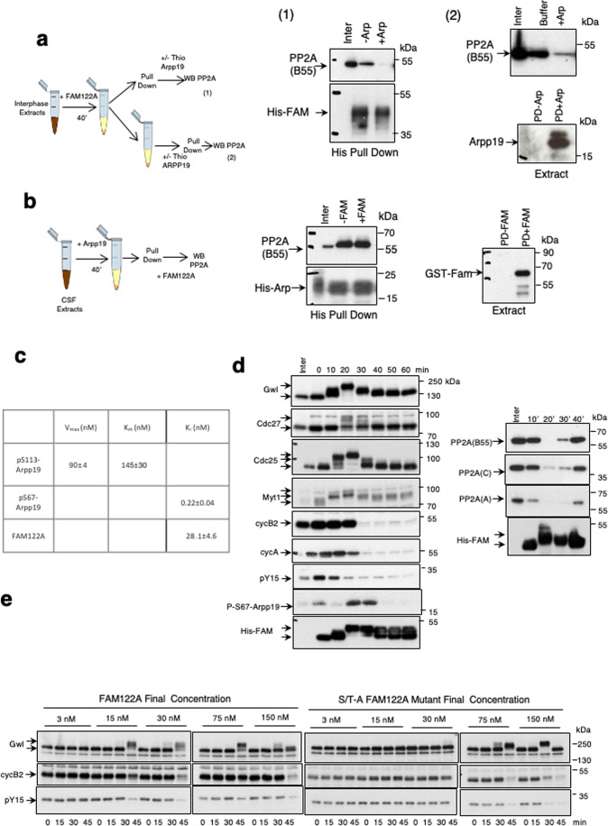
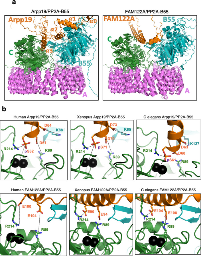
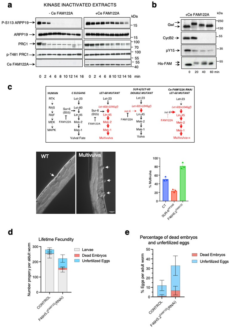
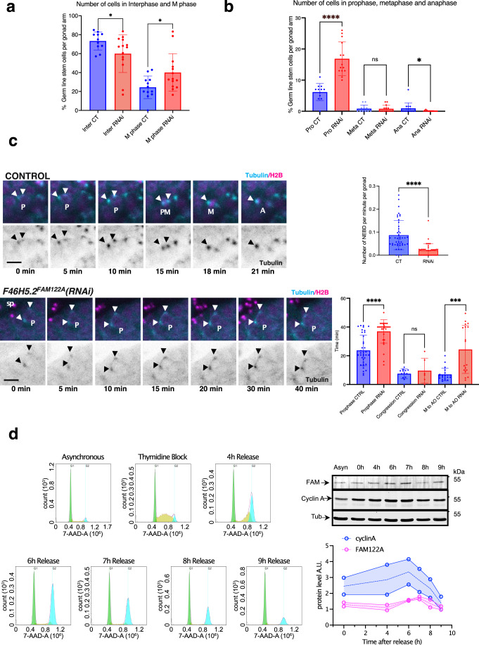
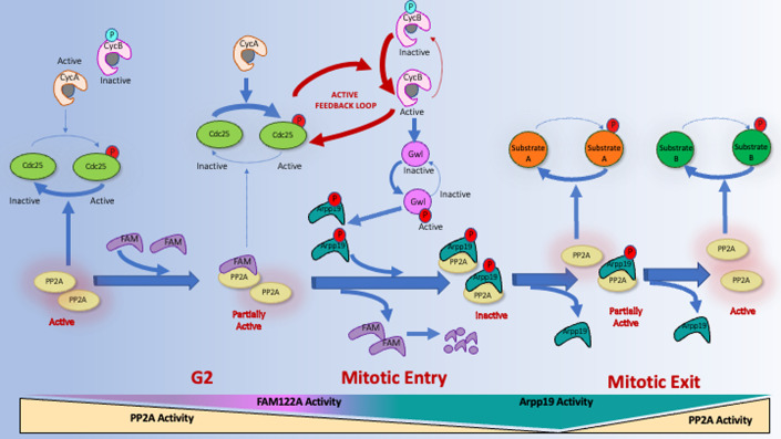
Entry into mitosis has been classically attributed to the activation of a cyclin B/Cdk1 amplification loop via a partial pool of this kinase becoming active at the end of G2 phase. However, how this initial pool is activated is still unknown. Here we discovered a new role of the recently identified PP2A-B55 inhibitor FAM122A in triggering mitotic entry. Accordingly, depletion of the orthologue of FAM122A in C. elegans prevents entry into mitosis in germline stem cells. Moreover, data from Xenopus egg extracts strongly suggest that FAM122A-dependent inhibition of PP2A-B55 could be the initial event promoting mitotic entry. Inhibition of this phosphatase allows subsequent phosphorylation of early mitotic substrates by cyclin A/Cdk, resulting in full cyclin B/Cdk1 and Greatwall (Gwl) kinase activation. Subsequent to Greatwall activation, Arpp19/ENSA become phosphorylated and now compete with FAM122A, promoting its dissociation from PP2A-B55 and taking over its phosphatase inhibition role until the end of mitosis.

Keywords: Arpp19; Cyclin A.; FAM122A; Mitosis; PP2A-B55.


Conflict of interest statement

The authors declare no competing interests.

Figures

Figure 1
Figure 1. PP2A-B55 inhibition by FAM122A does not involve changes in PP2A-C or Wee1 levels.
(A) 20 μl of interphase extracts were supplemented with a final concentration of 7.15 μM of Xenopus or of human FAM122A and 0,8 μl of the mix was recovered at different time points to measure the indicated proteins by western blot. (B) Arpp19 or PRC1 proteins “in vitro” phosphorylated by PKA or by cyclin A/Cdk1 respectively were supplemented at a final concentration of 1.65 μM together with Xenopus or human FAM122A (final concentration of 14.3 μM) to kinase inactivated interphase extracts and the dephosphorylation rate of S113 of Arpp19 and T481 of PRC1 as well as the levels of Xenopus and human FAM122A were analysed by western blot. (C) Interphase extracts were supplemented (Xe/HuFAM-PD) or not (CT-PD) with either Xenopus or human FAM122A as in (A) and used for histidine pull-down. The levels of PP2A B55, A and C subunits as well as the amount of FAM122A were checked by western blot. Source data are available online for this figure.
Figure 2
Figure 2. FAM122A triggers mitotic entry by inhibiting PP2A-B55 and permitting cyclin A/Cdk-dependent phosphorylation of mitotic substrates.
(A) Extracts were treated as for Fig. 1A and the levels of the indicated proteins as well as the phosphorylation of Cdk1 on tyrosine 15 were checked by western blot. (B) His-wildtype Arpp19, the Gwl phosphorylation site mutant (Serine 71 -to-Alanine) of this protein, His-human FAM122A and His-Xenopus FAM122A were phosphorylated “in vitro” by GST-human GwlK72M hyperactive mutant in a final phosphorylation reaction mixture of 10 μl. 5 μl were then used for western blot using anti-histidine antibodies to detect Arpp19 and human and Xenopus FAM122A levels or anti-Gwl antibodies to detect Gwl amount. The rest was used to detect γ33P by autoradiography. (C) Interphase extracts were depleted with a control or anti-Gwl antibodies and supplemented with Xe FAM122A. Samples were then analysed over time for the levels and phosphorylation of the indicated proteins. (D) Interphase extracts were supplemented with the Xenopus FAM122A protein in which all serine/threonine residues were mutated into alanine. Samples were recovered at the indicated time points and used for western blot. (E) Interphase extracts supplemented with a wildtype or a Xenopus FAM122A protein in which all serine/threonine residues were mutated into alanine and used for histidine pulldown and western blot to measure their association with B55, A and C subunits of PP2A. (F) Interphase extracts were depleted using control or anti-Cdc25 antibodies, or submitted to two consecutive depletions using firstly anti-Cdc25 and secondly anti-cyclin A antibodies. Depleted supernatants were then supplemented with Xe FAM122A protein. When indicated, purified cyclin A was added to the mix to a final concentration of 100 nM. Samples were recovered over time and used for western blot. Depletion of cyclin A and Cdc25 in the extracts was confirmed by western blot. Source data are available online for this figure.
Figure 3
Figure 3. Molecular determinants controlling FAM122A inhibitory activity.
(A) AlphaFold Monomer v2.0 predicts two structured αhelices (αHelix 1 and αHelix 2) in the human FAM122A protein and human. Xenopus laevis amino acid sequence alignment. αHelix 1 and αHelix 2 are indicated. (B) Interphase extracts were supplemented with the wildtype or the αH1 or αH2 mutants of Xe FAM122A and the phosphorylation and levels of the indicated proteins measured at the indicated times. (C) Interphase extracts supplemented with wildtype and αH1 or αH2 mutants were used for His-pulldown to measure the association of these proteins to B55 and C subunits of PP2A-B55. (D) The wildtype and the indicated mutant forms of Xe FAM122A were supplemented to interphase extracts and used for western blot. (E) His-pulldown was performed in extracts treated as in (D) to check the binding of the different forms of Xe FAM122A form to B55 and C. (F) Table indicating the capacity of the different single mutants of Xe FAM122A to induce mitotic entry and to bind to PP2A-B55. Mutants that have lost their inhibitory activity are depicted in red. The sequence of the αH1, the SLiM consensus sequence and the sequence of αH2 are shown. Residues essential to keep the inhibitory activity of Xe FAM122A are highlighted in red. Source data are available online for this figure.
Figure 4
Figure 4. Gwl activation at mitotic entry promotes the dissociation of FAM122A from PP2A-B55.
(A) Interphase and CSF extracts were supplemented with Xe FAM122A and Hu FAM122A and used for His-pulldown to measure the association of this protein with B55 and C subunits of PP2A-B55. The levels of His-Xe/HuFAM122A recovered in the pull down are also shown. Inter PD: Interphase Pull Down. CSF PD: CSF pull down. (B) Interphase extracts were depleted with control or with anti-Cdc27 antibodies and subsequently supplemented with Xe FAM122A and used for western blot or for His-pulldown to assess the binding of Xe FAM122A to PP2A-B55. (C) Interphase extracts were depleted of Cdc27, submitted to a second immunodepletion using control (lanes 1 & 3) or anti-Gwl antibodies (lanes 2 & 4) and supplemented with Xe FAM122A. 20 min later Gwl activity was restored (lanes 3 & 4) or not (lanes 1 & 2) by adding a hyperactive form of Gwl (GwlK72M). Samples were used for western blot and His-pulldown. Source data are available online for this figure.
Figure 5
Figure 5. Arpp19 dissociates FAM122A from PP2A-B55 during mitosis.
(A) Interphase extracts were supplemented with a final concentration of 7.15 μM of Xe FAM122A and 40 min later divided into two samples. One of these samples was used to perform a His-pulldown that was then supplemented with a final concentration of 2.7 μM of untagged Thio-Arpp19 protein and used to measure bound B55 protein. The second sample was firstly supplemented with untagged 69.3 pmol of Thio-Arpp19 and subsequently used for His-pulldown to measure His-Xe FAM122A bound B55 subunit of PP2A. A sample of the His-Xe FAM122A pull downs supplemented (PD+Arp) or not (PD-Arp) with untagged Thio-Arpp19 is shown. (B) CSF extracts were supplemented with a final concentration of 3,6 μM His-Arpp19 and, 40 min later, used for His-pulldown. Arpp19-pull downs were then supplemented with a final concentration of 10 μM GST-Xe FAM122A and the levels of B55 remaining were measured. A sample of the His-Arpp19 pull downs supplemented (PD + FAM) or not (PD-FAM) is shown. (C) Table indicating the kinetic parameters of the dephosphorylation of the PP2A-B55 substrate p-S113-Arpp19 and the inhibitory constant of FAM122A and Thio-S71-Arpp19 in kinase inactivated interphase extracts. Conditions used in these assays are detailed in Methods. (D) Interphase extracts were supplemented with His-Xe FAM122A and used for either western blot to measure the levels and the phosphorylation of the indicated proteins and or for His-pulldown determine Xe FAM122A binding to PP2A B55, A and C subunits at the indicated time points. (E) Interphase extracts were supplemented with His-FAM122A to the indicated final concentrations and mitotic entry was assessed by the phosphorylation of Gwl, dephosphorylation of tyrosine 15 of Cdk1 and the degradation of cyclin B2. Source data are available online for this figure.
Figure 6
Figure 6. Arpp19 and FAM122A differently bind PP2A-B55.
(A) Superposition of the models of Arpp19/ENSA and FAM122A in complex with ternary PP2A-B55 as predicted by Alphafold_multimer v 2.2 for three species (human, Xenopus and C. elegans). Arrp19/ENSA and truncated FAM122A are in orange ribbon while PP2A-B55 is in blue (B55), dark green (catalytic C subunit) and pink (the scaffold A subunit). (B) Zoom onto the entrance of the catalytic site of PP2A in complex with human Arpp19 and FAM122A. Manganese ions are shown as black spheres and sidechains are drawn as sticks with carbon colours as the corresponding polypeptide chains. Figure was drawn using Pymol (http://www.pymol.org).
Figure 7
Figure 7. FAM122A is required to correctly enter into mitosis in C elegans.
(A) Arpp19 or PRC1 “in vitro” phosphorylated by PKA or Cdk1 respectively were supplemented together with Ce FAM122A to interphase extracts and the levels and dephosphorylation rate of S113 of Arpp19 or T481 of PRC1, as well as the amount of Ce FAM122A, were analysed by western blot. (B) Ce FAM122A was added to interphase extracts and the levels and phosphorylation of the indicated proteins measured by western blot. (C) Scheme representing the genetic dependences of the gain of function let-60(gf) mutant and sur-6 (B55 orthologue) mutant on multivulva phenotype in C. elegans. sur-6 mutant (diminishes PP2A-B55 activity) on a let-60(gf) mutant background decreases the multivulva phenotype. Conversely, the depletion of F46H5.2 (FAM122A orthologue) in a let-60(gf) mutant background significantly increases this phenotype suggesting that, as expected, F46H5.2 protein would act as an inhibitor of PP2A-B55 in C. elegans. Big red arrows: increased multivulva phenotype. Shown are two representative images of a wildtype worm displaying one vulva or a mutant worm with a multivulva phenotype. The percentage of worms displaying multivulva phenotype in each mutant is represented as a mean +/− standard deviation. Significant differences calculated by non-parametric two tailed Mann-Whitney test are shown. (D) The number of larvae, dead embryos and unfertilized eggs were counted for control and F46H5.2(RNAi) worms and represented as mean values +/− standard deviation. (E) The percentage of dead embryos and unfertilized eggs were counted in worms at first day of adulthood and represented as for (D). Source data are available online for this figure.
Figure 8
Figure 8. FAM122A is required “in vivo” in C. elegans to permit mitotic entry and progression in germ stem cells.
(A) Nematode strains expressing histone and gamma-tubulin tagged with GFP were immobilised and immediately imaged by confocal microscopy for the quantification of interphase and mitotic cells as reported in the scheme of Appendix Fig. S6. Data are represented as mean ± standard deviation. (B) Prophase, metaphase and anaphase germ stem cells in gonads of worms were counted as in (A) and represented as mean ± standard deviation. (C) A GFP-tagged tubulin and with RFP-tagged histone nematode strain was followed by time lapse confocal microscopy and the timing of mitotic progression was determined in germ stem cells. Shown are representative images of stem cells performing mitotic division over time in control and RNAi treated worms. The number of cells performing nuclear envelope breakdown (NEBD) per minute and per gonad were counted and represented as the mean values ± standard deviation. Timing of cells to perform prophase (from first centrosome movement to nuclear envelope breakdown), congression (from nuclear envelope breakdown to metaphase plate) or anaphase onset were also recorded and represented as mean values ± standard deviation. Significant differences calculated by non-parametric two tailed Mann–Whitney test are shown. Arrowheads: centrosomes. P:prophase. Sp: Spermatozoids. PM: prometaphase. A: anaphase. Scale bar: 10 μm. *p < 0.01; ***p < 0.001; ****p < 0.0001. n ≥ 10. (D) RPE1 cells were (Thymidine Block) or not (Asynchronous) synchronized with thymidine treatment and released at the indicated timepoints. Released cells were used for western blot to measure FAM122A, cyclin A and tubulin levels at the different timepoints. Western blot signals were measured and FAM122A and cyclin A amounts were normalized and represented. n = 2. Source data are available online for this figure.
Figure 9
Figure 9. Hypothetic model representing the network of activation/inactivation of cyclin B/Cdk1 and mitotic entry/exit by the sequential inhibition of PP2A-B55 by FAM122A and Arpp19.
PP2A-B55 activity is maintained high during G2 preventing the phosphorylation and activation of Cdc25 by cyclin A/Cdk. A first phosphorylation and activation of Cdc25 phosphatase by cyclin A/Cdk is achieved when a critical FAM122A-dependent inhibition of PP2A-B55 is reached. Cdc25 will then dephosphorylate and activate the cyclin B/Cdk1 feedback loop resulting in the activation of Gwl, phosphorylation of Arpp19 and mitotic entry. Phospho-Arpp19 will then induce FAM122A dissociation of PP2A-B55 taking over the inhibition of this phosphatase and promoting the correct temporal dephosphorylation of mitotic substrates. Active and inactive pathways are represented as solid thick arrows and dashed thin arrows respectively. Activatory phosphorylation sites are represented in red whereas inhibitory phosphorylation of Cdk1 is shown in blue.

References

    1. Alvarez-Fernandez M, Sanchez-Martinez R, Sanz-Castillo B, Gan PP, Sanz-Flores M, Trakala M, Ruiz-Torres M, Lorca T, Castro A, Malumbres M. Greatwall is essential to prevent mitotic collapse after nuclear envelope breakdown in mammals. Proc Natal Acad Sci USA. 2013;110:17374–17379. doi: 10.1073/pnas.1310745110. - DOI - PMC - PubMed
    1. Blake-Hodek KA, Williams BC, Zhao Y, Castilho PV, Chen W, Mao Y, Yamamoto TM, Goldberg ML. Determinants for activation of the atypical AGC kinase Greatwall during M phase entry. Mol Cell Biol. 2012;32:1337–1353. doi: 10.1128/MCB.06525-11. - DOI - PMC - PubMed
    1. Booher RN, Holman PS, Fattaey A. Human Myt1 is a cell cycle-regulated kinase that inhibits Cdc2 but not Cdk2 activity. J Biol Chem. 1997;272:22300–22306. doi: 10.1074/jbc.272.35.22300. - DOI - PubMed
    1. Brautigan DL, Shenolikar S. Protein serine/threonine phosphatases: keys to unlocking regulators and substrates. Annu Rev Biochem. 2018;87:921–964. doi: 10.1146/annurev-biochem-062917-012332. - DOI - PubMed
    1. Brenner S. The genetics of Caenorhabditis elegans. Genetics. 1974;77:71–94. doi: 10.1093/genetics/77.1.71. - DOI - PMC - PubMed

MeSH terms