. 2024 Feb 20;14(1):4178.
doi: 10.1038/s41598-024-54867-3.

Stress-induced stenotic vascular remodeling via reduction of plasma omega-3 fatty acid metabolite 4-oxoDHA by noradrenaline

Affiliations

Stress-induced stenotic vascular remodeling via reduction of plasma omega-3 fatty acid metabolite 4-oxoDHA by noradrenaline

Makoto Nishimori et al. Sci Rep. .

Abstract

Stress has garnered significant attention as a prominent risk factor for inflammation-related diseases, particularly cardiovascular diseases (CVDs). However, the precise mechanisms underlying stress-driven CVDs remain elusive, thereby impeding the development of preventive and therapeutic strategies. To explore the correlation between plasma lipid metabolites and human depressive states, liquid chromatography-mass spectrometry (LC/MS) based analysis of plasma and the self-rating depression (SDS) scale questionnaire were employed. We also used a mouse model with restraint stress to study its effects on plasma lipid metabolites and stenotic vascular remodeling following carotid ligation. In vitro functional and mechanistic studies were performed using macrophages, endothelial cells, and neutrophil cells. We revealed a significant association between depressive state and reduced plasma levels of 4-oxoDHA, a specific omega-3 fatty acid metabolite biosynthesized by 5-lipoxygenase (LO), mainly in neutrophils. In mice, restraint stress decreased plasma 4-oxoDHA levels and exacerbated stenotic vascular remodeling, ameliorated by 4-oxoDHA supplementation. 4-oxoDHA enhanced Nrf2-HO-1 pathways, exerting anti-inflammatory effects on endothelial cells and macrophages. One of the stress hormones, noradrenaline, reduced 4-oxoDHA and the degraded 5-LO in neutrophils through the proteasome system, facilitated by dopamine D2-like receptor activation. Our study proposed circulating 4-oxoDHA levels as a stress biomarker and supplementation of 4-oxoDHA as a novel therapeutic approach for controlling stress-related vascular inflammation.


Conflict of interest statement

The authors declare no competing interests.

Figures
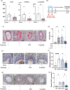
Figure 1
Figure 1
Restraint stress reduces plasma 4-oxoDHA levels and deteriorates vascular remodeling in mice. (a) Mice were treated with or without stress by physical restriction (2 h) for two consecutive days. DHA, 4-HDHA, and 4-oxoDHA levels in plasma were analyzed using LC/MS/MS on day 3. Data are shown as ng/mL in plasma, mean ± s.e. (n = 8). *Indicates p < 0.05. (b) Experimental protocol for carotid artery ligation model. Mice were treated with or without stress as above; then, the right common carotid artery was subjected to the carotid ligation model (see “Methods” section) on day 3. Specific mice were treated with 1 µg of DHA or 4-oxoDHA (intraperitoneal injection) on days of restraint stress. On day 10, stenotic vascular remodeling was histologically evaluated. (c) Representative sections at 300 µm from the carotid bifurcation are shown. Arrowheads indicate neointimal formation. Scales indicate 100 µm. (d) Stenosis rate (neointimal lesion area/lumen area). Data are expressed as mean ± s.e. (n = 9–12). *Indicates p < 0.05. (e) Infiltration of neutrophils (arrowheads) across endothelial cells was evaluated by immunohistochemistry with an anti-CD45 antibody. Scales indicate 50 µm. (f) Number of infiltrated neutrophils per section. *,****Indicate p < 0.05, p < 0.0001. (g) ICAM-1 expression in endothelial cells at 500 µm from the bifurcation was evaluated by immunohistochemistry with an anti-ICAM-1 antibody. Scales indicate 100 µm. (h) Relative ICAM-1 expression was quantified using Image-J software. Data are expressed as fold change compared to control, mean ± s.e. (n = 4–7). *Indicates p < 0.05.
Figure 2
Figure 2
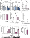
4-oxoDHA augments Nrf2-HO-1 pathways and anti-inflammatory properties in endothelial cells. (a) Pathway analysis of RNA-sequence data from the control (N/C) and 4-oxoDHA treated (4-oxoDHA) HUVECs. (b) HUVECs were treated with DHA (10 µM), 4-HDHA (0.1–10 µM), and 4-oxoDHA (0.1–10 µM) for 6 h at 37 °C. Whole-cell lysates were processed for western blot analysis to determine heme oxygenase-1 (HO-1) expression. β-actin was employed as the internal standard. Data are expressed as fold induction of HO-1 compared to control, mean ± s.e. (n = 3). ****Indicates p < 0.0001. (c) Endothelial cell line (EA.hy 926) was treated with DHA (1 µM), 4-HDHA (0.01–1 µM), and 4-oxoDHA (0.01–1 µM) for 6 h at 37 °C. Whole-cell lysates were processed for western blot analysis to determine HO-1 expression. β-actin was used as the internal standard. Data are expressed as fold induction of HO-1 compared to control, mean ± s.e. (n = 5). **Indicates p < 0.01. (d) HUVECs were treated with 10 µM of DHA, 4-HDHA, and 4-oxoDHA for 2 h at 37 °C. Nuclear fractions were processed for western blot analysis to determine Nrf2 expression. PCNA was used as the internal standard. Data are expressed as fold induction of Nrf2 compared to control (N/C), mean ± s.e. (n = 3) **,***Indicate p < 0.01, p < 0.001. (e) EA.hy 926 cells were treated with 1 µM of DHA, 4-HDHA, and 4-oxoDHA for 1 h, followed by stimulation of H2O2 (500 µM) for 2 h at 37 °C. After incubation, endothelial permeability was investigated with FITC-labeled dextran (MW: 10,000). Data are expressed as percent reduction compared to control, mean ± s.e. (n = 3–4). **Indicates p < 0.01. (f,g) HUVECs were treated with 10 µM DHA or 4-oxoDHA after stimulation with Kdo2 (0.5 µg/mL). ICAM-1 (f) and E-selectin (g) mRNA were analyzed using real-time RT-PCR. Data are expressed as fold change compared to control, mean ± s.e. (n = 7). *,**Indicate p < 0.05, p < 0.01.
Figure 3
Figure 3
4-oxoDHA augments Nrf2-HO-1 pathways and anti-inflammatory properties in macrophages. (a) RAW 264.7 cells were treated with DHA, 4-HDHA, and 4-oxoDHA (0.01–1 µM) or vehicle (N/C) for 6 h at 37 °C. Lysates were processed for western blot analysis to determine HO-1 expression. β-actin was used as the internal standard. Data are expressed as fold change compared to vehicle, mean ± s.e. (n = 4–5). ****Indicates p < 0.0001. (b) RAW 264.7 cells were incubated with Kdo2 for 1 h, followed by treatment of DHA (10 µM), 4-HDHA (0.1–10 µM), and 4-oxoDHA (0.1–10 µM) for 2 h at 37 °C. Nuclear fractions were processed for western blot analysis to determine Nrf2 expression. PCNA was used as the internal standard. Data are expressed as fold change compared to vehicle, mean ± s.e. (n = 4). **Indicates p < 0.01. (c–f) RAW 264.7 cells were incubated with Kdo2 for 1 h, followed by treatment with 1–10 µM of DHA, 4-HDHA, and 4-oxoDHA for 2 h at 37 °C. CCL2 (c), IL1-β (d), GCLM (e), and NQO1 (f) mRNA were analyzed using real-time PCR. Data are expressed as fold change compared to vehicle, mean ± s.e. (n = 3). *,**,***Indicate p < 0.05, p < 0.01, p < 0.001. (g) RAW 264.7 cells were incubated with 1 µM of DHA, 4-HDHA, and 4-oxoDHA for 6 h at 37 °C, and phagocytosis was evaluated with FITC-labeled zymosan particles. Data are expressed as fold change compared to vehicle, mean ± s.e. (n = 4). *Indicates p < 0.05.
Figure 4
Figure 4
Noradrenaline regulates 4-oxoDHA production and 5-LO protein expression via dopamine D2-like receptors in neutrophils. (a) Whole blood was stimulated with noradrenaline (NA) or vehicle for 1 h at 37 °C. Fatty acid metabolites were analyzed using LC/MS/MS. Data are expressed as fold change compared to the vehicle-treated group, mean ± s.e. (n = 3). *,**Indicate p < 0.05, p < 0.01 compared to vehicle. (b) Neutrophil cell line (HL-60) was treated with noradrenaline or vehicle for 2.5 h at 37 °C. Fatty acid metabolites were analyzed as above. *Indicate p < 0.05 compared to vehicle. (c) HL-60 cells were treated with noradrenaline for 2.5 h at 37 °C. Cell lysates were processed for western blot analysis to determine 5-LO expression. β-actin was used as the internal standard. Data are expressed as fold change compared to control, mean ± s.e. (n = 3). **,***Indicate p < 0.01, p < 0.001. (d) HL-60 cells were treated with noradrenaline for 2.5 h at 37 °C. Annexin-V and PI negative fractions are shown as percent of total cells, mean ± s.e. (n = 4). (e) HL-60 cells were treated with noradrenaline (1000 µM) for 2.5 h at 37°C, with and without 1 h pretreatment with MLN 2238 (proteasome inhibitor). Cell lysates were processed for western blot analysis to determine 5-LO expression. β-actin was used as the internal standard. Data are expressed as fold change compared to control, mean ± s.e. (n = 3). *,**Indicate p < 0.05, p < 0.01. (f) HL-60 cells were treated with noradrenaline (1000 µM) for 2.5 h at 37 °C, with and without 1-h pretreatment with phentolamine (alpha receptor inhibitor, 150 nM) and propranolol (beta receptor inhibitor, 120 nM). Cell lysates were processed for western blot analysis to determine 5-LO expression. β-actin was used as the internal standard. Data are expressed as fold change compared to control, mean ± s.e. (n = 4). (g) HL-60 cells were treated with noradrenaline (1000 µM) for 2.5 h at 37 °C, with or without pretreatment with SCH39166 (D1-like receptor antagonist, 10 nM) or raclopride (D2-like receptor antagonist, 20 nM). Data are expressed as fold change compared to control, mean ± s.e. (n = 4). ****Indicate p < 0.0001. (h) HL-60 cells were treated with noradrenaline (1000 µM) for 2.5 h at 37 °C, with and without 1-h pretreatment with Barbadian (β-arrestin inhibitor, 100 µM). Cell lysates were processed for western blot analysis to determine 5-LO expression. β-actin was used as the internal standard. Data are expressed as fold change compared to control, mean ± s.e. (n = 4). **Indicates p < 0.01. (i) Mice were treated with or without stress by physical restriction (2 h) for two consecutive days (as in Fig. 1b). On day 3, 5-LO protein expression in neutrophils was monitored using flow cytometry. Data are expressed as mean fluorescent intensity, mean ± s.e. (n = 5). **Indicate p < 0.01.
Figure 5
Figure 5
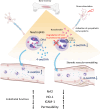
Circulating 4-oxoDHA was regulated by 5-LO mainly in neutrophils and activated Nrf2-HO-1 pathways, which maintained endothelial functions. However, under a stressed condition, a high concentration of noradrenaline was secreted in the bone marrow compartment, and D2-like receptor activation degraded 5-LO via the proteasome system in neutrophils. The decreased levels of circulating 4-oxoDHA would downregulate the Nrf2-HO-1 anti-inflammatory axis and increase ICAM-1 expression, vascular permeability, and remodeling.

References

    1. Yaribeygi H, Panahi Y, Sahraei H, Johnston TP, Sahebkar A. The impact of stress on body function: A review. EXCLI J. 2017;16:1057–1072. - PMC - PubMed
    1. McEwen BS, Bowles NP, Gray JD, Hill MN, Hunter RG, Karatsoreos IN, et al. Mechanisms of stress in the brain. Nat. Neurosci. 2015;18(10):1353–1363. doi: 10.1038/nn.4086. - DOI - PMC - PubMed
    1. Black PH. The inflammatory response is an integral part of the stress response: Implications for atherosclerosis, insulin resistance, type II diabetes and metabolic syndrome X. Brain Behav. Immun. 2003;17(5):350–364. doi: 10.1016/S0889-1591(03)00048-5. - DOI - PubMed
    1. Yusuf S, Hawken S, Ounpuu S, Dans T, Avezum A, Lanas F, et al. Effect of potentially modifiable risk factors associated with myocardial infarction in 52 countries (the INTERHEART study): Case–control study. Lancet. 2004;364(9438):937–952. doi: 10.1016/S0140-6736(04)17018-9. - DOI - PubMed
    1. Rosengren A, Hawken S, Ounpuu S, Sliwa K, Zubaid M, Almahmeed WA, et al. Association of psychosocial risk factors with risk of acute myocardial infarction in 11119 cases and 13648 controls from 52 countries (the INTERHEART study): Case–control study. Lancet. 2004;364(9438):953–962. doi: 10.1016/S0140-6736(04)17019-0. - DOI - PubMed