. 2024 Feb 6:16:1261217.
doi: 10.3389/fnmol.2023.1261217. eCollection 2023.

Exploring gene signatures and regulatory networks in a rat model of sciatica: implications and validation in neuropathic pain

Affiliations

Exploring gene signatures and regulatory networks in a rat model of sciatica: implications and validation in neuropathic pain

Mu Xu et al. Front Mol Neurosci. .

Abstract

Background: Sciatica (neuropathic pain [NP]) is a common disease characterized by pain from radiation along the sciatic nerve. The aim of this study was to study the genes associated with chronic systolic injury of sciatic nerve (SCN-CCI) in rats by RNA-Seq technique, and to explore their potential as therapeutic targets.

Methods: Sciatic nerve rat model was obtained by ligation of sciatic nerve and divided into two groups: SCN-CCI group and Sham group. Behavioral assessments were performed to evaluate pain sensitivity, following which their spinal cord dorsal horn were resected and RNA sequencing was conducted to identify differentially expressed genes (DEGs). Bioinformatics and functional enrichment analysis was performed to identify promising DEGs and their related biological processes and pathways associated with SCN-CCI. PPI network analysis and hub gene identification were conducted. QRT-PCR, western blot, ELISA, and immunofluorescence staining were performed on rat models to validate the expression of these hub genes and investigate related proteins and inflammatory markers.

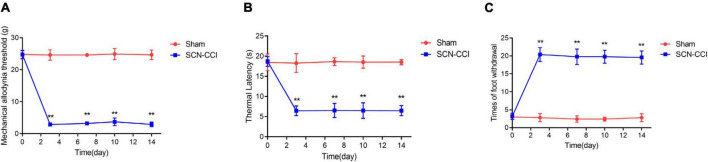
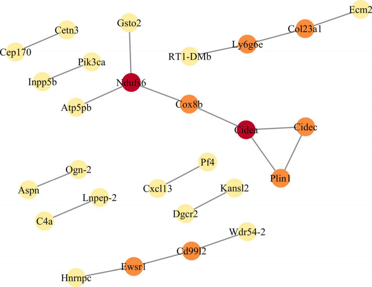
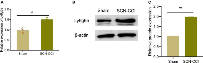
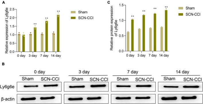
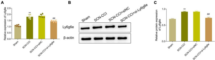
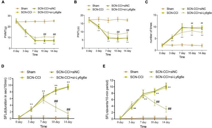
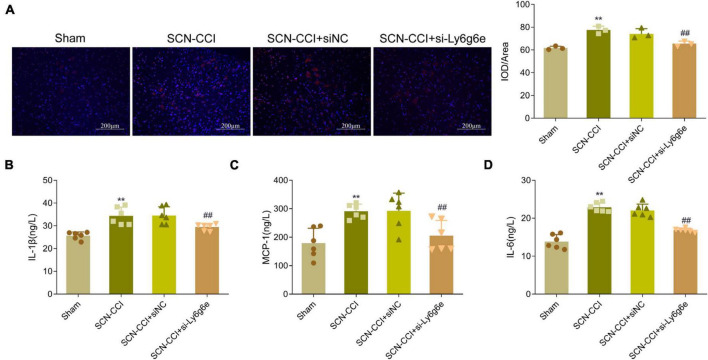
Results: The SCN-CCI rat model was successfully obtained, exhibiting increased pain sensitivity compared to the Sham group, as indicated by decreased mechanical allodynia thresholds, thermal latencies, and increased paw withdrawals. RNA-Seq analysis identified 117 DEGs in the SCN-CCI rat model, involved in various biological processes and pathways related to sciatica. PPI network analysis revealed hub genes, including Ly6g6e, which exhibited significant differential expression. QRT-PCR and Western blot analysis confirmed the expression patterns of these hub genes. Pain behavior assessment demonstrated reduced pain thresholds and increased paw flinching responses in the SCN-CCI group. Furthermore, the SCN-CCI group showed upregulated expression of Ly6g6e, increased protein levels of Ly6g6e, CGRP, and NGF, as well as elevated levels of IL-1β, MCP-1, and IL-6, and microglial cell activation in the spinal dorsal horn. ELISA results confirmed the increased levels of IL-1β, MCP-1, and IL-6 in the spinal dorsal horn.

Conclusion: These comprehensive findings provide valuable insights into the SCN-CCI rat model, DEGs associated with sciatica, hub genes (Ly6g6e as promising targets), pain behavior changes and molecular alterations.

Keywords: RNA sequencing (RNA-Seq); chronic systolic injury of sciatic nerve; differentially expressed genes (DEGs); neuropathic pain (NP); sciatica.


Conflict of interest statement

The authors declare that the research was conducted in the absence of any commercial or financial relationships that could be construed as a potential conflict of interest.

Figures

FIGURE 1
FIGURE 1
Establishment of sciatic nerve chronic constriction injury (SCN-CCI) rat model (n = 6 rats/group). (A–C) The mechanical allodynia threshold (A), thermal hyperalgesia latency (B) and times of paw withdrawal (C) of rats in the Sham and SCN-CCI group measured on days 0, 3, 7, 10, and 14 (n = 6 rats), **P < 0.01 vs. Sham group. SCN-CCI, chronic constriction injury. Student’s t-test is used for data analysis.
FIGURE 2
FIGURE 2
Screening of sciatica-associated differentially expressed genes (DEGs) (n = 2 rats/group). Volcano plot of sciatica-associated DEGs; SCN-CCI, sciatic nerve chronic constriction injury.
FIGURE 3
FIGURE 3
Constructs the protein-protein interaction (PPI) network module of differentially expressed genes (DEGs). The PPI network of DEGs is constructed by STRING database, and the PPI network module is obtained by MCODE plugin in Cytoscape. The shade of color indicates the score size of the node (red > orange > yellow). PPI, protein-protein interaction; DEGs, differentially expressed genes.
FIGURE 4
FIGURE 4
Identification of Hub Gene. (A) mRNA levels of Ly6g6e (A) in the left L4-L6 spinal cord tissue of the Sham and SCN-CCI groups were detected by qRT-PCR at day 14 (n = 6 rats/group); (B,C): protein expression levels of Ly6g6e in the left L4-L6 spinal cord tissue of the Sham and SCN-CCI groups were detected by western blot (n = 3 rats/group). **P < 0.01 vs. Sham, Sham surgery group; SCN-CCI, chronic constriction injury. Student’s t-test is used for data analysis.
FIGURE 5
FIGURE 5
After modeling, the expression level of Ly6g6e in the spinal dorsal horn tissue of rats was observed. (A) The mRNA expression level of Ly6g6e in spinal dorsal horn tissues of rats in different groups was detected by qRT-PCR at day 0, 3, 7 and 14 (n = 3 rats/group). (B,C) Western blot was used to detect Ly6g6e protein expression levels at day 0, 3, 7 and 14 in spinal dorsal horn tissues of rats in different groups (n = 3 rats/group). **P < 0.01 vs. Sham group. Sham, Sham surgery group; SCN-CCI, chronic constriction injury. Student’s t-test is used for data analysis.
FIGURE 6
FIGURE 6
Expression level of Ly6g6e siRNA in SCN-CCI rats. (A) The mRNA expression level of Ly6g6e in spinal dorsal horn tissues of rats in each group was detected by qRT-PCR at day 14 (n = 6 rats/group). (B,C) Western blot analysis of Ly6g6e protein expression in spinal dorsal horn tissues of rats in different groups at day 14 (n = 3 rats/group), **P < 0.01 vs. Sham group. ##P < 0.01 vs. SCN-CCI + siNC group. ANOVA followed Tukey post hoc test is used for data analysis.
FIGURE 7
FIGURE 7
The effect of Ly6g6e siRNA on pain behavior in SCN-CCI rats. (A–C) The induced pain behavior of rats in the sham group, SCN-CCI group, SCN-CCI + siNC group, and SCN-CCI + si-Ly6g6e group was evaluated at day 0, 3, 7, 10, and 14 (n = 6 rats/group), including: (A) mechanical dysodynia (mechanical threshold for claw withdrawal, PWMT), (B) thermal hypersensitivity (latent period of claw deheating, PWTL), (C) cold dysodynia. (D,E) The spontaneous pain behaviors of rats in the sham group, SCN-CCI group, SCN-CCI + siNC group, and SCN-CCI + si-Ly6g6e group was evaluated at day 0, 3, 7, 10, and 14 (n = 6 rats/group), including: (D) spontaneous foot-lifting (SFL) duration (SFLd), (E) spontaneous foot-lifting (SFL) number (SFLn),**P < 0.01 vs. Sham group. ##P < 0.01 vs. SCN-CCI + siNC group. Two-way ANOVA followed Tukey post hoc test is used for data analysis.
FIGURE 8
FIGURE 8
Knocking down Ly6g6e inhibits the protein expression levels of CRPG and NGF in the spinal dorsal horn tissue of SCN-CCI rats. (A,B) Western blot was used to detect the protein expression levels of CRPG and NGF in spinal dorsal horn tissues of rats in Sham group, SCN-CCI group, SCN-CCI + siNC group and SCN-CCI + si-Ly6g6e group at day 14 (n = 3 rats/group). **P < 0.01 vs. Sham group. ##P < 0.01 vs. SCN-CCI + siNC group. ANOVA followed Tukey post hoc test is used for data analysis.
FIGURE 9
FIGURE 9
Knockdown of Ly6g6e inhibits the secretion of inflammatory factors and the activation of Microglia in SCN-CCI rats. (A) The activation of microglial cells (CD68) in spinal dorsal horn tissues of rats in Sham group, SCN-CCI group, SCN-CCI + siNC group and SCN-CCI + si-Ly6g6e group was observed by immunofluorescence staining at day 14. (B–D) ELISA detection of IL-1β, MCP-1, and IL-6 levels in the spinal cord dorsal horn tissue of rats from the Sham group, SCN-CCI group, SCN-CCI + siNC group, and SCN-CCI + si-Ly6g6e group at day 14 (n = 6 rats/group). (B–D) Bar graphs showing the comparison of panel (B) IL-1β, (C) MCP-1 and (D) IL-6 levels in the spinal dorsal horn tissue between different experimental groups. Data are presented as mean ± standard deviation. **P < 0.01 vs. Sham group. ##P < 0.01 vs. SCN-CCI + siNC group. ANOVA followed Tukey post hoc test is used for data analysis.

Similar articles

Cited by

References

    1. Abbaszadeh A., Darabi S., Hasanvand A., Amini-Khoei H., Abbasnezhad A., Choghakhori R., et al. (2018). Minocycline through attenuation of oxidative stress and inflammatory response reduces the neuropathic pain in a rat model of chronic constriction injury. Iran J. Basic Med. Sci. 21 138–144. 10.22038/IJBMS.2017.24248.6053 - DOI - PMC - PubMed
    1. Banik R. K., Sia T., Ibrahim M. M., Sivanesan E., Uhelski M., Pena A., et al. (2023). Increases in local skin temperature correlate with spontaneous foot lifting and heat hyperalgesia in both incisional inflammatory models of pain. Pain Rep. 8:e1097. 10.1097/PR9.0000000000001097 - DOI - PMC - PubMed
    1. Bates D., Schultheis B. C., Hanes M. C., Jolly S. M., Chakravarthy K. V., Deer T. R., et al. (2019). A comprehensive algorithm for management of neuropathic pain. Pain Med. 20 S2–S12. 10.1093/pm/pnz075 - DOI - PMC - PubMed
    1. Boakye P. A., Tang S. J., Smith P. A. (2021). Mediators of neuropathic pain; focus on spinal microglia, CSF-1, BDNF, CCL21, TNF-α, Wnt ligands, and interleukin 1β. Front. Pain Res. 2:698157. 10.3389/fpain.2021.698157 - DOI - PMC - PubMed
    1. Bouhassira D. (2019). Neuropathic pain: Definition, assessment and epidemiology. Rev. Neurol. 175 16–25. 10.1016/j.neurol.2018.09.016 - DOI - PubMed