

A hybrid receptor binding protein enables phage F341 infection of Campylobacter by binding to flagella and lipooligosaccharides
- PMID: 38380094
- PMCID: PMC10877375
- DOI: 10.3389/fmicb.2024.1358909
A hybrid receptor binding protein enables phage F341 infection of Campylobacter by binding to flagella and lipooligosaccharides
Abstract
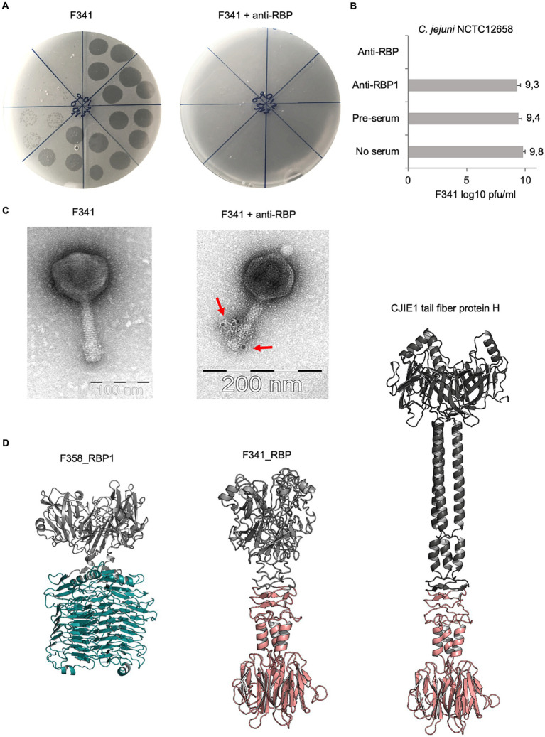
Flagellotropic bacteriophages are interesting candidates as therapeutics against pathogenic bacteria dependent on flagellar motility for colonization and causing disease. Yet, phage resistance other than loss of motility has been scarcely studied. Here we developed a soft agar assay to study flagellotropic phage F341 resistance in motile Campylobacter jejuni. We found that phage adsorption was prevented by diverse genetic mutations in the lipooligosaccharides forming the secondary receptor of phage F341. Genome sequencing showed phage F341 belongs to the Fletchervirus genus otherwise comprising capsular-dependent C. jejuni phages. Interestingly, phage F341 encodes a hybrid receptor binding protein (RBP) predicted as a short tail fiber showing partial similarity to RBP1 encoded by capsular-dependent Fletchervirus, but with a receptor binding domain similar to tail fiber protein H of C. jejuni CJIE1 prophages. Thus, C. jejuni prophages may represent a genetic pool from where lytic Fletchervirus phages can acquire new traits like recognition of new receptors.
Keywords: Campylobacter; Fletchervirus; flagella; flagellotropic phage; phage; phage receptor; phage resistance; receptor binding protein.
Copyright © 2024 Ostenfeld, Sørensen, Neve, Vitt, Klumpp and Sørensen.
Conflict of interest statement
The authors declare that the research was conducted in the absence of any commercial or financial relationships that could be construed as a potential conflict of interest.
Figures
Similar articles
-
Campylobacter jejuni motility is required for infection of the flagellotropic bacteriophage F341.Appl Environ Microbiol. 2014 Nov;80(22):7096-106. doi: 10.1128/AEM.02057-14. Epub 2014 Sep 26. Appl Environ Microbiol. 2014. PMID: 25261508 Free PMC article.
-
Binding of Phage-Encoded FlaGrab to Motile Campylobacter jejuni Flagella Inhibits Growth, Downregulates Energy Metabolism, and Requires Specific Flagellar Glycans.Front Microbiol. 2020 Mar 20;11:397. doi: 10.3389/fmicb.2020.00397. eCollection 2020. Front Microbiol. 2020. PMID: 32265863 Free PMC article.
-
Primary isolation strain determines both phage type and receptors recognised by Campylobacter jejuni bacteriophages.PLoS One. 2015 Jan 13;10(1):e0116287. doi: 10.1371/journal.pone.0116287. eCollection 2015. PLoS One. 2015. PMID: 25585385 Free PMC article.
-
Flagellotropic Bacteriophages: Opportunities and Challenges for Antimicrobial Applications.Int J Mol Sci. 2022 Jun 25;23(13):7084. doi: 10.3390/ijms23137084. Int J Mol Sci. 2022. PMID: 35806089 Free PMC article. Review.
-
Flagellotropic phages: common yet diverse host interaction strategies.Curr Opin Microbiol. 2024 Apr;78:102451. doi: 10.1016/j.mib.2024.102451. Epub 2024 Mar 6. Curr Opin Microbiol. 2024. PMID: 38452595 Review.
Cited by
-
Evolutionary consequences of bacterial resistance to a flagellotropic phage.bioRxiv [Preprint]. 2025 May 10:2025.05.06.652435. doi: 10.1101/2025.05.06.652435. bioRxiv. 2025. PMID: 40654869 Free PMC article. Preprint.
-
Campycins are novel broad-spectrum antibacterials killing Campylobacter jejuni.Appl Microbiol Biotechnol. 2024 Oct 9;108(1):484. doi: 10.1007/s00253-024-13317-w. Appl Microbiol Biotechnol. 2024. PMID: 39382702 Free PMC article.
-
fENko-Kae01 is a flagellum-specific jumbo phage infecting Klebsiella aerogenes.BMC Microbiol. 2024 Jul 1;24(1):234. doi: 10.1186/s12866-024-03387-1. BMC Microbiol. 2024. PMID: 38951769 Free PMC article.
-
A review of the fighting Acinetobacter baumannii on three fronts: antibiotics, phages, and nanoparticles.Mol Biol Rep. 2024 Oct 8;51(1):1044. doi: 10.1007/s11033-024-09979-4. Mol Biol Rep. 2024. PMID: 39377967 Review.
-
Global structural survey of the flagellotropic myophage φTE infecting agricultural pathogen Pectobacterium atrosepticum.Nat Commun. 2025 Apr 5;16(1):3257. doi: 10.1038/s41467-025-58514-x. Nat Commun. 2025. PMID: 40188083 Free PMC article.
References
-
- Ang C. W., Laman J. D., Willison H. J., Wagner E. R., Endtz H. P., De Klerk M. A., et al. . (2002). Structure of Campylobacter jejuni lipopolysaccharides determines antiganglioside specificity and clinical features of Guillain–Barré and Miller fisher patients. Infect. Immun. 70, 1202–1208. doi: 10.1128/IAI.70.3.1202-1208.2002, PMID: - DOI - PMC - PubMed
LinkOut - more resources
Full Text Sources