. 2024 Apr 1;131(4):626-637.
doi: 10.1152/jn.00037.2023. Epub 2024 Feb 21.

Maturation of glutamatergic transmission onto dorsal raphe serotonergic neurons

Affiliations

Maturation of glutamatergic transmission onto dorsal raphe serotonergic neurons

Alexandre Kisner et al. J Neurophysiol. .

Abstract

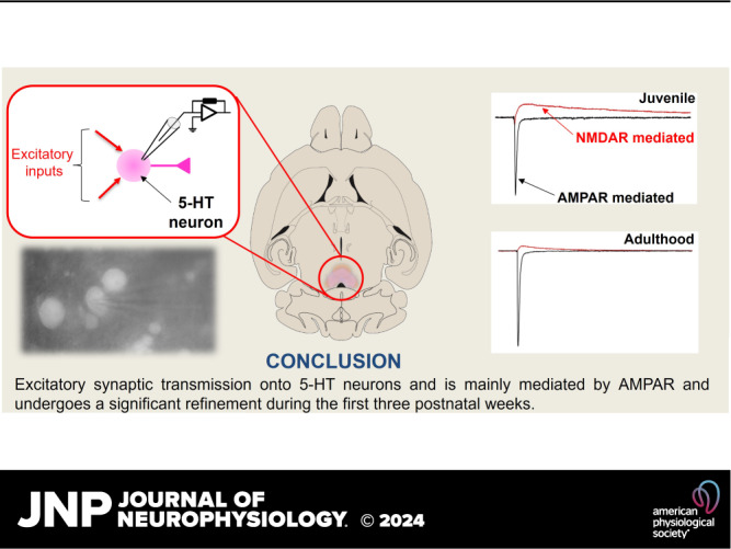
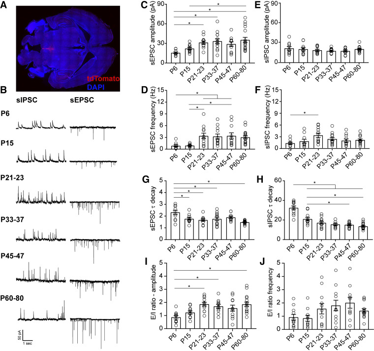
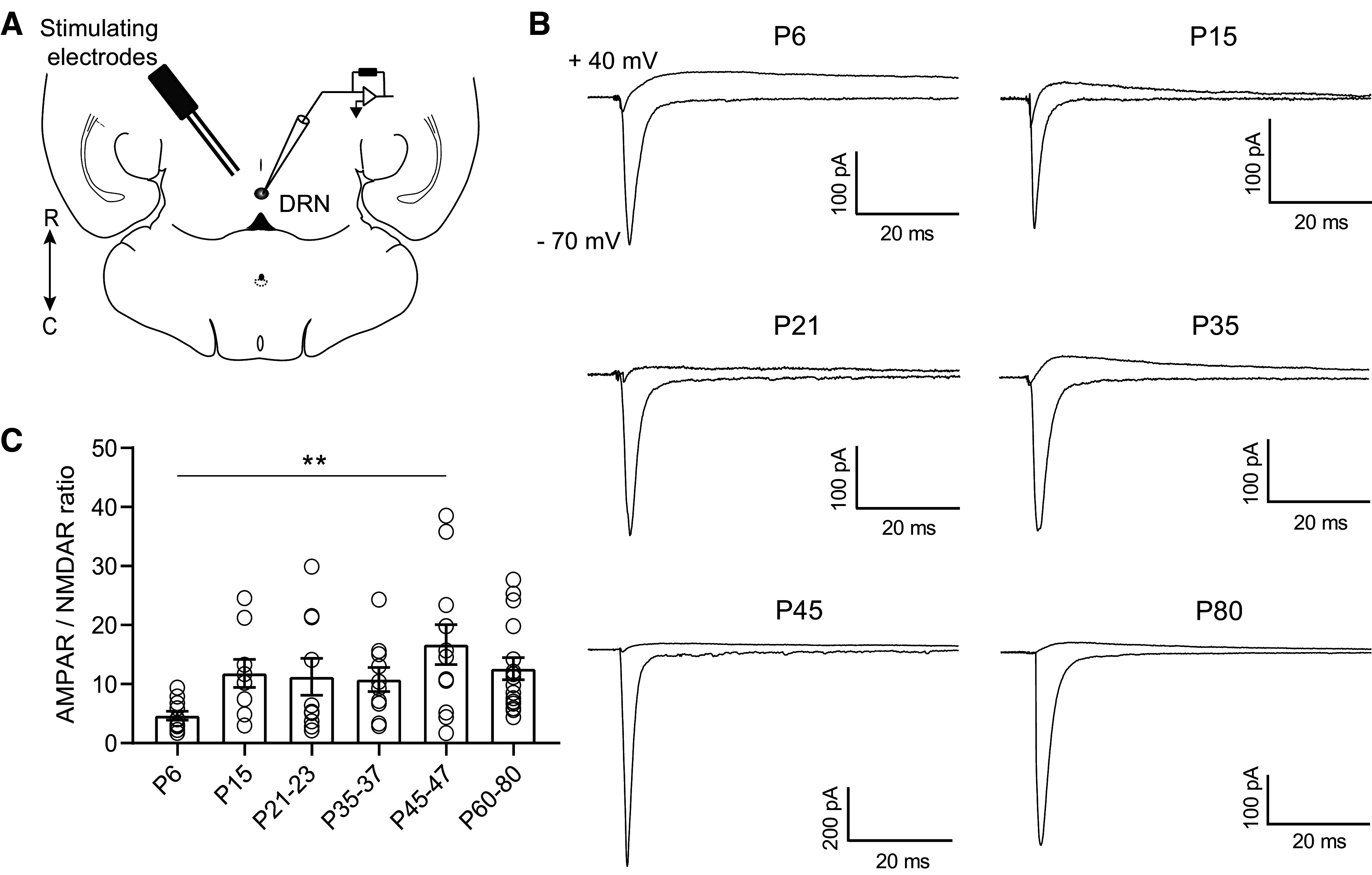
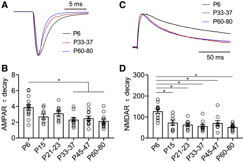
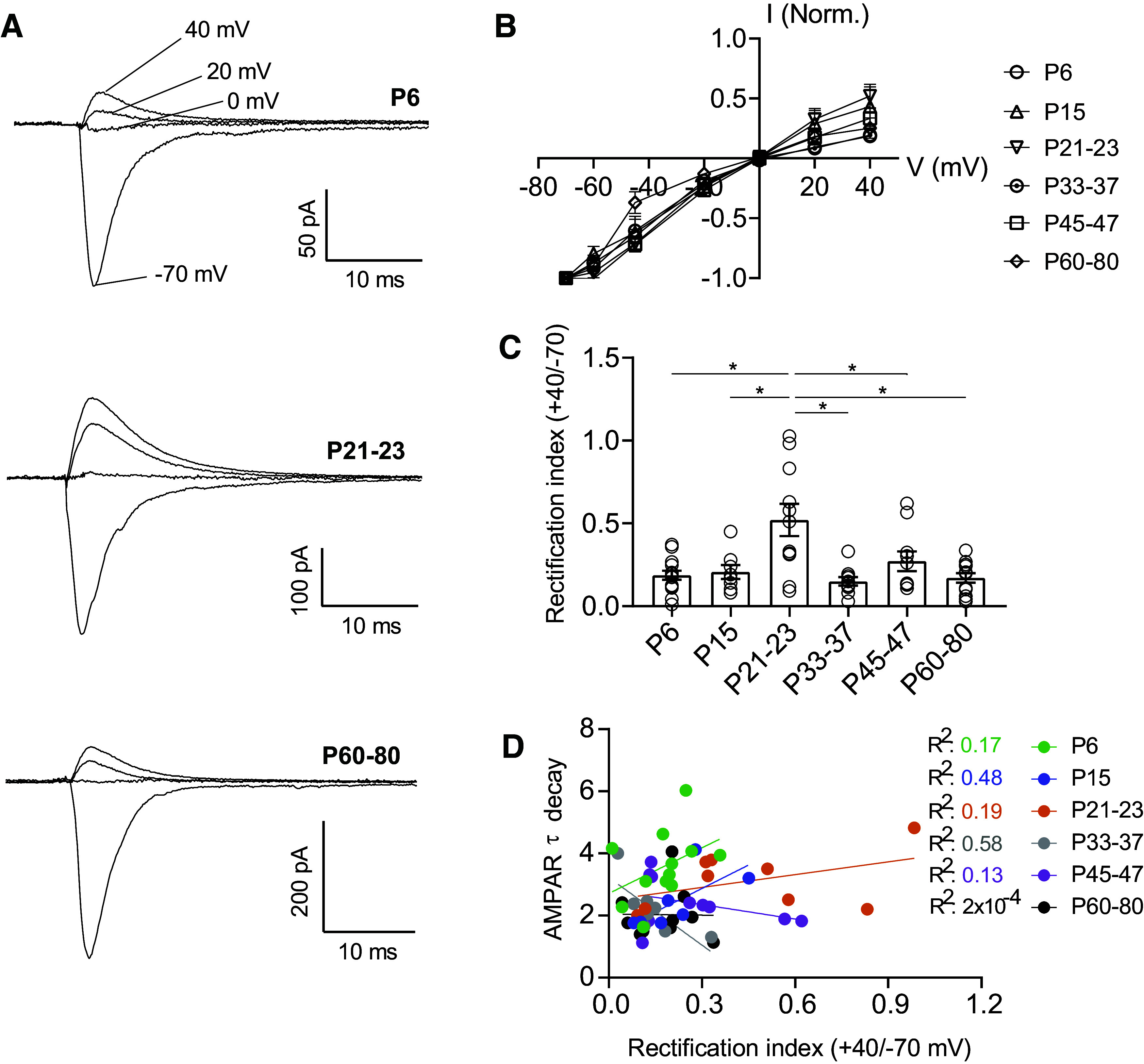
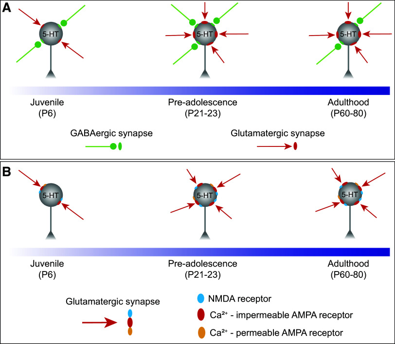
Serotonergic neurons in the dorsal raphe nucleus (DRN) play important roles early in postnatal development in the maturation and modulation of higher-order emotional, sensory, and cognitive circuitry. The pivotal functions of these cells in brain development make them a critical substrate by which early experience can be wired into the brain. In this study, we investigated the maturation of synapses onto dorsal raphe serotonergic neurons in typically developing male and female mice using whole cell patch-clamp recordings in ex vivo brain slices. We show that while inhibition of these neurons is relatively stable across development, glutamatergic synapses greatly increase in strength between postnatal day 6 (P6) and P21-23. In contrast to forebrain regions, where the components making up glutamatergic synapses are dynamic across early life, we find that DRN excitatory synapses maintain a very high ratio of α-amino-3-hydroxy-5-methyl-4-isoxazolepropionic acid (AMPA) to N-methyl-d-aspartate (NMDA) receptors and a rectifying component of the AMPA response until adulthood. Overall, these findings reveal that the development of serotonergic neurons is marked by a significant refinement of glutamatergic synapses during the first three postnatal weeks. This suggests this time is a sensitive period of heightened plasticity for the integration of information from upstream brain areas. Genetic and environmental insults during this period could lead to alterations in serotonergic output, impacting both the development of forebrain circuits and lifelong neuromodulatory actions.NEW & NOTEWORTHY Serotonergic neurons are regulators of both the development of and ongoing activity in neuronal circuits controlling affective, cognitive, and sensory processing. Here, we characterize the maturation of extrinsic synaptic inputs onto these cells, showing that the first three postnatal weeks are a period of synaptic refinement and a potential window for experience-dependent plasticity in response to both enrichment and adversity.

Keywords: development; dorsal raphe; excitatory synapse; glutamate; serotonin.


Conflict of interest statement

No conflicts of interest, financial or otherwise, are declared by the authors.

Figures

None
Graphical abstract
Figure 1.
Figure 1.
Maturation of spontaneous synaptic transmission onto DRN serotonergic neurons. A: representative 4×-tiled image of a horizontal slice of the dorsal raphe in an adult ePet-Cre:Ai14 animal. Red, tdTomato; blue, DAPI. B: representative traces of spontaneous synaptic transmission at developmental time points. For each time point, traces were recorded from the same 5-HT neuron at holding potentials of −70 mV (sEPSC) and 0 mV (sIPSC). C and D: developmental changes in the mean amplitude and frequency of sEPSCs. E and F: developmental changes in the mean amplitude and frequency of sIPSCs. G: time constant of decay (τdecay) for sEPSCs. H: τdecay for sIPSCs. I: excitation/inhibition (E/I) ratio for amplitude of sPSCs. J: E/I ratio for frequency of sPSCs. P6, n = 10 cells (5 mice); P15, n = 11 cells (7 mice); P21–23, n = 12 cells (8 mice); P33–37, n = 12 cells (8 mice); P45–47, n = 11 cells (8 mice); P60–80, n = 17 cells (10 mice). Bar graphs represent means ± SE. *P < 0.05, one-way ANOVA followed by Tukey’s multiple comparison test. DRN, dorsal raphe nucleus; sEPSC, spontaneous excitatory postsynaptic current; sIPSC, spontaneous inhibitory postsynaptic current.
Figure 2.
Figure 2.
Excitatory currents onto DRN serotonergic neurons are primarily mediated by AMPAR. A: schematic of the experiment illustrating the placement of the bipolar-stimulating electrode in the DRN and recording of evoked excitatory currents from 5-HT neurons. B: representative traces of electrically evoked excitatory synaptic currents recorded from 5-HT neurons at holding potentials of −70 mV and +40 mV. Recordings at +40 mV were performed in the presence of the AMPA antagonist NBQX (10 µM). C: quantification of AMPA/NMDA ratios across maturation. P6, n = 11 cells (6 mice); P15, n = 9 cells (6 mice); P21–23, n = 10 cells (9 mice); P33–37, n = 10 cells (7 mice); P45–47, n = 12 cells (8 mice); P60–80, n = 16 cells (11 mice). Bar graphs represent means ± SE. **P < 0.01, one-way ANOVA followed by Tukey’s multiple comparison test. DRN, dorsal raphe nucleus; NMDA, N-methyl-d-aspartate.
Figure 3.
Figure 3.
AMPA and NMDA receptor subunits are developmentally regulated in 5-HT neurons. A: scaled representative traces of evoked AMPAR currents. B: time constant of decay (τdecay) for evoked AMPAR currents recorded at −70 mV. C: scaled representative traces of evoked NMDAR currents. D: time constant of decay (τdecay) for evoked NMDAR currents recorded at +40 mV in the presence of the AMPA antagonist NBQX (10 µM). P6, n = 11 cells (6 mice); P15, n = 9 cells (6 mice); P21–23, n = 10 cells (9 mice); P33–37, n = 10 cells (7 mice); P45–47, n = 12 cells (8 mice); P60–80, n = 16 cells (11 mice). Bar diagrams represent means ± SE. *P < 0.05, one-way ANOVA followed by Tukey’s multiple comparison test. NMDA, N-methyl-d-aspartate.
Figure 4.
Figure 4.
AMPAR-mediated currents are inwardly rectifying throughout development in a subset of 5-HT neurons. A: representative traces of evoked excitatory synaptic currents recorded from 5-HT neurons at holding potentials of −70 mV to +40 mV. B: average peak current-voltage curves with slight deviation from a linear relationship. C: rectification index of AMPAR currents, calculated as the ratio of peak amplitude at +40 mV divided by the peak amplitude at −70 mV for each cell. D: correlation between AMPAR EPSC τ decay and rectification index for each time point. P6, n = 14 cells (7 mice); P15, n = 8 cells (7 mice); P21–23, n = 11 cells (6 mice); P33–37, n = 10 cells (5 mice); P45–47, n = 10 cells (7 mice); P60–80, n = 12 cells (7 mice). Bar graphs represent means ± SE. *P < 0.05, one-way ANOVA followed by Tukey’s multiple comparison test.
Figure 5.
Figure 5.
Schematic summarizing the maturation of synaptic inputs onto 5-HT neurons. A: the overall synaptic drive is led by glutamatergic transmission, which undergoes an abrupt enhancement between juvenile and the preadolescent period and then remained stable throughout adulthood. B: glutamatergic transmission is mainly carried by AMPA receptors and in a subset of 5-HT neurons, these receptors have a high permeability to calcium and are inwardly rectifying across the whole developmental period.

Update of

References

    1. Tau GZ, Peterson BS. Normal development of brain circuits. Neuropsychopharmacology 35: 147–168, 2010. doi:10.1038/npp.2009.115. - DOI - PMC - PubMed
    1. Suri D, Teixeira CM, Cagliostro MKC, Mahadevia D, Ansorge MS. Monoamine-sensitive developmental periods impacting adult emotional and cognitive behaviors. Neuropsychopharmacology 40: 88–112, 2015. doi:10.1038/npp.2014.231. - DOI - PMC - PubMed
    1. Hartley CA, Lee FS. Sensitive periods in affective development: nonlinear maturation of fear learning. Neuropsychopharmacology 40: 50–60, 2015. doi:10.1038/npp.2014.179. - DOI - PMC - PubMed
    1. Schoenfeld TJ, Cameron HA. Adult neurogenesis and mental illness. Neuropsychopharmacology 40: 113–128, 2015. doi:10.1038/npp.2014.230. - DOI - PMC - PubMed
    1. Guang S, Pang N, Deng X, Yang L, He F, Wu L, Chen C, Yin F, Peng J. Synaptopathology involved in autism spectrum disorder. Front Cell Neurosci 12: 470, 2018. doi:10.3389/fncel.2018.00470. - DOI - PMC - PubMed

Publication types

Substances

LinkOut - more resources