. 2024 Feb 23;10(8):eadi9379.
doi: 10.1126/sciadv.adi9379. Epub 2024 Feb 21.

Spontaneous, persistent, T cell-dependent IFN-γ release in patients who progress to Long Covid

Affiliations

Spontaneous, persistent, T cell-dependent IFN-γ release in patients who progress to Long Covid

Benjamin A Krishna et al. Sci Adv. .

Abstract

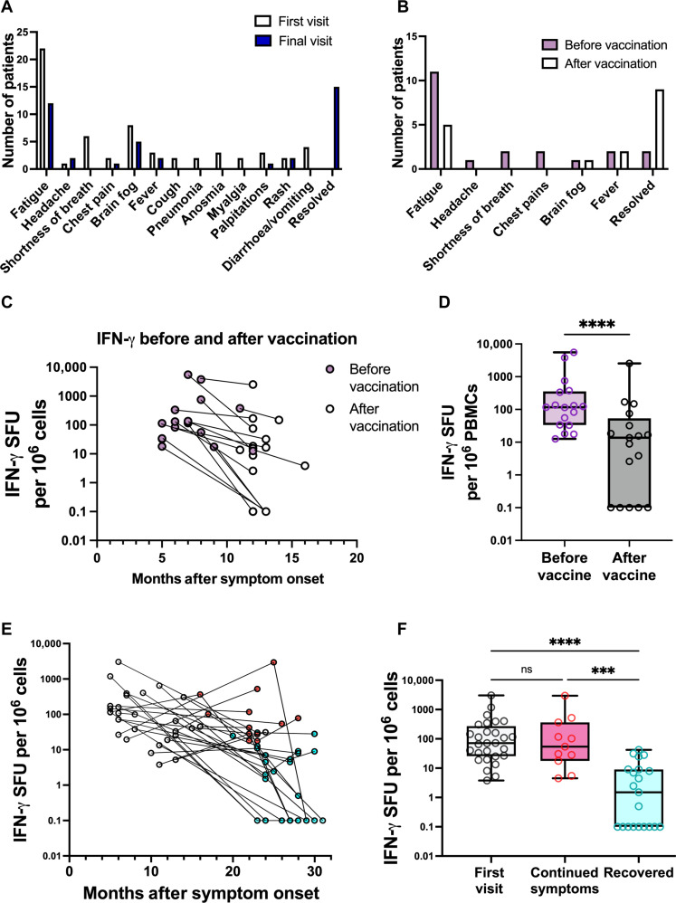
After acute infection with severe acute respiratory syndrome coronavirus-2 (SARS-CoV-2), a proportion of patients experience persistent symptoms beyond 12 weeks, termed Long Covid. Understanding the mechanisms that cause this debilitating disease and identifying biomarkers for diagnostic, therapeutic, and monitoring purposes are urgently required. We detected persistently high levels of interferon-γ (IFN-γ) from peripheral blood mononuclear cells of patients with Long Covid using highly sensitive FluoroSpot assays. This IFN-γ release was seen in the absence of ex vivo peptide stimulation and remains persistently elevated in patients with Long Covid, unlike the resolution seen in patients recovering from acute SARS-CoV-2 infection. The IFN-γ release was CD8+ T cell-mediated and dependent on antigen presentation by CD14+ cells. Longitudinal follow-up of our study cohort showed that symptom improvement and resolution correlated with a decrease in IFN-γ production to baseline levels. Our study highlights a potential mechanism underlying Long Covid, enabling the search for biomarkers and therapeutics in patients with Long Covid.


Figures

Fig. 1.
Fig. 1.. Spontaneous IFN-γ release is triggered by SARS-CoV-2 infection and fails to resolve to baseline in patients with diagnosed Long Covid.
(A to C) PBMCs were isolated from the blood of negative control unexposed donors (red, n = 54), patients diagnosed with Long Covid (burgundy, n = 87), and positive-control RT-qPCR–confirmed donors at 28, 90, or 180 days after PCR test (cyan, n = 51; green, n = 20; blue, n = 40). These PBMCs were not stimulated with any peptides ex vivo. After 48 hours of incubation, IFN-γ release was measured by FluoroSpot assay as spot forming units (SFU) per million PBMCs. Each point represents the mean from a single donor, where donor samples were run in duplicate, and zero results were set as 0.1 to allow their inclusion on a log scale. L.O.D., limit of detection. We compared unexposed and Long Covid cohorts (A), unexposed against each time point for the cohort who experienced acute COVID-19 (B), and then unexposed, acute COVID-19 at day 180 and the Long Covid cohort specifically (C). Significance calculated by two-way Kruskal-Wallis analysis of variance (ANOVA), with Dunn’s multiple comparisons test, ****P < 0.0001. ns, not significant.
Fig. 2.
Fig. 2.. Spontaneous IFN-γ production resolves in individuals with symptom resolution.
(A) Data from Fig. 1B were replotted for PBMCs from 10 donors who gave samples at 28 and 180 days after positive RT-qPCR result and reported symptom recovery. (B) IFN-γ release was measured by FluoroSpot assay for patients with Long Covid who did not report symptom resolution. Results are plotted at time after symptom onset. (A) and (B) IFN-γ release was quantified as spot forming units per million PBMCs. Each point represents the mean from a single donor, where donor samples were run in duplicate and zero results were set as 0.1 to allow their inclusion on a log scale. Significance calculated by or two-way Wilcoxon signed-rank test for (A) where **P < 0.01.
Fig. 3.
Fig. 3.. Spontaneous IFN-γ release from the PBMCs of patients with Long Covid is caused by CD8+ T cell interaction with CD14+ cells via MHC class I peptide presentation.
(A to C) MACS separation was used to isolate CD14+, CD56+, CD4+, or CD8+ cells from PBMC donations from patients with Long Covid. Whole PBMCs (red) or various combinations of depleted PBMCs, isolated cell populations, or depleted PBMCs with the isolated populations added back in from eight patients diagnosed with Long Covid were plated without peptide stimulation for 48 hours to measure IFN-γ release by FluoroSpot assay. (D) PBMCs from patients diagnosed with Long Covid (LC; n = 12) or healthy controls (HE; n = 8) were incubated for 24 hours with brefeldin A and monensin to allow intracellular accumulation of IFN-γ. Cells were then stained for CD3, CD4, CD8, CD14, and IFN-γ to compare IFN-γ production in these cells types between healthy controls and Long Covid. (E) CD14+ cells from eight donors were isolated by MACS separation and incubated for 1 hour with anti–MHC class I, anti–MHC class II, or isotype control antibodies. The cells were then washed and returned to the donor PBMCs, and IFN-γ release was measured by FluoroSpot assay after 24 hours of incubation. (A to C) IFN-γ release was quantified as in Figs. 1 and 2. Significance calculated by Kruskal-Wallis two-way ANOVA [(A) to (C) and (E)] or Friedman two-way ANOVA test (D) with Dunn’s multiple comparison test, *P < 0.05, **P < 0.01, and ****P < 0.0001. (D) Data are shown as percentage of cells that were positive for IFN-γ. Significance was calculated using multiple two-way Mann-Whitney U tests. (F) Activation markers, CD69 and Human Leukocyte Antigen–DR isotype (HLA-DR), on CD8+ T cells were measured in 18 patients with Long Covid and 8 healthy donors using the gating strategy shown in fig. S7. Significance, was measured by Kruskal-Wallis ANOVA.
Fig. 4.
Fig. 4.. PBMCs from patients with Long Covid produce IFN-γ and TNF-α but not IL-10.
PBMCs were isolated from the blood of patients with Long Covid (blue, n = 12) and those never infected with SARS-CoV-2 (unexposed, red, n = 33). Spontaneous production of IFN-γ (A), IL-10 (B), and TNF-α (C) was measured by FluoroSpot assay on unstimulated cells. After 48 hours of incubation, cytokine release was measured by FluoroSpot assay as spot forming units per million PBMCs. Each point represents the mean from a single donor, where donor samples were run in duplicate and zero results were set as 0.1 to allow their inclusion on a log scale. Significance calculated by two-way Mann-Whitney U test. (D) We used ELISA assays to confirm increased cytokine production in PBMCs from patients with Long Covid compared to unexposed controls. We tested TNF-α as was seen in (C), as well as GM-CSF, IL-1β, and IFN-γ that were detected by LEGENDplex analysis (fig. S6). Values below the limit of detection (4 pg/ml) were shown as 4 pg/ml to allow their inclusion on a logarithmic scale. Statistical significance was calculated by multiple Mann-Whitney tests, with P values showing significance for IL-1β and IFN-γ only.
Fig. 5.
Fig. 5.. Reduction in Long Covid symptoms correlated with loss of spontaneous IFN-γ release.
Longitudinal follow-up revealed that some patients either recovered spontaneously (A) or after vaccination (B). (C to F) PBMCs were isolated from patients with Long Covid at the indicated time points after infection, and unstimulated IFN-γ production was measured by FluoroSpot analysis. (C) Seventeen patients reported symptoms improved after vaccination; here, we have plotted IFN-γ release before (pink) and after (gray) vaccination. (D) Unstimulated IFN-γ production reduction was statistically significant after vaccination as calculated by two-way Wilcoxon ranked sign test, ****P < 0.0001. (E) and (F) A further 21 of 34 patients experienced spontaneous symptoms improvement over the course of the study. (E) As in (C), unstimulated IFN-γ production is plotted against time for these patients, where red dots indicate that the patient reported continued symptoms, while cyan dots indicate an improvement or resolution of symptoms. (F) Unstimulated IFN-γ production was significantly lower in patients who recovered from Long Covid symptoms compared to those who had ongoing symptoms, as calculated by Kruskal-Wallis test. ***P < 0.001.

References

    1. Scherlinger M., Felten R., Gallais F., Nazon C., Chatelus E., Pijnenburg L., Mengin A., Gras A., Vidailhet P., Arnould-Michel R., Bibi-Triki S., Carapito R., Trouillet-Assant S., Perret M., Belot A., Bahram S., Arnaud L., Gottenberg J. E., Fafi-Kremer S., Sibilia J., Refining “long-COVID” by a prospective multimodal evaluation of patients with long-term symptoms attributed to SARS-CoV-2 infection. Infect. Dis. Ther. 10, 1747–1763 (2021). - PMC - PubMed
    1. Huang C., Huang L., Wang Y., Li X., Ren L., Gu X., Kang L., Guo L., Liu M., Zhou X., Luo J., Huang Z., Tu S., Zhao Y., Chen L., Xu D., Li Y., Li C., Peng L., Li Y., Xie W., Cui D., Shang L., Fan G., Xu J., Wang G., Wang Y., Zhong J., Wang C., Wang J., Zhang D., Cao B., 6-month consequences of COVID-19 in patients discharged from hospital: A cohort study. Lancet 397, 220–232 (2021). - PMC - PubMed
    1. Ayoubkhani D., Khunti K., Nafilyan V., Maddox T., Humberstone B., Diamond I., Banerjee A., Post-covid syndrome in individuals admitted to hospital with covid-19: Retrospective cohort study. BMJ 372, n693 (2021). - PMC - PubMed
    1. Gupta A., Madhavan M. V., Sehgal K., Nair N., Mahajan S., Sehrawat T. S., Bikdeli B., Ahluwalia N., Ausiello J. C., Wan E. Y., Freedberg D. E., Kirtane A. J., Parikh S. A., Maurer M. S., Nordvig A. S., Accili D., Bathon J. M., Mohan S., Bauer K. A., Leon M. B., Krumholz H. M., Uriel N., Mehra M. R., Elkind M. S. V., Stone G. W., Schwartz A., Ho D. D., Bilezikian J. P., Landry D. W., Extrapulmonary manifestations of COVID-19. Nat. Med. 26, 1017–1032 (2020). - PMC - PubMed
    1. Thompson E. J., Williams D. M., Walker A. J., Mitchell R. E., Niedzwiedz C. L., Yang T. C., Huggins C. F., Kwong A. S. F., Silverwood R. J., di Gessa G., Bowyer R. C. E., Northstone K., Hou B., Green M. J., Dodgeon B., Doores K. J., Duncan E. L., Williams F. M. K., OpenSAFELY Collaborative, Walker A. J., MacKenna B., Inglesby P., Rentsch C. T., Curtis H. J., Morton C. E., Morley J., Mehrkar A., Bacon S., Hickman G., Bates C., Croker R., Evans D., Ward T., Cockburn J., Davy S., Bhaskaran K., Schultze A., Williamson E. J., Hulme W. J., McDonald H. I., Tomlinson L., Mathur R., Eggo R. M., Wing K., Wong A. Y. S., Forbes H., Tazare J., Parry J., Hester F., Harper S., Douglas I. J., Evans S. J. W., Smeeth L., Goldacre B., Steptoe A., Porteous D. J., McEachan R. R. C., Tomlinson L., Goldacre B., Patalay P., Ploubidis G. B., Katikireddi S. V., Tilling K., Rentsch C. T., Timpson N. J., Chaturvedi N., Steves C. J., Long COVID burden and risk factors in 10 UK longitudinal studies and electronic health records. Nat. Commun. 13, 3528 (2022). - PMC - PubMed

Publication types