. 2024 Apr;17(2):272-287.
doi: 10.1016/j.mucimm.2024.02.005. Epub 2024 Feb 19.

IL-1α is required for T cell-driven weight loss after respiratory viral infection

Affiliations

IL-1α is required for T cell-driven weight loss after respiratory viral infection

Ziyin Wang et al. Mucosal Immunol. 2024 Apr.

Abstract

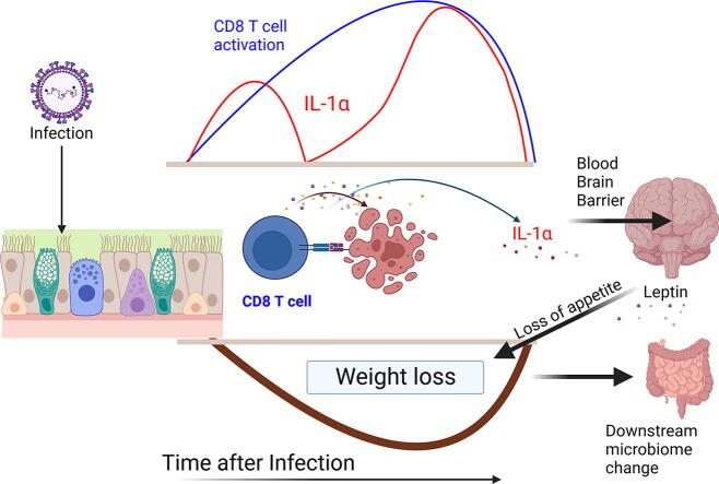
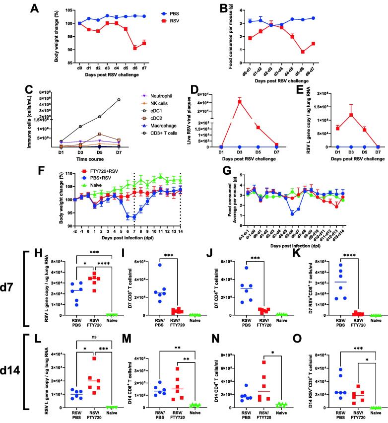
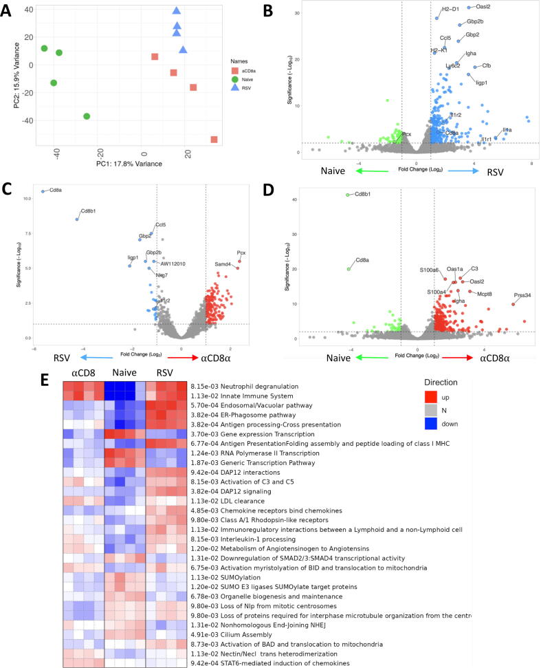
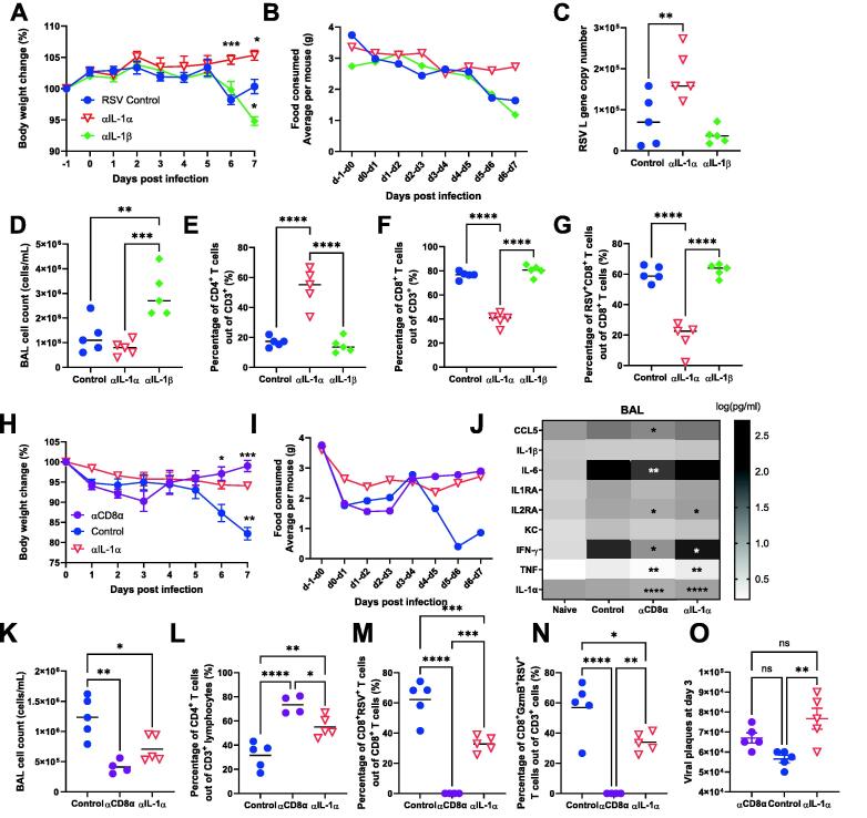
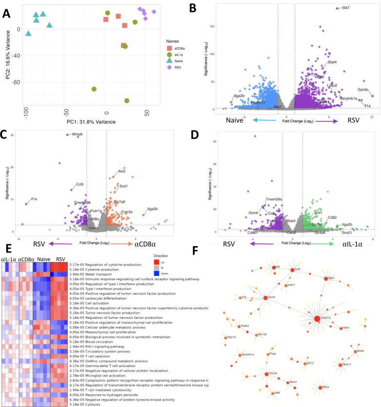
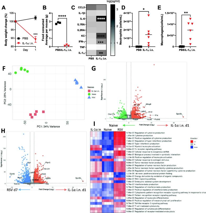
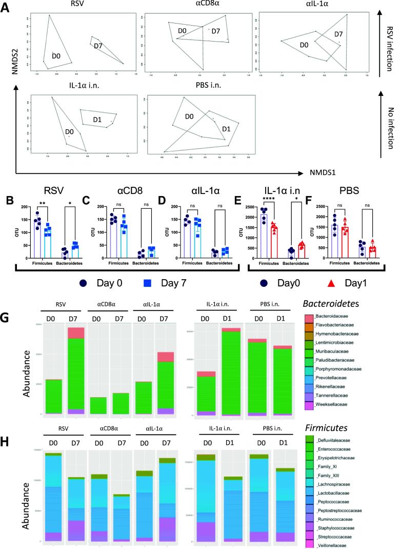
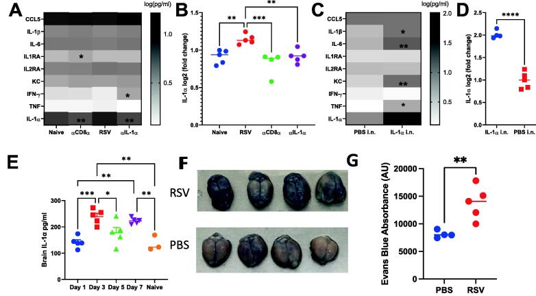
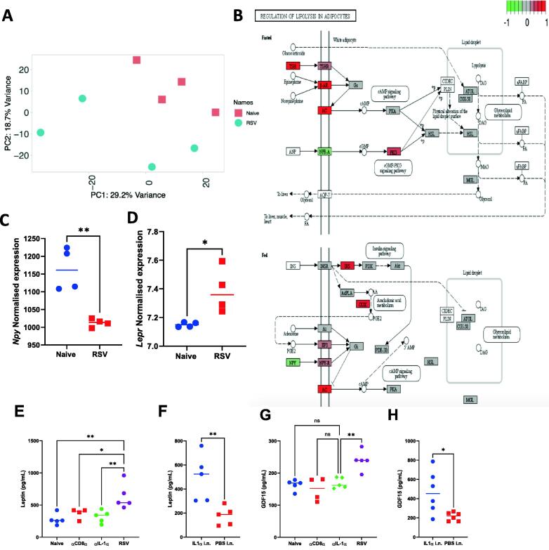
Respiratory viral infections remain a major cause of hospitalization and death worldwide. Patients with respiratory infections often lose weight. While acute weight loss is speculated to be a tolerance mechanism to limit pathogen growth, severe weight loss following infection can cause quality of life deterioration. Despite the clinical relevance of respiratory infection-induced weight loss, its mechanism is not yet completely understood. We utilized a model of CD 8+ T cell-driven weight loss during respiratory syncytial virus (RSV) infection to dissect the immune regulation of post-infection weight loss. Supporting previous data, bulk RNA sequencing indicated significant enrichment of the interleukin (IL)-1 signaling pathway after RSV infection. Despite increased viral load, infection-associated weight loss was significantly reduced after IL-1α (but not IL-1β) blockade. IL-1α depletion resulted in a reversal of the gut microbiota changes observed following RSV infection. Direct nasal instillation of IL-1α also caused weight loss. Of note, we detected IL-1α in the brain after either infection or nasal delivery. This was associated with changes in genes controlling appetite after RSV infection and corresponding changes in signaling molecules such as leptin and growth/differentiation factor 15. Together, these findings indicate a lung-brain-gut signaling axis for IL-1α in regulating weight loss after RSV infection.


Figures

None
Graphical abstract
Fig. 1
Fig. 1
Timing of T cell recruitment to lungs affects disease timing in RSV. 6-7 weeks old BALB/c SPF mice were intranasally infected 7.7 x 10⁶ PFU/ml RSV A2 subtype and culled on day 1, 3, 5, and 7 post infection. Body weight (A) and food consumed (B) were monitored daily. Immune cell populations were measured using flow cytometry (C) at each time course, and live viral plaques (D) and RSV L gene (E) were quantified. 6-7 weeks old BALB/c SPF mice were injected with 25µg of FTY720 or PBS only daily from day -2 to day 6. Mice were intranasally infected with 7.7 x 10⁶ PFU/ml RSV infection on day 0. Body weight (F) and food (G) were monitored daily from day -2 to day 14. At day 7, viral load was determined using RSV L gene qPCR on RNA extracted from the left lung lobe (H) and flow cytometry was used to analyze the number of CD8 T cell (I), CD4 T cells (J) and antigen-specific CD8+ T cells (K). At day 14, viral load was determined using RSV L gene qPCR (L) and flow cytometry was used to analyze the number of CD8 T cell (M), CD4 T cells (N) and antigen-specific CD8 T cells (O). N = 5, each dot represents an individual mouse (H-O); or mean +/-SEM –A - E, F-G). Significance calculated by ordinary one-way ANOVA and post test. *P  ≤  0.05, **P  ≤  0.01, ***P  ≤  0.001. Experiment was repeated twice.
Fig. 2
Fig. 2
CD8 depletion significantly alters the blood transcriptome following RSV infection. Blood RNA isolated from RSV infected mice with and without CD8 α monoclonal antibody (and naïve control) was analysed by RNASeq. Principle component analysis was carried out to identify batch effects (A). Differential gene expression in mice infected with RSV compared with naïve (B), α CD8 treated mice compared to RSV infection control (C), and α CD8 monoclonal treated mice compared to naïve (D). PGSEA analysis of the groups using Reactome curated data (E). Data from N=4 mice in each group.
Fig. 3
Fig. 3
IL-1 α but not IL-1 β blockade reduces weight loss following RSV infection. 6-7 weeks old BALB/c SPF mice were injected intraperitoneally with antibody neutralizing IL-1 α , IL-1β or control IgG on day -1, 1, 3 and 5 post infection. Body weight (A) and food (B) were monitored daily throughout infection. (C) Viral load was determined using RNA extracted from the left lung lobe. Cell count from bronchoalveolar lavage (D) and flow cytometry was performed to determine the percentage of CD4+ T cells (E), CD8+ T cells (F) and CD8+ RSV+ T cells (G). 6-7 weeks old BALB/c SPF mice were injected intraperitoneally with antibody depleting CD8 α or neutralizing IL-1 α , or control IgG on day -1, 2 and 5; or -1, 3 and 5 post infection. Body weight (H) and food (I) were monitored daily throughout infection. BAL supernatant was analysed for cytokine by multiplex bead assay (J). Cell counts from BAL (K) and flow cytometry was performed to determine the percentage of CD4+ T cells (L), CD8+ T cells (M) and CD8+ RSV+ T cells (N). Viral load measured using viral plaques were performed on day 3 post infection (O). N≥4 per group each dot represents an individual mouse (C-G, K-0); or mean +/-SEM (A, B, H-I). Significance calculated by ordinary one-way ANOVA and post test. *P  ≤  0.05, **P  ≤  0.01, ***P  ≤  0.001, ****P  ≤  0.0001. Experiment was repeated three times.
Fig. 4
Fig. 4
IL-1 α and CD8 depletion induce similar changes in lung transcriptomic signatures following RSV infection. Lung RNA extracted from mice receiving α CD8 α , α IL-1 α or IgG monoclonal antibody infected with RSV (RSV) as well as naïve (no treatment) underwent bulk RNASeq. Principal component analysis was carried out to identify any batch effect using the package pca and function prcomp in R (A). Volcano plot showing genes differentially expressed in mice infected with RSV compared with naïve (B), α CD8 α monoclonal treated mice compared to RSV infected control (C), α IL-1 α monoclonal antibody treated mice compared to RSV infected control (D) All plots generated using package Enhancedvolcano packagIn R). (E) PGSEA analysis of the groups using Reactome curated dataset in R version 1.30.0. (F) 102 significant differentially expressed genes (DEGs) important for inducing weight loss response were identified as up regulated in RSV compared to naïve and downregulated in both α CD8 α treated and α IL-1 α treated compared to RSV. The significant DEGs were used for Network analysis using NetworkAnalyst (version 3.0) using InnateDB curated interactions. Red colors indicate an increasing number of connections.
Fig. 5
Fig. 5
Intranasal administration of IL-1 α induces weight loss and induces similar global signatures in the lung as RSV infection.6-7 weeks old BALB/c SPF mice had 3 mg of recombinant IL-1 α protein or PBS introduced intranasally on day 0. Body weight (A) and food (B) were monitored. Lung supernatant analysed by multiplex cytokine bead assay. Cells were collected from bronchoalveolar lavage (BAL) (C) and flow cytometry was performed to determine the number of neutrophils (D) and macrophages (E). RNASeq was performed on lung RNA extracted from mice day 7 after intranasal administration, RSV or control. Principal component analysis of three conditions (F). Differentially expressed genes in mice administered with IL-1 α compared with naïve (G), IL-1 α administered mice compared to RSV infection control (H). PGSEA analysis of the groups using Reactome curated dataset (I). N≥5, each dot represents an individual mouse (D, E, F, I); or mean +/-SEM (A, G, H); or mean of 2 co-housed mice (B). Significance calculated by ordinary one-way ANOVA and post test. *P  ≤  0.05, **P  ≤  0.01, ***P  ≤  0.001, ****P  ≤  0.0001. Experiment was repeated twice.
Fig. 6
Fig. 6
Depletion of IL-1 α reverses the changes in the gut microbiome induced by RSV infection. Change in beta diversity visualized using NMDS on Brays-Curtis dissimilarity matrix for mice received RSV, α CD8 α , α IL-1 α antibody treatment, as well as IL-1 α intranasally (i.n.) administered or PBS (A). ASV abundance of Bacteroidetes or Firmicutes at phyla level for RSV (B), α CD8 α treated (C), α IL-1 α treated (I IL-1 α i.n. (E) or PBS i.n. (F) . *P  ≤  0.05, **P  ≤  0.01, ***P  ≤  0.001, ****P  ≤  0.0001. Abundances of Bacteroidetes at family level for antibody treated mice and IL-1 α intranasally administered mice (G). Abundances of Firmicutes at family level for antibody treated mice and IL-1 α intranasally administered mice (H).
Fig. 7
Fig. 7
RSV infection alters increases the levels of cytokines in the brain by increasing permeability. Brian supernatants were tested against a panel of cytokines (A, C), with a focus on the IL-1 α level (B, D). For RSV study, brain collected from naïve mice, or α CD8 α , α IL-1 α and or control Ig treated during RSV infection (A, B). For IL-1 α intranasal study, brain collected from PBS i.n. and IL-1 α i.n. treated mice. RNAseq was performed on brains from RSV infected and control (naïve) mice. Brain supernatant was collected on day 1, 3, 5, and 7 following infection and an IL-1 α ELISA was performed (E). 6-8 weeks old mice were infected with RSV and 200μl 2% Evans blue was injected intravenously on day 7 of infection 1 hour before the brains were extracted (F) and Evans blue absorbance was quantified using florescence absorbance excitation 485, emission 680 (G). N≥4, each dot represents an individual mouse (B, D-E, G); or mean +/-SEM (A, C). Significance calculated by ordinary one-way ANOVA and post test. *P  ≤  0.05, **P  ≤  0.01, ***P  ≤  0.001, ****P  ≤  0.0001. Experiment was repeated twice.
Fig. 8
Fig. 8
Brain transcriptomics revealed a role of leptin and GDF15 in weight loss following RSV infection. Principal component analysis was carried out to identify batch effects (A) and KEGG pathway analysis of differentially expressed genes termed Regulation of lipolysis in adipocytes (B). Normalised expression values taken from the Day 7 post RSV comparison for RNA levels of Npy (C) and Lepr (D) . Leptin (E, F) and GDF15 (G. H) measured in serum by ELISA from Naïve, α CD8 α , α IL-1 α and RSV (E, G) and PBS i.n. or IL-1 α i.n. treated mice (F, H). N≥5, each dot represents an individual mouse (C-H). Significance calculated by ordinary one-way ANOVA and post test. *P  ≤  0.05, **P  ≤  0.01, ***P  ≤  0.001, ****P  ≤  0.0001.

References

    1. Dapat C., et al. Gene signature of children with severe respiratory syncytial virus infection. Pediatr. Res. 2021;89:1664–1672. - PMC - PubMed
    1. Walsh E., et al. RSV-associated hospitalization in adults in the USA: A retrospective chart review investigating burden, management strategies, and outcomes. Health Sci. Rep. 2022;5:e556. - PMC - PubMed
    1. Falsey A.R., Walsh E.E. Respiratory syncytial virus infection in elderly adults. Drugs Aging. 2005;22:577–587. - PMC - PubMed
    1. García C.G., et al. Risk factors in children hospitalized with RSV bronchiolitis versus non-RSV bronchiolitis. Pediatrics. 2010;126:e1453–e1460. - PMC - PubMed
    1. Radigan K.A., et al. Influenza A virus infection induces muscle wasting via IL-6 regulation of the E3 ubiquitin ligase Atrogin-1. J. Immunol. 2019;202:484–493. - PMC - PubMed