

ZNF692 regulates nucleolar morphology by interacting with NPM1 and modifying its self-assembly properties
- PMID: 38382671
- PMCID: PMC10956046
- DOI: 10.1016/j.jbc.2024.105773
ZNF692 regulates nucleolar morphology by interacting with NPM1 and modifying its self-assembly properties
Abstract
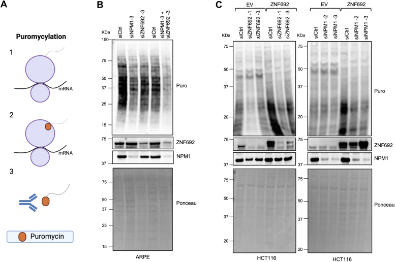
The nucleolus, a membrane-less organelle, is responsible for ribosomal RNA transcription, ribosomal RNA processing, and ribosome assembly. Nucleolar size and number are indicative of a cell's protein synthesis rate and proliferative capacity, and abnormalities in the nucleolus have been linked to neurodegenerative diseases and cancer. In this study, we demonstrated that the nucleolar protein ZNF692 directly interacts with nucleophosmin 1 (NPM1). Knocking down ZNF692 resulted in the nucleolar redistribution of NPM1 in ring-like structures and reduced protein synthesis. Purified NPM1 forms spherical condensates in vitro but mixing it with ZNF692 produces irregular condensates more closely resembling living cell nucleoli. Our findings indicate that ZNF692, by interacting with NPM1, plays a critical role in regulating nucleolar architecture and function in living cells.
Keywords: NPM1; ZNF692; condensates; nucleolus; protein assembly.
Copyright © 2024 The Authors. Published by Elsevier Inc. All rights reserved.
Conflict of interest statement
Conflict of interest The authors declare that they have no conflicts of interest with the contents of this article.
Figures


Similar articles
-
The interaction between nucleophosmin/NPM1 and the large ribosomal subunit precursors contribute to maintaining the nucleolar structure.Biochim Biophys Acta Mol Cell Res. 2021 Jan;1868(1):118879. doi: 10.1016/j.bbamcr.2020.118879. Epub 2020 Oct 8. Biochim Biophys Acta Mol Cell Res. 2021. PMID: 33039556
-
Elucidation of motifs in ribosomal protein S9 that mediate its nucleolar localization and binding to NPM1/nucleophosmin.PLoS One. 2012;7(12):e52476. doi: 10.1371/journal.pone.0052476. Epub 2012 Dec 20. PLoS One. 2012. PMID: 23285058 Free PMC article.
-
Nucleophosmin integrates within the nucleolus via multi-modal interactions with proteins displaying R-rich linear motifs and rRNA.Elife. 2016 Feb 2;5:e13571. doi: 10.7554/eLife.13571. Elife. 2016. PMID: 26836305 Free PMC article.
-
Phase separation via protein-protein and protein-RNA networks coordinates ribosome assembly in the nucleolus.Biochim Biophys Acta Gen Subj. 2025 Sep;1869(10):130835. doi: 10.1016/j.bbagen.2025.130835. Epub 2025 Jul 16. Biochim Biophys Acta Gen Subj. 2025. PMID: 40675030 Review.
-
Ribosomal RNA and nucleolar proteins from the oocyte are to some degree used for embryonic nucleolar formation in cattle and pig.Theriogenology. 2007 Sep 1;68 Suppl 1:S63-70. doi: 10.1016/j.theriogenology.2007.03.015. Epub 2007 Apr 26. Theriogenology. 2007. PMID: 17466364 Review.
Cited by
-
Immunocytochemical detection of proteins within cellular structures inaccessible to specific antibodies.Histochem Cell Biol. 2025 May 17;163(1):53. doi: 10.1007/s00418-025-02387-0. Histochem Cell Biol. 2025. PMID: 40381002 Review.
References
-
- Pelletier J., Thomas G., Volarevic S. Ribosome biogenesis in cancer: new players and therapeutic avenues. Nat. Rev. Cancer. 2018;18:51–63. - PubMed
-
- Donmez-Altuntas H., Akalin H., Karaman Y., Demirtas H., Imamoglu N., Ozkul Y. Evaluation of the nucleolar organizer regions in Alzheimer's disease. Gerontology. 2005;51:297–301. - PubMed
Publication types
MeSH terms
Substances
Grants and funding
LinkOut - more resources
Full Text Sources
Molecular Biology Databases
