Review
. 2024 Apr;1871(4):119700.
doi: 10.1016/j.bbamcr.2024.119700. Epub 2024 Feb 19.

Flipping the script: Advances in understanding how and why P4-ATPases flip lipid across membranes

Affiliations
Review

Flipping the script: Advances in understanding how and why P4-ATPases flip lipid across membranes

Adriana C Norris et al. Biochim Biophys Acta Mol Cell Res. 2024 Apr.

Abstract

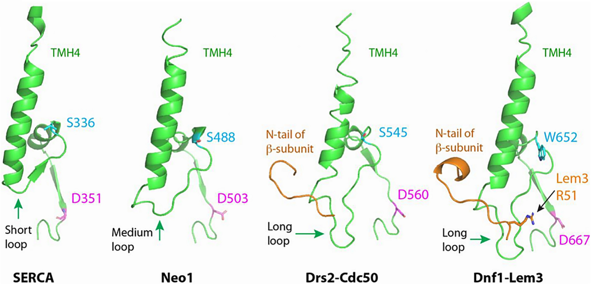
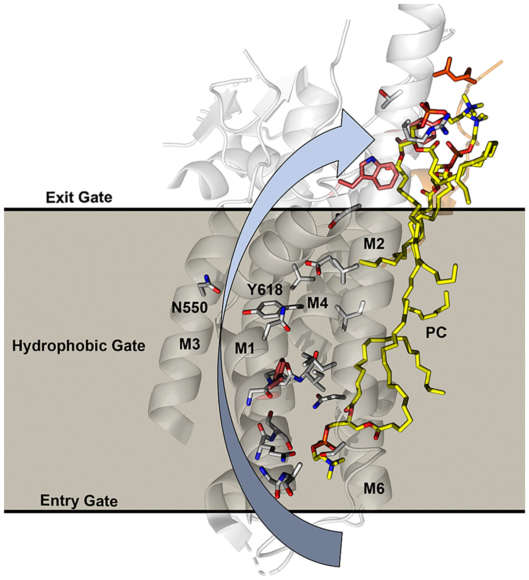
Type IV P-type ATPases (P4-ATPases) are a family of transmembrane enzymes that translocate lipid substrates from the outer to the inner leaflet of biological membranes and thus create an asymmetrical distribution of lipids within membranes. On the cellular level, this asymmetry is essential for maintaining the integrity and functionality of biological membranes, creating platforms for signaling events and facilitating vesicular trafficking. On the organismal level, this asymmetry has been shown to be important in maintaining blood homeostasis, liver metabolism, neural development, and the immune response. Indeed, dysregulation of P4-ATPases has been linked to several diseases; including anemia, cholestasis, neurological disease, and several cancers. This review will discuss the evolutionary transition of P4-ATPases from cation pumps to lipid flippases, the new lipid substrates that have been discovered, the significant advances that have been achieved in recent years regarding the structural mechanisms underlying the recognition and flipping of specific lipids across biological membranes, and the consequences of P4-ATPase dysfunction on cellular and physiological functions. Additionally, we emphasize the requirement for additional research to comprehensively understand the involvement of flippases in cellular physiology and disease and to explore their potential as targets for therapeutics in treating a variety of illnesses. The discussion in this review will primarily focus on the budding yeast, C. elegans, and mammalian P4-ATPases.

Keywords: Flippase; Glucosylceramide; Membrane asymmetry; P-type ATPase; Phosphatidylserine; Vesicular transport.


Conflict of interest statement

Declaration of competing interest The authors declare the following financial interests/personal relationships which may be considered as potential competing interests: Todd R. Graham reports financial support was provided by the National Institutes of Health.

Figures

Fig. 1.
Fig. 1.
Membrane asymmetry is controlled by flippases, floppases and scramblases. A few examples of lipid transporter structures with Protein Data Bank Identifiers. Substrate lipid is shown in yellow for Dnf1-Lem3 and MsbA.
Fig. 2.
Fig. 2.
Structure of ATP8A1-CDC50 in the E2P state bound to phosphatidylserine (PS). (A) Structure of ATP8A1-CDC50 with PS occluded in the entry gate (PDB: 6K7M). The P, N, and A domains are highlighted. The phospholipid binding site view in (B), depicts key residues in M1, M4, and M6 interacting with the phospholipid head group. αTM6 is shown in the foreground and M2 in the background. Hydrogen bond interactions are shown as dashed lines with corresponding distances labeled. (C) Alignment of M1 and M4 sequences that help form the lipid binding site. The position of residues relative to the highly conserved proline (at position 0 in M4) is indicated by the −4 to +4 values.
Fig. 3.
Fig. 3.
Extension of the cytosolic loop connecting TM4 to the P-domain in P4 ATPases relative to SERCA (P2-ATPase). A portion of each TM helix 4 (TMH4) is shown and the cytosolic extension of this helix is below the membrane border. The loop connecting TMH4 to the P domain in the monomeric Neo1 is intermediate in length between SERCA and the heterodimeric P4-ATPases. The functionally essential Neo1-S488 and Dnf1-W652 exit-gate residues (cyan) are positioned at the beginning of the P domain first helix and seem to form a substrate backstop in the exit gate that is closely linked to the phosphorylated aspartate (magenta residue). The N-terminal tail of the β-subunit (orange) associates with this loop and in the case of Dnf1 helps position Lem3-R51 for substrate interaction. Adapted from reference .
Fig. 4.
Fig. 4.
Model of the Post-Albers cycle for phospholipid transport by Dnf1/2-Lem3. The movement of cytosolic domains and lipid substrate during the Post-Albers cycle for Dnf1/2-Lem3 based on surface renditions. (1) ATP binds the N domain of E1 (Apo) relaxed state (7KY6). (2) A and N domains move towards each other. (3) The ATP γ-phosphate is transferred to an aspartate in the P domain. (4) A and N domains move clockwise to release ADP. (5) TM1 and TM2 tilting movements form the entry and exit gate binding sites (shown as halos). (6) Phospholipid binds to the entry and exit gate region of the E2P BeF3 conformation (7KYC) with the entry gate open to the exoplasmic leaflet. The source of the lipid in the exit gate at this stage is unclear but may derive from the cytosolic leaflet (dotted arrow). The dotted box encloses presumed structural states for which experimentally determined structures are lacking for Dnf1/2-Lem3. Structures of PS flippases stabilized with AlF4 have substrate occluded in the entry gate. (7) The N and A domains become more flexible as the aspartyl-phosphate is hydrolyzed and Pi is released. (8) Phospholipid substrate flips to the cytosolic side at some point in the E2P to E2 steps.(9–10) TM1 and TM2 tilts back, closing the entry gate and widening the exit gate. (11) Phospholipid is ejected from the flippase. Domain coloring: Yellow – actuator (A) domain, Blue – nucleotide-binding (N) domain, Green – phosphorylation (P) domain, Muted red shade – transmembrane domain, Bright red shade – linkers from transmembrane segments to A, N, and P Domains, Salmon – Lem3.
Fig. 5.
Fig. 5.
Structure of Dnf1-Lem3 with lipid bound. (A) Structure of the Dnf1-Lem3 in E2P state with phosphatidylcholine (PC) at the entry gate and exit gate (PDB: 7KYC). The major domains are highlighted. A close-up view of the proposed exit gate binding site (B) and entry gate binding site (C) is shown.
Fig. 6.
Fig. 6.
Potential substrate translocation path in the Dnf1-Lem3 membrane domain. The side chains are shown for residues where mutations alter the substrate preference of Dnf1-Lem3. N550 and Y618 are highlighted because these side chains do not line the TM2,4,6 groove, along which the headgroup is thought to slide during transport. However, N550 and Y618 could define part of the translocation pathway if TM1,2 partially separates from TM3,4,6 to allow the substrate headgroup to slide between them.
Fig. 7.
Fig. 7.
Involvement of P4-ATPases in human and mouse pathologies. The contribution of P4-ATPases to various pathologies in humans and mice can affect several locations in the body; including the lungs, brain, liver, skin, kidney, pancreas, GI tract, ears, blood and lymph, as well as female and male reproductive organs. Refer to the text for further description and citations. Schematic created using Biorender.com. NSCLC = Non-small cell lung cancer, LUSC = lung squamous cell carcinoma, PFIC1 = progressive familial intrahepatic cholestasis type 1, BRIC1 = benign recurrent intrahepatic cholestasis type 1, MCD = minimal change disease, MN = Membranous nephropathy, CRC = Colorectal cancer, ALL = Acute lymphocytic leukemia, CLL = Chronic lymphocytic leukemia, AML = acute myeloid leukemia, IPF = Idiopathic pulmonary fibrosis, CAMRQ = Cerebellar ataxia, impaired intellectual development, and disequilibrium syndrome.

References

    1. Alberts B, et al. , Molecular Biology of the Cell, 7th ed., W.W. Norton & Co., Chapters 10–12, 2022.
    1. Weibel ER, et al. , Correlated morphometric and biochemical studies on the liver cell. I. Morphometric model, stereologic methods, and normal morphometric data for rat liver, J. Cell Biol 42 (1) (1969) 68–91. - PMC - PubMed
    1. Bolender RP, Stereological analysis of the guinea pig pancreas. I. Analytical model and quantitative description of nonstimulated pancreatic exocrine cells, J. Cell Biol 61 (2) (1974) 269–287. - PMC - PubMed
    1. Levental I, Lyman E, Regulation of membrane protein structure and function by their lipid nano-environment, Nat. Rev. Mol. Cell Biol 24 (2) (2023) 107–122. - PMC - PubMed
    1. Harayama T, Riezman H, Understanding the diversity of membrane lipid composition, Nat. Rev. Mol. Cell Biol 19 (5) (2018) 281–296. - PubMed

Publication types

LinkOut - more resources