. 2024 Jun 17;95(7):682-690.
doi: 10.1136/jnnp-2023-332696.

Genotype-specific spinal cord damage in spinocerebellar ataxias: an ENIGMA-Ataxia study

Affiliations

Genotype-specific spinal cord damage in spinocerebellar ataxias: an ENIGMA-Ataxia study

Thiago Junqueira Ribeiro Rezende et al. J Neurol Neurosurg Psychiatry. .

Abstract

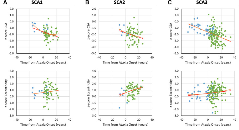
Background: Spinal cord damage is a feature of many spinocerebellar ataxias (SCAs), but well-powered in vivo studies are lacking and links with disease severity and progression remain unclear. Here we characterise cervical spinal cord morphometric abnormalities in SCA1, SCA2, SCA3 and SCA6 using a large multisite MRI dataset.

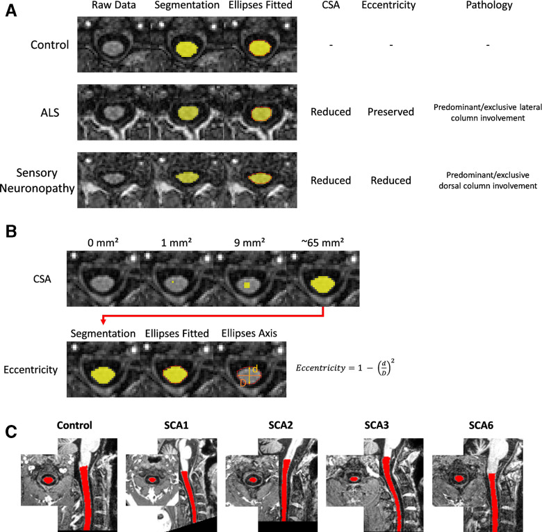
Methods: Upper spinal cord (vertebrae C1-C4) cross-sectional area (CSA) and eccentricity (flattening) were assessed using MRI data from nine sites within the ENIGMA-Ataxia consortium, including 364 people with ataxic SCA, 56 individuals with preataxic SCA and 394 nonataxic controls. Correlations and subgroup analyses within the SCA cohorts were undertaken based on disease duration and ataxia severity.

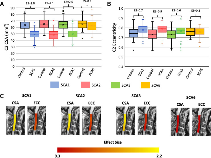
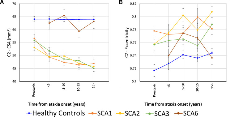
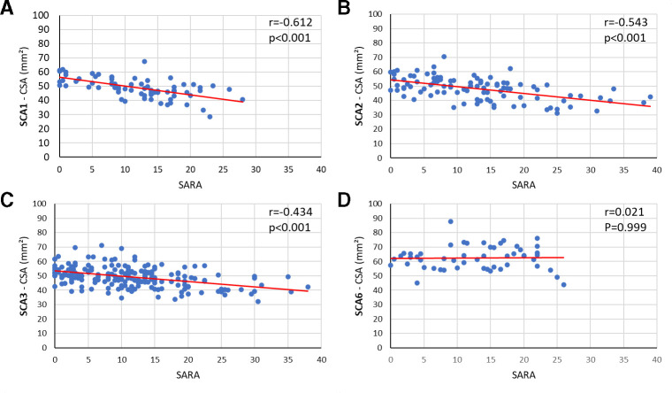
Results: Individuals in the ataxic stage of SCA1, SCA2 and SCA3, relative to non-ataxic controls, had significantly reduced CSA and increased eccentricity at all examined levels. CSA showed large effect sizes (d>2.0) and correlated with ataxia severity (r<-0.43) and disease duration (r<-0.21). Eccentricity correlated only with ataxia severity in SCA2 (r=0.28). No significant spinal cord differences were evident in SCA6. In preataxic individuals, CSA was significantly reduced in SCA2 (d=1.6) and SCA3 (d=1.7), and the SCA2 group also showed increased eccentricity (d=1.1) relative to nonataxic controls. Subgroup analyses confirmed that CSA and eccentricity are abnormal in early disease stages in SCA1, SCA2 and SCA3. CSA declined with disease progression in all, whereas eccentricity progressed only in SCA2.

Conclusions: Spinal cord abnormalities are an early and progressive feature of SCA1, SCA2 and SCA3, but not SCA6, which can be captured using quantitative MRI.

Keywords: MRI; cerebellar ataxia; image analysis; neurogenetics.


Conflict of interest statement

Competing interests: TJRR, FC, ARMM, JLP, OB, BKM, IHH, AD, DT, SLG, ID, IA, GO, CM, LN, AN, MG, LC, HAGT, SEO, CRHR, JFR, FM, AD, BW, JG, MS, PMT, SIT: none. The authors declare no competing interests. KR received honoraria for presentations or advisory boards from Biogen and Roche as well as clinical trial grants from Pfizer, Merck, Minoryx, Biogen and Roche. BB is cofounder, shareholder and CTO of AIRAmed GmbH. CL received research grants from Minoryx Therapeutics and research support from Biogen Inc.

Figures

Figure 1
Figure 1
Schematic illustration of the spinal cord morphometric parameters used in this study. (A) Clinical correlates of these parameters. In diseases characterised by selective lateral column/corticospinal tract degeneration, there is CSA reduction, but preserved eccentricity (middle lane, patient with amyotrophic lateral sclerosis,ALS). In diseases characterised by selective dorsal column degeneration, there is combined CSA and eccentricity reduction (lower lane, patient with autoimmune sensory neuronopathy). All segmentations are shown in axial slices of the same spinal cord level (C2). (B) Computation of CSA and eccentricity. (C) An exemplar MRI with spinal cord mask from each cohort. CSA, cross-sectional area.
Figure 2
Figure 2
Box plots showing group differences at the C2 spinal cord level in each disease group relative to a matched control group (age-adjusted, sex-adjusted and site-adjusted). (A) C2 cross-sectional area in square millimetres; (B) C2 eccentricity; (C) visualisation of effect sizes overlaid on a spinal cord template image.
Figure 3
Figure 3
Results showing the progressive atrophy of the (A) C2 CSA and (B) C2 eccentricity in participants with SCA1 (orange), SCA2 (yellow), SCA3 (green) and SCA6 (brown). Subgroups are defined based on time since ataxia onset. Preataxic: subjects with Scale for Assessment and Rating of Ataxia score <3 at the time of MRI assessment. The healthy controls were age-matched, sex-matched and site-matched for each SCA subgroup, with the plotted datapoint representing the mean cervical spinal cord area or eccentricity of all controls included in each subgroup; error bars=SE error of the mean. CSA, cross-sectional area; SCA, spinocerebellar ataxia.
Figure 4
Figure 4
Correlations between CSA at the C2 level and SARA score for (A) SCA1, (B) SCA2, (C) SCA3 and (D) SCA6. CSA, cross-sectional area; SARA, Scale for Assessment and Rating of Ataxia; SCA, spinocerebellar ataxia.
Figure 5
Figure 5
Graphs of z-transformed CSA or eccentricity at the C2 vertebral level versus time from ataxia onset (green). The negative values (blue) for disease duration indicate the predicted time to ataxia onset calculated using Tezenas formulas for SCA1 and SCA2 and Peng formula for SCA3, based on CAG repeat length and current participant age. CSA, cross-sectional area; SCA, spinocerebellar ataxia.

References

    1. Coutelier M, Jacoupy M, Janer A, et al. . NPTX1 mutations trigger endoplasmic reticulum stress and cause autosomal dominant cerebellar ataxia. Brain 2022;145:1519–34. 10.1093/brain/awab407 - DOI - PubMed
    1. Erichsen AK, Koht J, Stray-Pedersen A, et al. . Prevalence of hereditary ataxia and spastic paraplegia in southeast Norway: a population-based study[published Online First: 20090331]. Brain 2009;132(Pt 6):1577–88. 10.1093/brain/awp056 - DOI - PubMed
    1. Seidel K, Siswanto S, Brunt ERP, et al. . Brain pathology of spinocerebellar ataxias[published Online First: 20120609]. Acta Neuropathol 2012;124:1–21. 10.1007/s00401-012-1000-x - DOI - PubMed
    1. Rüb U, Schöls L, Paulson H, et al. . Clinical features, neurogenetics and neuropathology of the polyglutamine spinocerebellar ataxias type 1, 2, 3, 6 and 7[published Online First: 20130221]. Prog Neurobiol 2013;104:38–66. 10.1016/j.pneurobio.2013.01.001 - DOI - PubMed
    1. Rüb U, Bürk K, Timmann D, et al. . Spinocerebellar ataxia type 1 (SCA1): new pathoanatomical and clinico-pathological insights. Neuropathol Appl Neurobiol 2012;38:665–80. 10.1111/j.1365-2990.2012.01259.x - DOI - PubMed