. 2024 Mar 5;18(9):7148-7160.
doi: 10.1021/acsnano.3c11820. Epub 2024 Feb 21.

Sensing the Spin State of Room-Temperature Switchable Cyanometallate Frameworks with Nitrogen-Vacancy Centers in Nanodiamonds

Affiliations

Sensing the Spin State of Room-Temperature Switchable Cyanometallate Frameworks with Nitrogen-Vacancy Centers in Nanodiamonds

Bradley T Flinn et al. ACS Nano. .

Abstract

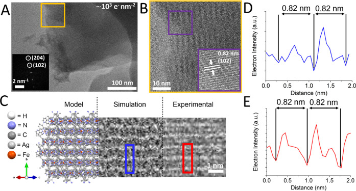
Room-temperature magnetically switchable materials play a vital role in current and upcoming quantum technologies, such as spintronics, molecular switches, and data storage devices. The increasing miniaturization of device architectures produces a need to develop analytical tools capable of precisely probing spin information at the single-particle level. In this work, we demonstrate a methodology using negatively charged nitrogen vacancies (NV-) in fluorescent nanodiamond (FND) particles to probe the magnetic switching of a spin crossover (SCO) metal-organic framework (MOF), [Fe(1,6-naphthyridine)2(Ag(CN)2)2] material (1), and a single-molecule photomagnet [X(18-crown-6)(H2O)3]Fe(CN)6·2H2O, where X = Eu and Dy (materials 2a and 2b, respectively), in response to heat, light, and electron beam exposure. We employ correlative light-electron microscopy using transmission electron microscopy (TEM) finder grids to accurately image and sense spin-spin interacting particles down to the single-particle level. We used surface-sensitive optically detected magnetic resonance (ODMR) and magnetic modulation (MM) of FND photoluminescence (PL) to sense spins to a distance of ca. 10-30 nm. We show that ODMR and MM sensing was not sensitive to the temperature-induced SCO of FeII in 1 as formation of paramagnetic FeIII through surface oxidation (detected by X-ray photoelectron spectroscopy) on heating obscured the signal of bulk SCO switching. We found that proximal FNDs could effectively sense the chemical transformations induced by the 200 keV electron beam in 1, namely, AgI → Ag0 and FeII → FeIII. However, transformations induced by the electron beam are irreversible as they substantially disrupt the structure of MOF particles. Finally, we demonstrate NV- sensing of reversible photomagnetic switching, FeIII + (18-crown-6) ⇆ FeII + (18-crown-6)+ •, triggered in 2a and 2b by 405 nm light. The photoredox process of 2a and 2b proved to be the best candidate for room-temperature single-particle magnetic switching utilizing FNDs as a sensor, which could have applications into next-generation quantum technologies.

Keywords: Nitrogen-vacancy sensing; metal−organic framework; nanodiamond; photomagnetism; spin-crossover; transmission electron microscopy.


Conflict of interest statement

The authors declare no competing financial interest.

Figures

Figure 1
Figure 1
(A) Schematic experimental inverted fluorescence microscope setup. FNDs are adsorbed directly onto TEM grids or glass coverslips. A custom-made PCB with a wire antenna was used for microwave delivery and an off NV axis electromagnet placed directly above the experimental setup was used to modulate magnetic field. Light illumination and detection pathways are also shown. Schematic Jablonski diagrams show the excitation and decay pathways of the NV center (B) without and (C) with the application of the off-axis magnetic field. Transitions between ground and excited triplet states (solid arrows), as well as microwave stimulated relaxations, are shown with nonradiative singlet state pathways, weak (− • –) and strong () transitions, highlighted.
Figure 2
Figure 2
(A) Variable-temperature XPS Fe(II) 2p photoelectron spectra for compound 1 showing surface chemical changes upon SCO. (B) A high-resolution XPS spectrum of the Fe 2p3/2 region at 363 and 183 K, showing HS and LS photoelectron behavior, respectively. Transition from the LS to HS state corresponds to a greater contribution of the satellite component. (C) Overlayed bright-field and fluorescence optical image, FNDs are shown as bright green spots. (D) TEM image showing a single microparticle of SCO compound 1 in direct contact with a small FND cluster over the hole of the carbon film. (E, F) Room-temperature ODMR and MM NV sensing, respectively, from the FND cluster highlighted with a white arrow in panels (C) and (D)). Upon addition of 1 (in the LS state) ODMR and MM contrast is reduced. (G) ODMR of 1 in both the LS (295 K) and HS (321 K) states. The NV resonant frequency was used to estimate temperature which was compared against a thermocouple attached to the GCS (thermocouple reading 328 K). Inset shows photographs of a section of the powder on the GCS at both 295 and 321 K (red low- spin and orange high spin, respectively). Singal to noise in panel (G) was lower due to the lower NA air-coupled objective used in heating measurements.
Figure 3
Figure 3
(A) Thermal variation of χmT for “aged” compound 1 showing hysteretic switching between LS (diamagnetic) and HS (paramagnetic) spin states. Field-cooled and zero-field-cooled susceptibility indicate the cooling and heating cycles, respectively. (“Aged” powder, SQUID: LS → HS at 338 K, HS → LS at 288 K). The spin-only (SO) magnetic moment for an Fe(II)-HS center is shown by the dashed black line (3 emu K mol–1). Inset: FC ZFC dχ/dT(T). (B) Raman C≡N band ratio analysis between LS and HS states (Figure S4 for full Raman spectra and more information on C≡N band ratio calculation) for “fresh” and “aged” powders of compound 1 illustrating the structural changes accompanying SCO. (“Aged” powder, Raman: LS → HS at 338 K, HS → LS at 287 K. “Fresh” Powder, Raman: LS → HS at 294 K, HS → LS at 287 K.)
Figure 4
Figure 4
(A) HRTEM image at low e beam fluence (∼103 e nm–2) of a thin area of a microparticle of compound 1. Inset of (A) shows a fast Fourier transform (FFT) image of the area marked in orange, first- and second-order diffraction is identified for the (102) and (204) planes, respectively. (B) Zoomed-in orange area shown in panel (A). Lattice fringes in HRTEM images correspond to the (102) planes (d-spacing is identified, marked with arrows and indexed). (C) A molecular model (left), simulated TEM image (middle) and experimental HRTEM image (right) of the (102) plane. (D, E) Line profiles of the simulated (blue box and trace) and experimental image (red box and trace), respectively.
Figure 5
Figure 5
(A) Overlayed bright-field and fluorescence images before (white square) e beam irradiation, indicating the FND cluster chosen for analysis (white asterisk) and the interacting particles of compound 1. (B) ODMR spectra and MM traces taken from the target FND cluster (labeled in panel (A) as a white asterisk) taken before and after the addition of compound 1, as well as after e beam irradiation of the microcube. (C) TEM image of the interacting microparticle of 1 and the FND cluster at low e fluence (stated at the bottom of images). (D) TEM image of the interacting microcube and FND cluster at higher e fluence (the FND cluster labeled with a white triangle illustrates the distance relationship between clusters shown in panel (A)). At relatively moderate flux (>103 e nm–2 s–1), we observed the beam sensitivity of compound 1. Insets of panels (C) and (D) also show EDX atomic percentage ratios taken for pristine and irradiated particles, respectively (Figure S13 for EDX spectra). (E) HR-TEM image from the area marked in panel (D) (red square) showing Ag0 metal NPs formed as a product of e beam-induced transformations on the surface of the SCO cube. Inset of (E) shows a common d-spacing found on the Ag0 NPs, shown to correspond to the (111) plane. Note as a control measure: At the e fluence used to induce transformations in compound 1, the sensing properties of the NV centers within FND particles remained unchanged (Figure S23).
Figure 6
Figure 6
(A) Mechanism of reversible PET. Light (hv) is used to initiate the forward transformation of the powder, whereas either time (t, >1 week) at room temperature, or heat (Δ, 80 °C for 2 h) can be used to reverse the phototransformation. (B) Solid-state EPR spectra of as-synthesized and illuminated powders of 2a. (C) Thermal variation of the value χmT for compound 2a in both the as-synthesized (green-yellow) and illuminated state (orange). Photodemagnetization at 300 K gives a change in susceptibility of 57.4%. Inset photographs show powders of 2a in both states. (D) M(H) curve for the as-synthesized and illuminated states of 2a at 2 K. Inset shows M(H) at 300 K.
Figure 7
Figure 7
(A, B) High-resolution XPS spectra of the Fe 2p3/2 region (panel (A)) and O 1s region (panel (B)) for as-synthesized and illuminated states of 2a. (C) ODMR spectra (inset show MM traces) for the FND cluster highlighted in panel (D) by a white circle, including illuminated states by 405 nm light (blue traces) on a GCS. (D) Overlayed bright-field and fluorescence image showing an example of a crystal of 2a that is in contact/close proximity to clusters of FNDs on a glass coverslip. (E) ODMR spectra probing the reversibility of the photoinduced electron transfer mechanism of 2a, including an annealing step to reverse the phototransformation. Set of measurements for (E) were conducted on TEM finder grids for ease of location after annealing. Inset of panel (E) shows the averaged normalized PL ODMR double minima values from spectra in panel (E) as a bar chart.

References

    1. Sanvito S. Molecular spintronics. Chem. Soc. Rev. 2011, 40 (6), 3336–3355. 10.1039/c1cs15047b. - DOI - PubMed
    1. Bogani L.; Wernsdorfer W. Molecular Spintronics Using Single-Molecule Magnets. Nat. Mater. 2008, 7 (3), 179–186. 10.1038/nmat2133. - DOI - PubMed
    1. Wang J. H.; Li Z. Y.; Yamashita M.; Bu X. H. Recent Progress on Cyano-bridged Transition-metal-based Single-Molecule Magnets and Single-chain Magnets. Coord. Chem. Rev. 2021, 428, 213617.10.1016/j.ccr.2020.213617. - DOI
    1. Hiiuk V. M.; Shova S.; Rotaru A.; Ksenofontov V.; Fritsky I. O.; Gural’skiy I. A. Room Temperature Hysteretic Spin Crossover in a New Cyanoheterometallic Framework. Chem. Commun. 2019, 55 (23), 3359–3362. 10.1039/C8CC10260K. - DOI - PubMed
    1. Cai L. Z.; Chen Q. S.; Zhang C. J.; Li P. X.; Wang M. S.; Guo G. C. Photochromism and Photomagnetism of a 3d-4f Hexacyanoferrate at Room Temperature. J. Am. Chem. Soc. 2015, 137 (34), 10882–10885. 10.1021/jacs.5b05320. - DOI - PubMed