

Myoid gonadal stromal tumor of the testis-the novel subtype of testicular gonadal stromal tumors: a case report and review of the literature
- PMID: 38383445
- PMCID: PMC10882757
- DOI: 10.1186/s13256-024-04393-7
Myoid gonadal stromal tumor of the testis-the novel subtype of testicular gonadal stromal tumors: a case report and review of the literature
Abstract
Background: Sex cord gonadal stromal tumors compose less than 10% of all testicular neoplasms and consist of a variety of histological subtypes. In 2016, the World Health Organization introduced a novel subtype, the myoid gonadal stromal tumor, that consists of spindle-shaped cells with immunohistologic features of muscle cells. Only few cases have been reported to date. Due to its rarity and owing to its only recent introduction, the current knowledge about myoid gonadal stromal tumor is limited, and particularly, appropriate clinical management is still ill-defined.
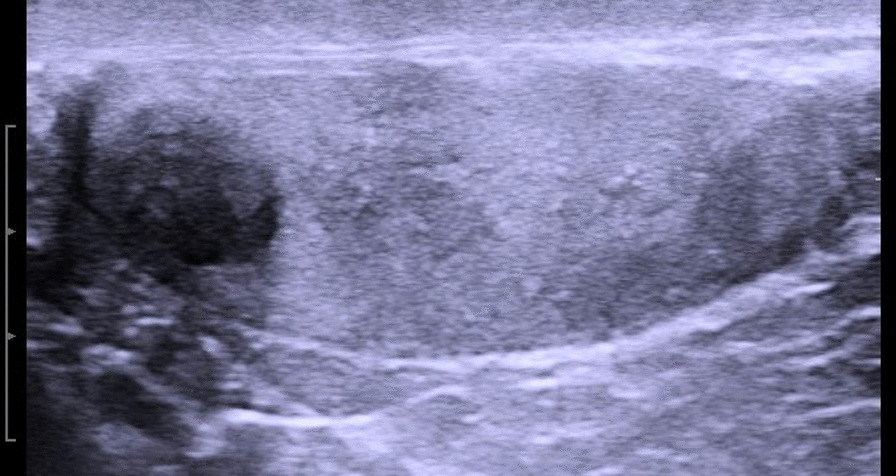
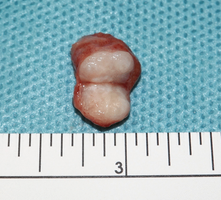
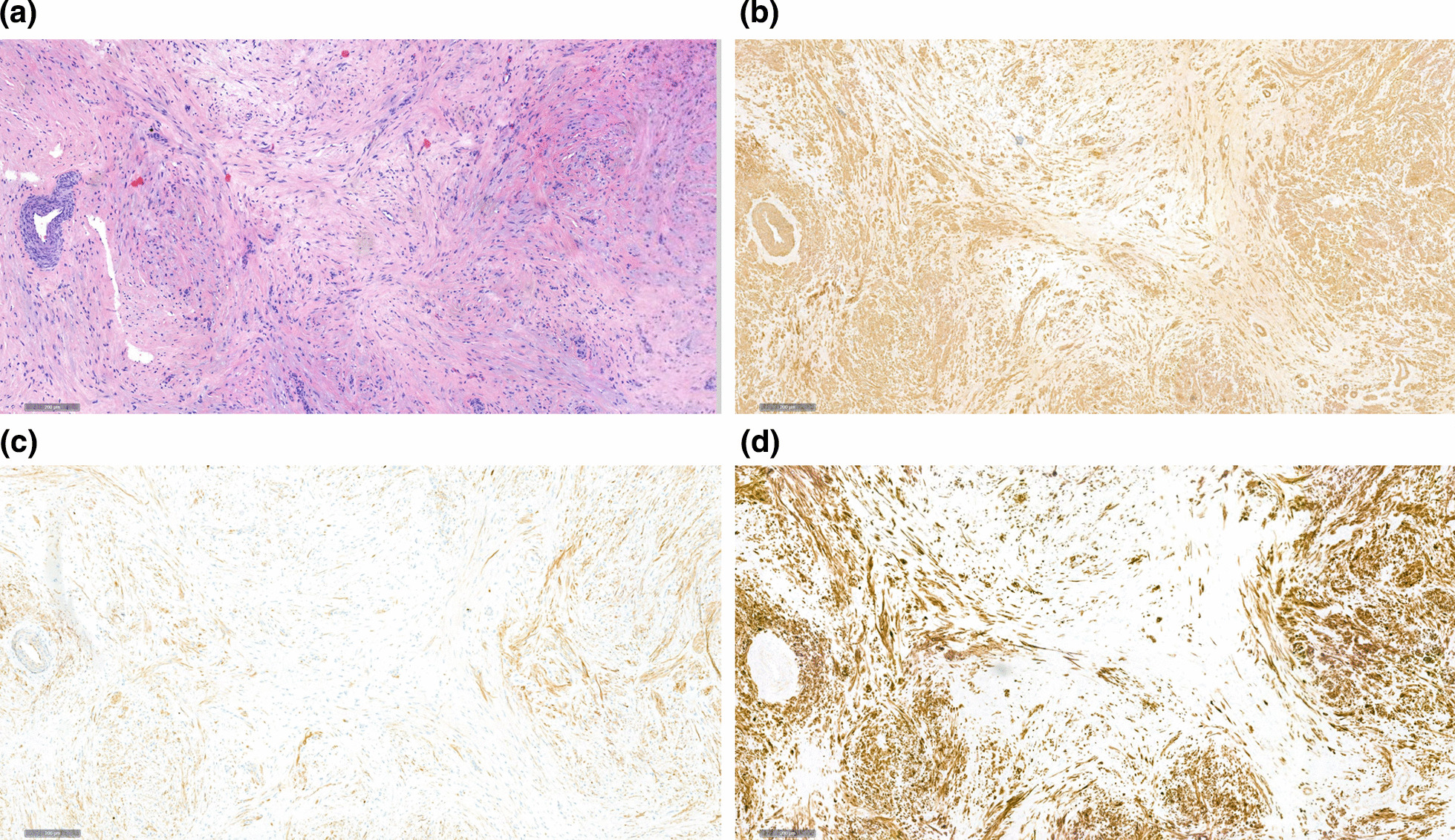
Case presentation: A 47-year-old man of Caucasian descent presented with nonspecific scrotal discomfort. A roundish and well demarcated hypoechoic mass of 8.5 mm in diameter was detected in the cranial region of the left testis. Serum tumor marker levels were within normal ranges. Testis-sparing surgery revealed a 9-mm whitish, hard mass with sharp surgical margin. Histologically, the neoplasm consisted of microfibrillar tissue with spindle-shaped cells harboring elongated nuclei. Immunohistochemical work-up disclosed expression of desmin, small muscle actin, and S100 protein giving evidence for the myogenic nature of the neoplastic cells. There was no indication of malignancy, neither histologically nor clinically. Follow-up of 1 year was uneventful.
Conclusion: A literature survey revealed 22 previous cases of myoid gonadal stromal tumor. The median age was 37 years, the median size of the neoplasm was 20 mm, and there was no side-preponderance. Myoid gonadal stromal tumor is not much different from other subtypes of gonadal stromal tumors nor from testicular gem cell tumors regarding age and laterality; however, tumor size is smaller in myoid gonadal stromal tumors than in germ cell tumors. Although rarely performed so far, testis-sparing surgery probably constitutes an appropriate treatment of this neoplasm. Myoid gonadal stromal tumor represents an emerging novel entity of benign testicular new growths that caregivers of patients with testicular tumors should be aware of.
Keywords: Desmin; Gonadal stromal tumor; Inhibin; S100 protein; Smooth muscle actin; Testicular neoplasm; Testis-sparing surgery.
© 2024. The Author(s).
Conflict of interest statement
The authors declare that they have no competing interests.
Figures
References
-
- Mikuz G. Update on the pathology of testicular tumors. Anal Quant Cytopathol Histpathol. 2015;37(1):75–85. - PubMed
-
- Grogg J, Schneider K, Bode PK, Kranzbühler B, Eberli D, Sulser T, Lorch A, Beyer J, Hermanns T, Fankhauser CD. Sertoli cell tumors of the testes: systematic literature review and meta-analysis of outcomes in 435 patients. Oncologist. 2020;25(7):585–590. doi: 10.1634/theoncologist.2019-0692. - DOI - PMC - PubMed
Publication types
MeSH terms
Substances
LinkOut - more resources
Full Text Sources
Medical
Research Materials
