. 2024 Feb 21;7(5):e202302132.
doi: 10.26508/lsa.202302132. Print 2024 May.

Breast implant surface topography triggers a chronic-like inflammatory response

Affiliations

Breast implant surface topography triggers a chronic-like inflammatory response

Valeriano Vinci et al. Life Sci Alliance. .

Abstract

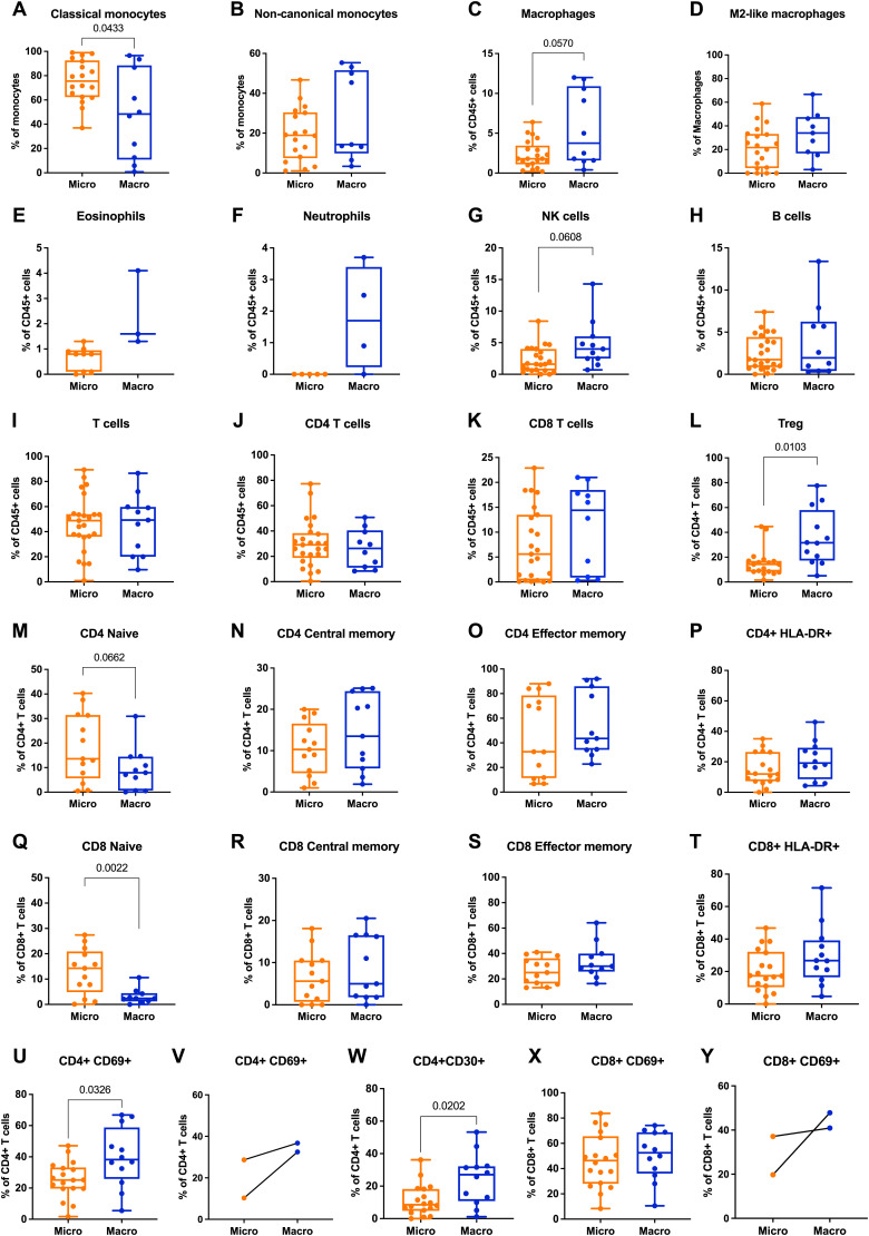
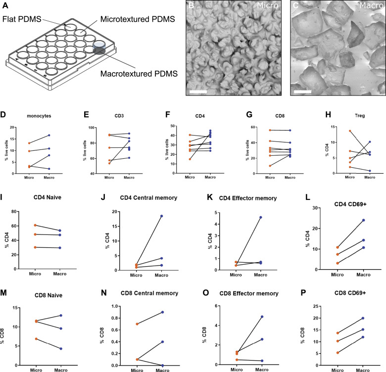
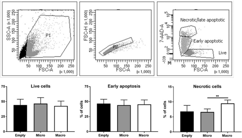
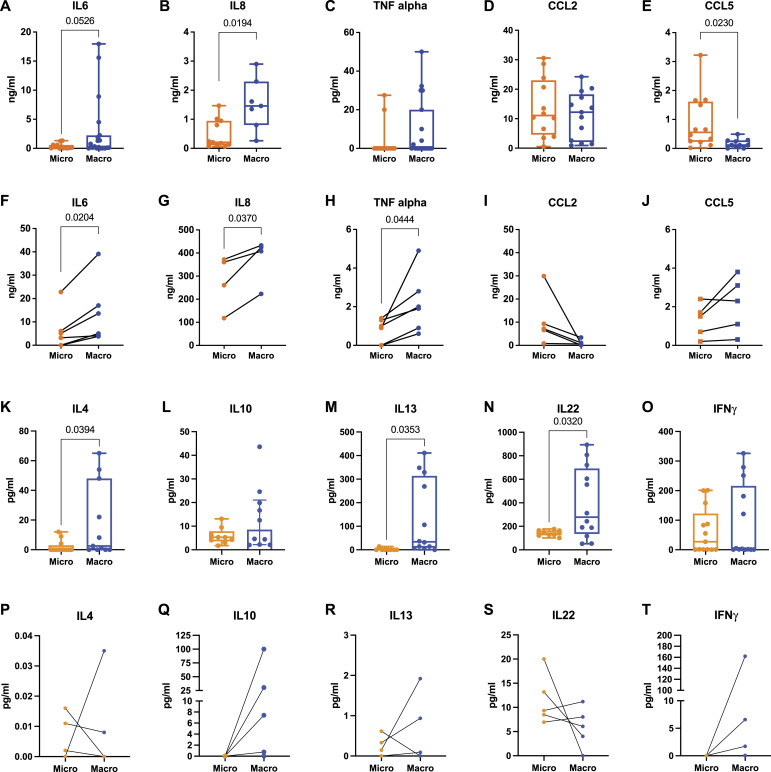
Breast implants are extensively employed for both reconstructive and esthetic purposes. However, the safety of breast implants with textured surfaces has been questioned, owing to a potential correlation with anaplastic large-cell lymphoma and the recurrence of breast cancer. This study investigates the immune response elicited by different prosthetic surfaces, focusing on the comparison between macrotextured and microtextured breast implants. Through the analysis of intraoperatively harvested periprosthetic fluids and cell culture experiments on surface replicas, we demonstrate that macrotextured surfaces elicit a more pronounced chronic-like activation of leucocytes and an increased release of inflammatory cytokines, in contrast to microtextured surfaces. In addition, in vitro fluorescent imaging of leucocytes revealed an accumulation of lymphocytes within the cavities of the macrotextured surfaces, indicating that the physical entrapment of these cells may contribute to their activation. These findings suggest that the topography of implant surfaces plays a significant role in promoting a chronic-like inflammatory environment, which could be a contributing factor in the development of lymphomas associated with a wide range of implantable devices.


Conflict of interest statement

The authors declare that they have no conflict of interest.

Figures

Figure S1.
Figure S1.. Taxonomic analysis of bacteria in periprosthetic fluids derived from patients with microtextured (Micro) or macrotextured (Macro) implants assessed through shotgun metagenomic sequencing.
(A, B) Distribution of bacterial families and bacterial species richness found in the periprosthetic fluid. (C, D) Comparison between microbiota of same patients implanted with different textured prosthesis in terms of taxonomic analysis and species richness.
Figure 1.
Figure 1.. Flow cytometry analysis of periprosthetic leukocytes from patients with microtextured and macrotextured breast prostheses.
Frequencies of the different immune subsets calculated as (A, B) frequency of viable monocytes, (C, E, F, G, H, I, J, K) frequency of viable CD45+ cells, (D) frequency of viable macrophages, (L, M, N, O, P, U, V, W) frequency of viable CD4+ T cells, (Q, R, S, T, X, Y) frequency of viable CD8+ T cells. Each histogram in the figure represents the mean ± SD of the measured parameters. The statistical significance of differences between microtextured and macrotextured prostheses was determined using an unpaired t test with Welch’s correction.
Figure 2.
Figure 2.. Flow cytometry analysis of PBMCs cultured on model surface textures.
(A) Schematic representation of the in vitro experimental model used to assess the immune response of PBMCs to different surface textures. (B) Scanning electron microscopy image of a PDMS replica of a microtextured surface (Mentor Siltex), showing rounded bumps and cavities, with dimensions ranging from 10 to 50 μm, created using the coating emulation technique. (C) Scanning electron microscopy image of a PDMS replica of a macrotextured surface (Allergan Biocell), showing cubic cavities characteristic of this texture, with dimensions ranging from 100 to 400 μm, obtained by the “salt loss” technique. (D, E, F, G, H, I, J, K, L, M, N, O, P) Results of flow cytometry analysis depicting various immune cell populations and their activation status in PBMCs cultured on these model surfaces.
Figure S2.
Figure S2.. Topographic features of model surface replicas.
(A) Scanning electron microscopy (SEM) image of a polydimethylsiloxane (PDMS) model of a microtextured surface showing measurements of topographic features. (B) Cross section of a microtextured model surface. (C) SEM image of the PDMS model of a macrotextured surface showing measurements of topographic features. (D) Cross section of a macrotextured model surface.
Figure S3.
Figure S3.. Gating strategy applied for assessing PBMC viability by flow cytometry.
Physical parameters were used to sequentially exclude debris and doublets from the analysis. On singlets, PBMCs were defined alive based on 7AAD fluorescence. A representative analysis is shown in the upper row. In vitro cell viability assays: live, early apoptotic, and necrotic cells were analyzed (lower row). Histograms represent the mean ± SD. The statistical significance was determined using an unpaired t test with Welch’s correction. **P < 0.001.
Figure 3.
Figure 3.. Quantification of soluble mediators in periprosthetic fluids and supernatants of PBMCs cultured on microtextured or macrotextured surfaces.
(A, B, C, D, E) ELISA quantification of IL6, IL8, TNF-alpha, CCL2, and CCL5 in the periprosthetic fluid of patients with either microtextured or macrotextured implants. (F, G, H, I, J) ELISA quantification of the same set of cytokines and chemokines (as in (A, B, C, D, E)) released from PBMCs cultured on microtextured or macrotextured surfaces. (K, L, M, N, O) ELLA quantification of IL4, IL10, IL13, IL22, and INFɣ in the periprosthetic fluid of patients with microtextured and macrotextured implants. (P, Q, R, S, T) ELLA quantification of the same set of cytokines (as in (F, G, H, I, J)) released from PBMCs cultured on microtextured or macrotextured surfaces. Histograms represent the mean ± SD, based on data from at least four independent donors. (A, B, C, D, E, F, G, H, I, J, K, L, M, N, O, P, Q, R, S, T) Statistical significance was determined using an unpaired t test with Welch’s correction (A, B, C, D, E, K, L, M, N, O) and a paired t test (F, G, H, I, J, P, Q, R, S, T).
Figure S4.
Figure S4.. ELLA quantification of IL6 and IL8.
(A, B) Cytokine levels in periprosthetic fluid derived from patients with microtextured (Micro) or macrotextured (Macro) implants. Histograms represent the mean ± SD. Statistical significance was assessed using an unpaired t test with Welch’s correction. (C, D) Cytokine levels in supernatants of PBMCs cultured on model surface textures.
Figure 4.
Figure 4.. Leukocyte distribution on model surface textures.
(A, B, C, D) Overlay of phase-contrast (blue) and fluorescent images (red indicating PBMCs) acquired 12 h after plating cells on different surfaces: flat (A), microtextured (B), and macrotextured ((C) for donor 1, (D) for donor 2). Scale bar, 200 μm. (E) Overlay of phase-contrast and fluorescent images (green for monocytes, red for lymphocytes) acquired at intervals of 0, 5, 10, and 15 h from the beginning of the experiment. Here, cells were plated on a macrotextured surface and co-cultured with S. epidermidis. Scale bar, 100 μm.

References

    1. Alessandri-Bonetti M, Jeong T, Vaienti L, De La Cruz C, Gimbel ML, Nguyen VT, Egro FM (2023) The role of microorganisms in the development of breast implant-associated anaplastic large cell lymphoma. Pathogens 12: 313. 10.3390/pathogens12020313 - DOI - PMC - PubMed
    1. Arber DA, Orazi A, Hasserjian R, Thiele J, Borowitz MJ, Le Beau MM, Bloomfield CD, Cazzola M, Vardiman JW (2016) The 2016 revision to the World Health Organization classification of myeloid neoplasms and acute leukemia. Blood 127: 2391–2405. 10.1182/blood-2016-03-643544 - DOI - PubMed
    1. Barnsley GP, Sigurdson LJ, Barnsley SE (2006) Textured surface breast implants in the prevention of capsular contracture among breast augmentation patients: A meta-analysis of randomized controlled trials. Plast Reconstr Surg 117: 2182–2190. 10.1097/01.prs.0000218184.47372.d5 - DOI - PubMed
    1. Barr S, Hill EW, Bayat A (2017) Functional biocompatibility testing of silicone breast implants and a novel classification system based on surface roughness. J Mech Behav Biomed Mater 75: 75–81. 10.1016/j.jmbbm.2017.06.030 - DOI - PubMed
    1. Belgiovine C, Digifico E, Anfray C, Ummarino A, Torres Andón F (2020) Targeting tumor-associated macrophages in anti-cancer therapies: Convincing the traitors to do the right thing. J Clin Med 9: 3226. 10.3390/jcm9103226 - DOI - PMC - PubMed