. 2024 Feb 21;15(2):160.
doi: 10.1038/s41419-024-06507-x.

Modification of BCLX pre-mRNA splicing has antitumor efficacy alone or in combination with radiotherapy in human glioblastoma cells

Affiliations

Modification of BCLX pre-mRNA splicing has antitumor efficacy alone or in combination with radiotherapy in human glioblastoma cells

Zhihui Dou et al. Cell Death Dis. .

Abstract

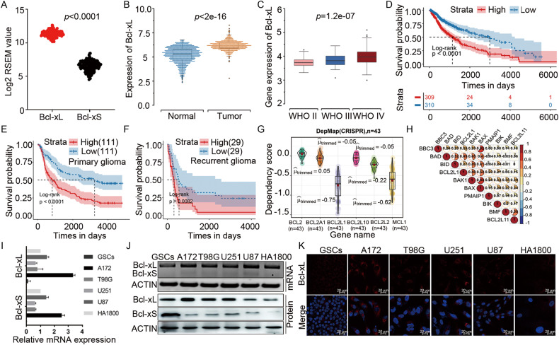
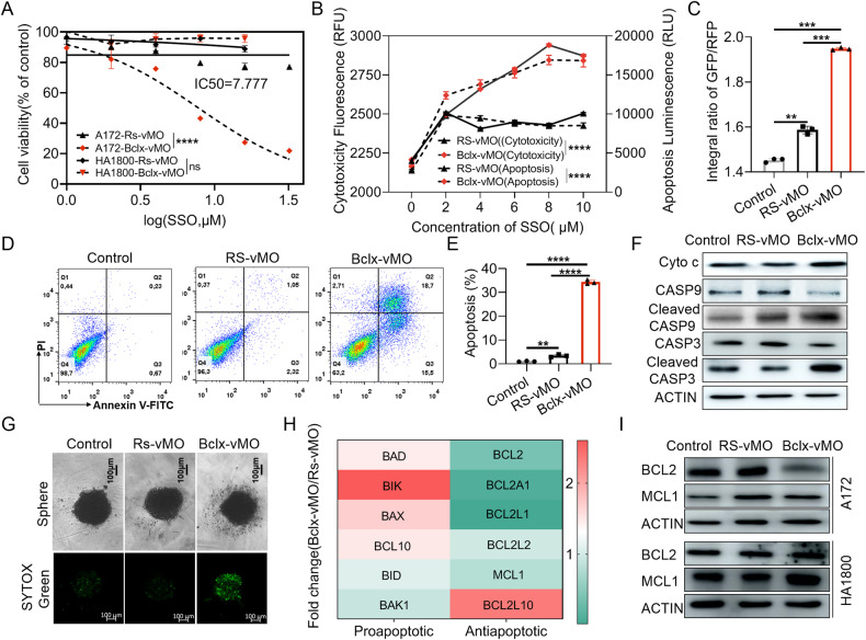
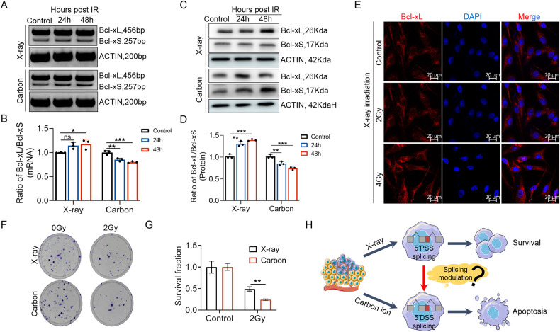
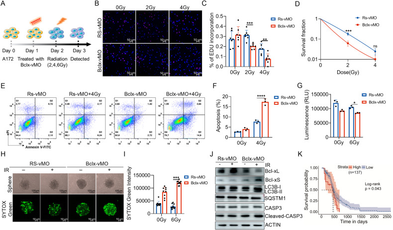
Dysregulation of anti-apoptotic and pro-apoptotic protein isoforms arising from aberrant splicing is a crucial hallmark of cancers and may contribute to therapeutic resistance. Thus, targeting RNA splicing to redirect isoform expression of apoptosis-related genes could lead to promising anti-cancer phenotypes. Glioblastoma (GBM) is the most common type of malignant brain tumor in adults. In this study, through RT-PCR and Western Blot analysis, we found that BCLX pre-mRNA is aberrantly spliced in GBM cells with a favored splicing of anti-apoptotic Bcl-xL. Modulation of BCLX pre-mRNA splicing using splice-switching oligonucleotides (SSOs) efficiently elevated the pro-apoptotic isoform Bcl-xS at the expense of the anti-apoptotic Bcl-xL. Induction of Bcl-xS by SSOs activated apoptosis and autophagy in GBM cells. In addition, we found that ionizing radiation could also modulate the alternative splicing of BCLX. In contrast to heavy (carbon) ion irradiation, low energy X-ray radiation-induced an increased ratio of Bcl-xL/Bcl-xS. Inhibiting Bcl-xL through splicing regulation can significantly enhance the radiation sensitivity of 2D and 3D GBM cells. These results suggested that manipulation of BCLX pre-mRNA alternative splicing by splice-switching oligonucleotides is a novel approach to inhibit glioblastoma tumorigenesis alone or in combination with radiotherapy.


Conflict of interest statement

The authors declare no competing interests.

Figures

Fig. 1
Fig. 1. Anti-apoptotic Bcl-xL is highly expressed in GBM samples and GBM cell lines, and correlated with worse prognosis.
A Comparison of Bcl-xL and Bcl-xS mRNA expression levels between GBM tissues based on TSVdb (http://tsvdb.com/index.html) database. B Expression of Bcl-xL in normal brains and GBM from TCGA. C Correlation between Bcl-xL expression and pathologic grades of GBM. DF Kaplan–Meier survival analysis for Bcl-xL variant expression in patients with GBM from TCGA and CGGA datasets. G The dependency profile of pro-survival BCL2 family genes (BCL2L1, BCL2L10, BCL2, BCL2A1, BCL2L2, and MCL1) in GBM cell lines based on genome-wide CRISPR. Data were downloaded from the DepMap datasets (DepMap; https://depmap.org/portal/). H Correlation analysis of the expression of pro-apoptotic BCL2 family genes and anti-apoptotic BCL2 family genes from TCGA datasets. I The mRNA expression of Bcl-xL and Bcl-xS were analyzed by q-PCR. J The expression of BCLX splicing isoforms in GBM cell lines and HA1800 cells were analyzed by RT-PCR and Western Blot. K Immunofluorescence staining of Bcl-xL protein in GBM cells and HA1800 cells. Data in vitro represent at least three independent experiments. Anti-apoptotic Bcl-xL is highly expressed in GBM samples and GBM cell lines, and correlated with worse prognosis.
Fig. 2
Fig. 2. Bclx-vMO regulated Bcl-x splicing mode in GBM cells effectively.
A Alternative splicing mode and splicing regulation by SSOs in cancer cells. B Localization and delivery efficiencies of Bclx-vMO using Laser confocal microscopy. Fluorescence signal represented the Bclx-vMO delivered by Endo-Porter combined to the target RNA and increased with the amount of Bclx-vMO used. CF A172 cells were transfected with 4, 8, μM vMO. The expression of Bcl-xL and Bcl-xS were analyzed by PCR and western blot. Bclx-vMO elevated Bcl-xS at the expense of Bcl- xL both at mRNA (C, D) and protein levels (E, F), and significantly decreased the ratio of Bcl-xL/Bcl-xS. G Data are shown as mean values ± S.D. from three independent experiments. One-Way ANOVA followed by Dunnett’s multiple comparisons test are reported. For all panels “**” indicates p < 0.01, “***” indicates p < 0.001, “****” indicates p < 0.0001, “ns” indicates no significance.
Fig. 3
Fig. 3. Cytotoxicity of GBM cell lines after shift of Bcl-x pre-mRNA alternative splicing from Bcl-xL to Bcl-xS.
A The cell viability of different concentrations of Rs-vMO and Bclx-vMO on A172 cells and normal astrocyte HA1800 after 48 h transfection (Two-way Anova). B Cell cytotoxicity and apoptosis assay of A172 cells after 48 h transfection (Two-way Anova). C Fluorescence of A172 cells stained using JC-10 was ascertained through high content analysis system. D, E Apoptosis rate of A172 cell lines received 4 μM doses of vMO were detected by flow cytometer. F Western blot analysis of activated apoptin expression in A172 cell lines treated with vMO for 48 h. G 3D Cell viability of A172 cells treated with vMO was measured using SYTOX green and images were visualized and captured using optical microscope and confocal scanning microscope. H PCR array analysis of pro-apoptotic and anti-apoptotic BCL2 family members. I Western blot analysis of anti-apoptotic BCL2 family protein (BCL2, MCL1) expression in A172 cell lines treated with vMO for 48 h. The concentration of Rs-vMO and Bclx-vMO used was 4 μM when not specified. Data are shown as mean values ± S.D. from three independent experiments. One-Way ANOVA followed by Dunnett’s multiple comparisons test are reported. For all panels, “***” indicates p < 0.001, “****” indicates p < 0.0001.
Fig. 4
Fig. 4. Correction of BCLX pre-mRNA alternative splicing from Bcl-xL to Bcl-xS induces autophagy in A172 cancer cells.
A Volcano plot of Spearmans’ rank correlation coefficient between Bcl-xL (BCL2L1) and tested proteins. Gene expression data were downloaded from TCGA-GBM. The “corrplot” package in R was used to obtain the correlation coefficient and p-value of genes correlated with Bcl-xL expression. B, C The autophagosomes induced by Bclx-vMO were visualized and quantified. D, E The expression of BECN1, SQSTM1/p62, and conversion of LC3I to LC3II were detected and analyzed through western blotting. F, G A172 cells were treated with 4 μM Bclx-vMO for 48 h. Representative microscopy images were obtained by transmission electron microscopy. The red arrow indicates autophagic vacuoles containing cytoplasmic context (F) or damaged mitochondrial membranes (G). The concentration of Rs-vMO and Bclx-vMO used was 4 μM when not specified. Data are shown as mean values ± S.D. from three independent experiments. One-Way ANOVA followed by Dunnett’s multiple comparisons test are reported. For all panels “*” indicates p < 0.05, “****” indicates p < 0.0001, “ns” indicates no significance.
Fig. 5
Fig. 5. Blocking BCLX splicing regulation-induced autophagy significantly attenuates apoptosis in A172 cells.
A A172 cells were co-incubated with CQ or 3-MA and Bclx-vMO for 48 h. Cell viability was assessed by MTT assay. B, C A172 cells were co-incubated with CQ or 3-MA and Bclx-vMO for 48 h. Then, the apoptosis rate of A172 cells detected by flow cytometer. Quantification of apoptosis in cells treated is presented. D, E 3D Cell viability of A172 combined-treated with Bclx-vMO and CQ was measured using SYTOX Green. F, G Total proteins of A172 cells were extracted and the expression of proteins related to apoptosis and autophagy was detected by western blot. The concentration of Rs-vMO and Bclx-vMO used was 4 μM when not specified. Data are shown as mean values ± S.D. from ≥three independent experiments. One-Way ANOVA followed by Dunnett’s multiple comparisons test are reported. For all panels “*” indicates p < 0.05, “**” indicates p < 0.01, “***” indicates p < 0.001, “****” indicates p < 0.0001, “ns” indicates no significance.
Fig. 6
Fig. 6. Effect of X-ray and heavy ion irradiation on the alternative splicing and expression of BCLX gene.
A, B The splicing patterns of BCLX gene after X-ray and carbon ion irradiation was assessed by RT-PCR analysis. C, D Effect of X-ray and carbon ion irradiation on the expression of Bcl-xL and Bcl-xS at protein level. E Effect of X-ray irradiation on the spatial distribution and expression of Bcl-xL isoforms. F, G Clonal survival of A172 cells after X-ray and carbon ion irradiation. H Schematic diagram of the different splicing patterns of BCLX after X-ray and carbon ion irradiation. Data are shown as mean values ± S.D. from three independent experiments. One-way ANOVA followed by Dunnett’s multiple comparisons test are reported. For all panels “*” indicates p < 0.05, “**” indicates p < 0.01, “***” indicates p < 0.001, “ns” indicates no significance.
Fig. 7
Fig. 7. Correction of BCLX splicing from anti-apoptotic Bcl-xL to Bcl-xS sensitizes GBM cells to IR.
A Schematic illustration of experiment design and treatment plan. B, C EdU assay demonstrated that Bclx-vMO could sensitize A172 cells to X-ray irradiation. Typical photos of the EdU assay were captured with confocal microscopy. D Colony formation assays using A172 cells with inhibition of Bcl-xL or control combined with or without IR treatment. E, F Apoptosis of A172 cells with BCLX splicing correction or control combined with or without IR were evaluated by flow cytometry. G The cell viability of 3D spheroid cultures of A172 cells was determined using Cell Titer-Glo 3D according to the instructions. H, I The dead cells in 3D spheroid cultures of A172 with BCLX splicing correction or control combined with or without IR were measured using SYTOX Green. J Western blot was performed to measure the levels of proteins related to apoptosis and autophagy in A172 cells with splicing modulation upon IR. K Kaplan–Meier survival analysis of GBM patients who received radiation based on Bcl-xL expression (TCGA datasets). Data are shown as mean values ± S.D. from ≥three independent experiments. One-way ANOVA followed by Dunnett’s multiple comparisons test are reported. For all panels “***” indicates p < 0.001, “****” indicates p < 0.0001.
Fig. 8
Fig. 8
Schematic representation showing the proposed mechanisms through which the BCLX splicing modulation regulates apoptosis and radiosensitivity in GBM cells.

Similar articles

Cited by

References

    1. Kahles A, Lehmann KV, Toussaint NC, Hüser M, Stark SG, Sachsenberg T, et al. Comprehensive analysis of alternative splicing across tumors from 8,705 patients. Cancer Cell. 2018;34:211–224.e216. doi: 10.1016/j.ccell.2018.07.001. - DOI - PMC - PubMed
    1. Bradley RK, Anczuków O. RNA splicing dysregulation and the hallmarks of cancer. Nat Rev Cancer. 2023;23:135–55. doi: 10.1038/s41568-022-00541-7. - DOI - PMC - PubMed
    1. Urbanski LM, Leclair N, Anczuków O. Alternative-splicing defects in cancer: Splicing regulators and their downstream targets, guiding the way to novel cancer therapeutics. Wiley Interdiscip Rev RNA. 2018;9:e1476. doi: 10.1002/wrna.1476. - DOI - PMC - PubMed
    1. Dvinge H, Kim E, Abdel-Wahab O, Bradley RK. RNA splicing factors as oncoproteins and tumour suppressors. Nat Rev Cancer. 2016;16:413–30. doi: 10.1038/nrc.2016.51. - DOI - PMC - PubMed
    1. Crews LA, Balaian L, Delos Santos NP, Leu HS, Court AC, Lazzari E, et al. RNA splicing modulation selectively impairs leukemia stem cell maintenance in secondary human AML. Cell Stem Cell. 2016;19:599–612. doi: 10.1016/j.stem.2016.08.003. - DOI - PMC - PubMed

Publication types

MeSH terms