. 2024 Mar;627(8002):204-211.
doi: 10.1038/s41586-024-07089-6. Epub 2024 Feb 21.

Targeted protein degradation via intramolecular bivalent glues

Affiliations

Targeted protein degradation via intramolecular bivalent glues

Oliver Hsia et al. Nature. 2024 Mar.

Abstract

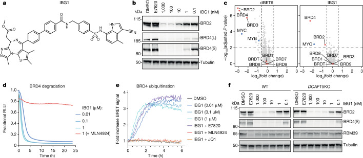
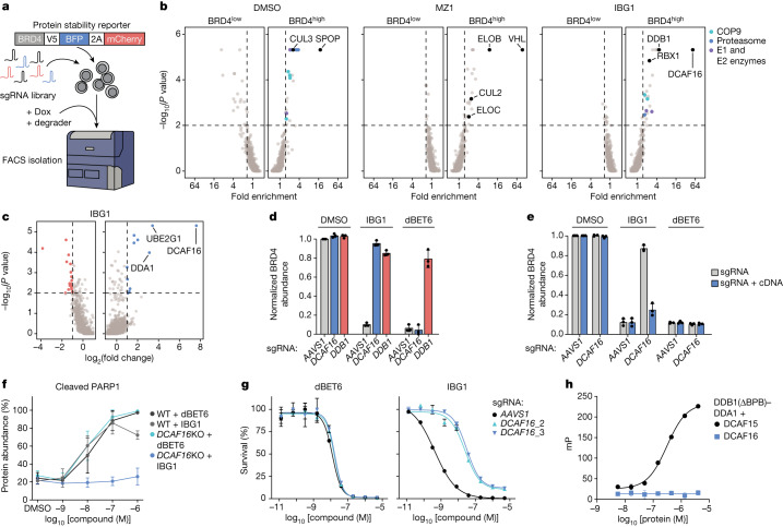
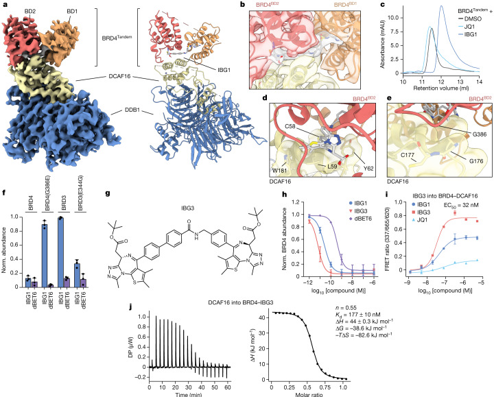
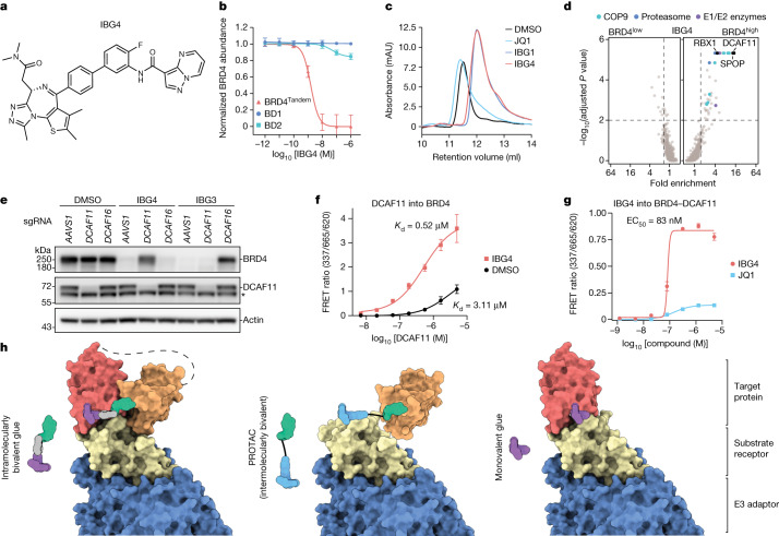
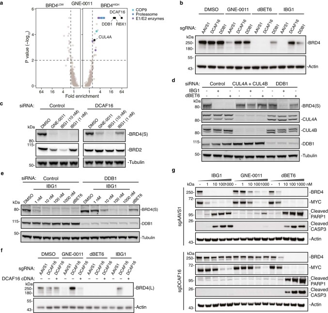
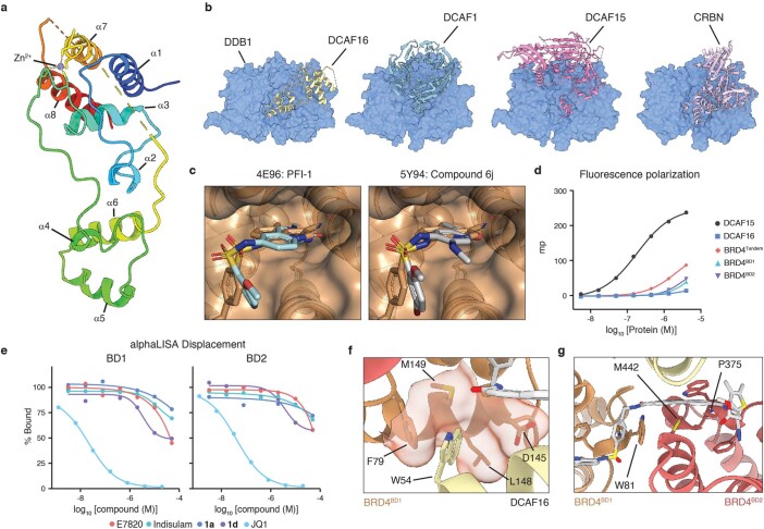
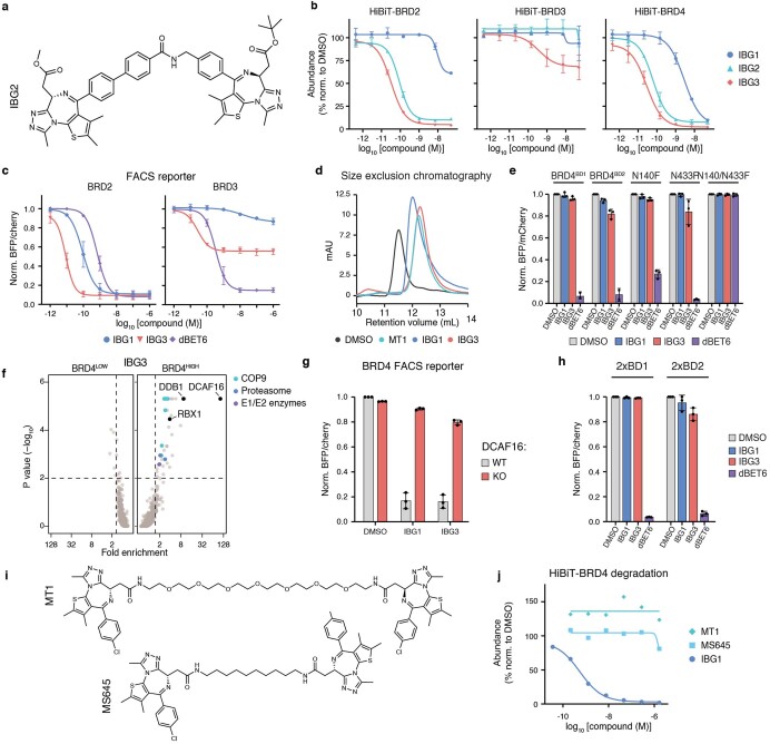
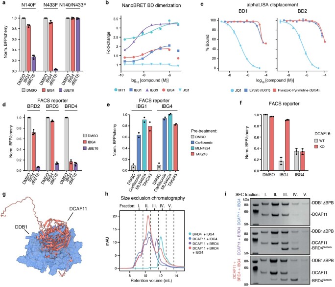
Targeted protein degradation is a pharmacological modality that is based on the induced proximity of an E3 ubiquitin ligase and a target protein to promote target ubiquitination and proteasomal degradation. This has been achieved either via proteolysis-targeting chimeras (PROTACs)-bifunctional compounds composed of two separate moieties that individually bind the target and E3 ligase, or via molecular glues that monovalently bind either the ligase or the target1-4. Here, using orthogonal genetic screening, biophysical characterization and structural reconstitution, we investigate the mechanism of action of bifunctional degraders of BRD2 and BRD4, termed intramolecular bivalent glues (IBGs), and find that instead of connecting target and ligase in trans as PROTACs do, they simultaneously engage and connect two adjacent domains of the target protein in cis. This conformational change 'glues' BRD4 to the E3 ligases DCAF11 or DCAF16, leveraging intrinsic target-ligase affinities that do not translate to BRD4 degradation in the absence of compound. Structural insights into the ternary BRD4-IBG1-DCAF16 complex guided the rational design of improved degraders of low picomolar potency. We thus introduce a new modality in targeted protein degradation, which works by bridging protein domains in cis to enhance surface complementarity with E3 ligases for productive ubiquitination and degradation.


Conflict of interest statement

A.C. is a scientific founder, shareholder and advisor of Amphista Therapeutics, a company that is developing targeted protein degradation therapeutic platforms. The Ciulli laboratory receives or has received sponsored research support from Almirall, Amgen, Amphista Therapeutics, Boehringer Ingelheim, Eisai, Merck KaaG, Nurix Therapeutics, Ono Pharmaceutical and Tocris-Biotechne. A.T. is currently an employee of Amphista Therapeutics. G.E.W. is scientific founder and shareholder of Proxygen and Solgate. The Winter laboratory has received research funding from Pfizer. The other authors declare no competing interests.

Figures

Fig. 1
Fig. 1. IBG1 degrades BRD2 and BRD4 independently of DCAF15.
a, Structure of IBG1. b, BET protein degradation activity of IBG1. HEK293 cells were treated for 6 h with DMSO, E7820 (1 μM) or increasing concentrations of IBG1. BET protein was quantified by immunoblot. Data representative of n = 3 independent experiments. c, Whole-proteome changes after degrader treatment. Quantitative proteomics in KBM7 cells was performed after 6 h of treatment with DMSO, IBG1 (1 nM) or dBET6 (10 nM). log2-transformed fold change and −log10-transformed Benjamini–Hochberg adjusted one-way analysis of variance (ANOVA) P value compared with DMSO treatment. n = 3 biological replicates. d, NanoBRET kinetic degradation assay. BromoTag–HiBiT–BRD4 knock-in HEK293 cells were treated with IBG1 with or without MLN4924 (10 µM) pre-treatment for 1 h. Mean of n = 3 biological replicates. RLU, relative light units. e, NanoBRET kinetic ubiquitination assay. LgBiT-transfected HiBiT–BromoTag–BRD4 knock-in HEK293 cells were treated with IBG1 at indicated concentrations or at 10 nM following pre-treatment with JQ1, E7820 (both 10 µM) or MLN4924 (1 µM) for 1 h. Mean of n = 4 biological replicates. f, DCAF15-independent BET protein degradation. Wild-type (WT) and DCAF15-knockout (KO) HCT-116 cells were treated with increasing concentrations of IBG1 for 6 h and BET protein was quantified by immunoblot. Data representative of n = 3 independent experiments.
Fig. 2
Fig. 2. IBG1-induced degradation of BRD2 and BRD4 is dependent on CRL4–DCAF16.
a, Schematic of FACS-based CRISPR–Cas9 screens. Doxycycline (Dox)-inducible Cas9 (iCas9) KBM7 BRD4–BFP reporter cells were transduced with a CRL-focused sgRNA library, treated with BET degraders and sorted based on BRD4–BFP/mCherry ratios. b, FACS-based CRISPR screens for BRD4 stability. KBM7 iCas9 BRD4 reporter cells were treated with DMSO, MZ1 (10 nM) or IBG1 (1 nM) for 6 h before sorting. 20S proteasome subunits, COP9 signalosome subunits and E1 and E2 ubiquitin enzymes inside the scoring window (one-sided MAGeCK P value < 0.01, fold change > 1.5) are highlighted. c, CRISPR–Cas9 viability screen. HCT-116 cells were transduced with Cas9 and a ubiquitin–proteasome system-focused sgRNA library and treated with IBG1 (58 nM; fourfold half-maximal inhibitory concentration (IC50)) for 6 days. Genes with a fold change > 2 and one-sided MAGeCK P value < 0.01 are highlighted. d, Screen validation. KBM7 iCas9 BRD4–BFP reporter cells were transduced with AAVS1, DCAF16 or DDB1-targeting sgRNAs, treated with DMSO, IBG1 (1 nM) or dBET6 (10 nM) for 6 h, and BRD4–BFP was quantified by FACS. e, DCAF16 knockout and rescue. KBM7 iCas9 BRD4–BFP reporter cells were transduced with AAVS1 or DCAF16-targeting sgRNAs, with or without sgRNA-resistant DCAF16 cDNA. After knockout of endogenous DCAF16, cells were treated for 6 h as in b and BRD4–BFP was quantified by FACS. f, Apoptosis induction. Wild-type or DCAF16-knockout KBM7 cells were treated with indicated concentrations of dBET6 or IBG1 for 16 h. Cleaved PARP1 was evaluated by immunoblotting. g, Viability assay. Wild-type or DCAF16-knockout KBM7 cells were treated with IBG1 or dBET6 for 72 h and cell viability was evaluated by CellTiterGlo assay. Mean ± s.d. of n = 3 biological replicates. h, Fluorescence polarization binary binding assay. FITC-labelled sulfonamide probe (Supplementary Methods) was titrated into DCAF15–DDB1(ΔBPB)–DDA1 or DCAF16–DDB1(ΔBPB)–DDA1. DDB1(ΔBPB) lacks the cullin-binding domain (BPB). n = 3 technical replicates. df, n = 3 independent experiments. dh, Mean ± s.d.
Fig. 3
Fig. 3. IBG1 enhances the intrinsic interaction between the tandem bromodomain region of BRD4 and DCAF16.
a, ITC measurement of DCAF16–DDB1(ΔBPB)–DDA1 binding to pre-incubated BRD4Tandem–IBG1 complex (1:1.1 molar ratio). Data representative of n = 2 independent experiments. DP, differential power. b, TR-FRET ternary complex-formation assay. Europium-labelled anti-His bound to BRD4Tandem was incubated with equimolar Cy5-labelled DCAF16–DDB1(ΔBPB)–DDA1 and increasing concentrations of IBG1 or JQ1. Mean ± s.d. of n = 3 technical replicates. c, TR-FRET complex-stabilization assay. His-tagged BRD4Tandem- or BRD4BD1 (200 nM) bound to anti-His–europium was incubated with increasing concentrations of Cy5-labelled DCAF16–DDB1(ΔBPB)–DDA1 in the presence or absence of 1 µM IBG1. Mean ± s.d. of n = 2 independent experiments, each with 2 technical replicates. The asterisk denotes a datapoint that was excluded from non-linear regression fitting. d,e, UV chromatograms from SEC analysis. DCAF16–DDB1(ΔBPB)–DDA1 and BRD4Tandem alone or mixed at a 2:1 molar ratio in the presence of excess IBG1 (d), DCAF16–DDB1(ΔBPB)–DDA1 and BRD4Tandem mixed at a 1:1 molar ratio in the absence or presence of excess IBG1 (e), or DCAF16–DDB1(ΔBPB)–DDA1 mixed with BRD4BD1 and BRD4BD2 at a molar ratio of 1:1:1 with excess IBG1 (e) were run on an S200 10/300 column. Data representative of n = 2 independent experiments. mAU, milli-absorbance units. f, BET protein stability reporter assay. Tandem mTagBFP fusions with BRD2, BRD3 or BRD4 bromodomains, isolated BRD4 bromodomains or bromodomain chimeras were expressed in KBM7 cells and protein stability was quantified by FACS following treatment with DMSO, IBG1 (1 nM) or dBET6 (10 nM) for 6 h. Mean ± s.d. of n = 3 independent experiments.
Fig. 4
Fig. 4. IBG1 engages both BRD4 bromodomains simultaneously and glues BRD4 to DCAF16.
a, Electron density (left) and model (right) of the complex formed between DCAF16, DDB1(ΔBPB), BRD4Tandem (BD1 and BD2) and IBG1. b, Electron density at the DCAF16–IBG1–BRD4 interface. The JQ1 moiety binds to BD2, and the sulfonamide engages BD1. c, UV chromatograms from SEC analysis. Recombinant BRD4Tandem was incubated with DMSO, JQ1 or IBG1 at a 1:2 molar ratio and run on an S200 10/300 column. Data representative of n = 2 independent experiments. d, A hydrophobic cage formed by DCAF16 residues C58, L59, Y62 and W181 encloses the JQ1 moiety and linker phenyl ring of IBG1. e, Selectivity-determining residue G386 of BD2 at the interface with DCAF16. Colours in b,d,e as in a. f, FACS reporter assay. KBM7 reporter cells expressing wild-type BRD3, BRD4 or indicated single-point mutant bromodomain tandems were treated with IBG1 (1 nM) or dBET6 (10 nM) for 6 h and BET protein stability was evaluated by FACS. Mean ± s.d. of n = 3 independent experiments. gj, Structure (g) and mechanistic characterization (hj) of the dual-JQ1-containing BET degrader IBG3. h, BRD4 degradation. KBM7 reporter cells expressing BRD4Tandem were treated for 6 h with increasing concentrations of IBG1, IBG3 or dBET6, and BRD4 protein stability was assessed by FACS. Mean ± s.d. of n = 3 independent experiments. i, TR-FRET ternary complex-formation assay. Anti-His–europium bound to BRD4Tandem was incubated with equimolar Cy5-labelled DCAF16–DDB1(ΔBPB)–DDA1 and increasing concentrations of IBG1, IBG3 or JQ1. Data for JQ1 and IBG1 as in Fig. 3b. Mean ± s.d. of n = 3 technical replicates. j, ITC measurements of DCAF16–DDB1(ΔBPB)–DDA1 complex binding to pre-incubated BRD4Tandem–IBG3 (1:1.1 molar ratio). Data representative of n = 2 independent experiments.
Fig. 5
Fig. 5. IBG4 is a DCAF11-dependent intramolecular bivalent glue degrader.
a, Structure of IBG4. b, Tandem bromodomain requirement of IBG4. KBM7 reporter cells expressing BRD4Tandem, BD1 or BD2 were treated for 6 h with increasing concentrations of IBG4, and protein degradation was evaluated by FACS. Mean ± s.d. of n = 3 independent experiments. c, UV chromatograms from SEC analysis of BRD4Tandem incubated with DMSO, JQ1, IBG1 or IBG4. Data representative of n = 2 independent experiments. Data for DMSO, JQ1 and IBG1 as in Fig. 4c. d, CRISPR–Cas9 BRD4 stability screen. KBM7 iCas9 BRD4 reporter cells expressing a CRL-focused sgRNA library were treated with IBG4 (100 nM) for 6 h before FACS as in Fig. 2b. The 20S proteasome subunits, COP9 signalosome subunits and E1 and E2 ubiquitin enzymes inside the scoring window (one-sided MAGeCK P value < 0.01, fold change > 1.5; dashed lines) are highlighted. e, Immunoblot-based screen validation. AAVS1 control, DCAF11- or DCAF16-knockout KBM7 cells were treated with DMSO, IBG4 (100 nM) or IBG3 (0.1 nM) for 6 h and BRD4 was quantified by immunoblotting. The asterisk denotes a nonspecific band. Data representative of n = 3 independent experiments. f, TR-FRET complex-stabilization assay. His-tagged BRD4Tandem (100 nM) bound to anti-His–europium was incubated with increasing concentrations of Cy5-labelled DCAF11–DDB1(ΔBPB)–DDA1 and 500 nM IBG4 or DMSO. Mean ± s.d. of n = 2 independent experiments, each with 3 technical replicates. g, TR-FRET complex-formation assay. His-tagged BRD4Tandem bound to anti-His–europium was incubated with equimolar Cy5-labelled DCAF11–DDB1(ΔBPB)–DDA1 and increasing concentrations of IBG4 or JQ1. Mean ± s.d. of n = 3 technical replicates. h, Schematic model of the different modes of molecular recognition with traditional monovalent glues and bivalent PROTACs versus intramolecularly bivalent glues revealed in this work.
Extended Data Fig. 1
Extended Data Fig. 1. IBG1 degrades BRD2 and BRD4 independent of DCAF15.
a,b, Structure (a) and BET protein degradation (b) of sulfonamide-based PROTAC DAT389. HeLa cells were treated with increasing concentrations of MZ1 or DAT389 for 16 h and BET protein levels were analysed by immunoblot (n = 1). c, Cytotoxicity of IBG1 and VHL-based PROTAC MZ1. MV4;11 and HCT-116 cells were treated with increasing concentrations of compounds for 24 or 96 h, respectively, and cell viability was assessed via CellTiterGlo assay. Dose-response curves were fitted using non-linear regression. n = 2 biological replicates, mean +/− s.d. d, End-point HiBiT protein degradation. BRD2, BRD3 or BRD4 HiBiT knock-in HEK293 cells were treated with the indicated compounds for 5 h and levels of HiBiT-tagged proteins were quantified via the HiBiT lytic detection system. Dose-response curves were fitted using non-linear regression. n = 3 independent experiments, mean +/− s.d. e, Degradation activities of IBG1. BET protein levels were quantified by immunoblotting after compound treatment in HEK293, HCT-116 WT and DCAF15 KO cells. n = 3 independent experiments, mean +/− s.d. Source data, Supplementary Fig. 1. f,g, In-cell mechanistic evaluation of IBG1. HCT-116 WT (f) or DCAF15 knockdown (g) cells were treated for 2 h with E7820 (1 µM) or IBG1 (10 nM) alone, or after 1 h pre-treatment with JQ1 (10 µM), MG132 (50 µM) or MLN4924 (3 µM). Western blot representative of 3 (f) or 2 (g) independent experiments.
Extended Data Fig. 2
Extended Data Fig. 2. IBG1 degrades BRD2/4 via CRL4DCAF16.
a, BRD4 stability CRISPR screen. KBM7 iCas9 BRD4 dual fluorescence reporter cells expressing a CRL-focused sgRNA library were treated with GNE-0011 (1 µM) for 6 h before flow cytometric cell sorting into BRD4low, BRD4mid and BRD4high fractions as in Fig. 2b. 20 S proteasome subunits (blue), COP9 signalosome subunits (cyan) and E1 or E2 ubiquitin enzymes (purple) inside the scoring window (one-sided MAGeCK p-value < 0.01, fold-change > 1.5; dashed lines) are highlighted. bf, Immunoblot-based CRISPR/Cas9 screen validation. b, CRISPR-based validation. KBM7 iCas9 cells were lentivirally transduced with sgRNAs targeting AAVS1, DCAF16 or DDB1 and 3 days after Cas9 induction, cells were treated with GNE-0011 (1 µM), dBET6 (10 nM) or IBG1 (1 nM) for 6 h and BRD4 levels were analysed via immunoblot. Data are representative of n = 2 independent experiments. ce, siRNA-based validation. HCT-116 cells were transfected with siRNA pools targeting the indicated genes and treated with DMSO, IBG1, GNE-0011 or dBET6 for 2 h at the indicated concentrations and BET protein levels were analysed via immunoblotting. Data are representative of n = 2 independent experiments. f, DCAF16 knockout/rescue. KBM7 iCas9 cells were lentivirally transduced with DCAF16-targeting or AAVS1 control sgRNAs, as well as a DCAF16 cDNA in which the sgRNA target sites were removed by synonymous mutations. After knockout of endogenous DCAF16 and compound treatment for 6 h as above, BRD4 expression levels were assessed via immunoblotting (n = 1). g, Induction of apoptosis. KBM7 iCas9 WT or DCAF16 knockout cells were treated with increasing concentrations of IBG1, GNE-0011 or dBET6 for 16 h and levels of BRD4, MYC, cleaved PARP1 and cleaved caspase 3 were analysed via immunoblotting as in Fig. 2g. Data are representative of n = 3 independent experiments.
Extended Data Fig. 3
Extended Data Fig. 3. Mechanistic evaluation of IBG1 mechanism of action.
a, Competitive degradation assay. HCT-116 cells were pre-treated for 1 h with 10 µM of sulfonamide-containing truncations of IBG1 (compounds 1ad), followed by 2-hour treatment with IBG1 (10 nM) and immunoblot analysis. Data is representative of n = 2 independent experiments. b, Degradation activities of JQ1-containing truncations of IBG1. HCT-116 cells were treated with indicated concentrations of JQ1-containing truncations of IBG1 (compounds 1eg) for 6 h and analysed by immunoblotting. Data is representative of n = 2 independent experiments. c, alphaLISA displacement assay. His-BRD4BD2 preincubated with a biotinylated JQ1 probe was titrated against increasing concentrations of IBG1 or truncated compounds 1eg. n = 3 technical replicates, mean +/− s.d. d, Isothermal titration calorimetry measurement of DCAF16-DDB1ΔBPB-DDA1 binding to BRD4Tandem (n = 1). e, alphaLISA displacement assay. His-BRD4Tandem or His-BRD4BD1 were preincubated with a biotinylated JQ1 probe and titrated against increasing concentrations of IBG1 in the presence or absence of DCAF16. n = 2 independent experiments each with 3 technical replicates, mean +/− s.d. f, Protein stability reporter assay. WT or truncated forms of BRD4 fused to mTagBFP (left) were stably expressed in KBM7 cells and after 6-hour treatment with DMSO, IBG1 (1 nM) or dBET6 (10 nM) protein stability was quantified via flow cytometric evaluation of the mTagBFP/mCherry ratio (right). BD, bromodomain; NPS, N-terminal phosphorylation sites; BID, basic residue-enriched interaction domain; ET, extraterminal domain; SEED, Serine/Glutamic acid/Aspartic acid-rich region. n = 3 independent experiments, mean +/− s.d. g,h, BromoTag degradation. HEK293 cells stably expressing BromoTag-MCM4 were treated for 5 h with DMSO, BromoTag degrader AGB1 and non-degrader cis-AGB1, IBG1, or ‘bumped’ IBG1 analogue bIBG1 (g) and BromoTag-MCM4 levels were analysed by immunoblotting (h). Data representative of n = 2 independent experiments.
Extended Data Fig. 4
Extended Data Fig. 4. Cryo-EM data processing.
a, Workflow for Cryo-EM data processing. b, Gold-standard Fourier shell correlation at a cut-off of 0.143. c, Local resolution estimation on the unsharpened map. d,e, Angular distribution plot (d) and posterior position directional distribution plot (e) for the final local refinement.
Extended Data Fig. 5
Extended Data Fig. 5. Structure-based characterization of the ternary BRD4-IBG1-DCAF16 complex.
a, Structure of DCAF16 coloured rainbow from N- to C-terminus. b, Comparison of DCAF16 and structurally distinct CRL4 substrate receptors DCAF1, DCAF15, and CRBN (PDB entries 5JK7, 6UD7, and 5FQD, respectively) bound to DDB1 (blue). c, Comparison of binding mode in the acetyl-lysine pocket of BRD4BD1 (orange surface and cartoon) between IBG1 (orange) and known sulfonamide BET inhibitors PFI-1 (left, light blue; PDB 4E96) and compound 6j (right, grey; PDB 5Y94). The cyano group of IBG1 overlays close to a conserved water molecule found in both crystal structures and other published BD1 structures. d, Fluorescence polarization binary binding assay. Proteins were titrated into 20 nM FITC-sulfonamide probe, as in Fig. 2g. n = 3 technical replicates, mean +/− s.d. e, alphaLISA displacement assay. Competition of a biotinylated-JQ1 probe following titration of compounds 1a, 1d, E7820, Indisulam, or JQ1 into His-BRD4BD1 (left) or His-BRD4BD2 (right). Data, mean of n = 2 technical replicates. f, Detailed view of DCAF16-BD1 interface. Residue W54 of DCAF16 binds to a hydrophobic pocket on the surface of BD1. g, Detail view of BD1-BD2 interface. Residue M442 of BD2 is sandwiched between residues W81 and P375 of the BD1 and BD2 WPF shelves, respectively, as well as the linker of IBG1.
Extended Data Fig. 6
Extended Data Fig. 6. Rational design of improved intramolecular bivalent glue BET degraders.
a, Structure of double JQ1 containing intramolecular bivalent glue degrader IBG2. b, HiBiT degradation assay. HEK293 HiBiT knock-in cells were treated with IBG1, IBG2 or IBG3 for 5 h and levels of BRD2-, BRD3- and BRD4-HiBiT proteins were quantified via HiBiT lytic detection system. Data, n = 3 independent experiments, mean +/− s.d. c, BET protein degradation specificity. KBM7 cells expressing BRD2Tandem or BRD3Tandem dual fluorescence reporters were treated with increasing concentrations of IBG1, IBG3 or dBET6 for 6 h and BET protein levels were quantified via flow cytometry. d, Size exclusion chromatograms of BRD4Tandem incubated with DMSO, MT1, IBG1 or IBG3. Data for DMSO and IBG1 as in Fig. 4c, data representative of n = 2 independent experiments. e, Bromodomain tandem selectivity. KBM7 cells expressing isolated BRD4 bromodomains or mutated BRD4Tandem constructs were treated with IBG1 (1 nM), IBG3 (0.1 nM) or dBET6 (10 nM) for 6 h and protein levels were evaluated via flow cytometry. f, BRD4 stability CRISPR screen. KBM7 iCas9 BRD4 dual fluorescence reporter cells expressing a CRL-focused sgRNA library were treated with IBG3 (0.1 nM) for 6 h before flow cytometric cell sorting as in Fig. 2b. 20 S proteasome subunits (blue), COP9 signalosome subunits (cyan) and E1 or E2 ubiquitin enzymes (purple) inside the scoring window (one-sided MAGeCK p-value < 0.01, fold-change > 1.5; dashed lines) are highlighted. g, DCAF16 dependency. BRD4(S) dual fluorescence reporter KBM7 iCas9 cells were lentivirally transduced with a DCAF16-targeting sgRNA and 3 days post Cas9 induction cells were treated with DMSO, IBG1 (1 nM) or IBG3 (0.1 nM) for 6 h before FACS-based quantification of BRD4 levels. h, Bromodomain arrangement. KBM7 cells expressing dual fluorescence reporters harbouring tandems of either BD1 or BD2 of BRD4 were treated with DMSO, IBG1 (1 nM), IBG3 (0.1 nM) or dBET6 (10 nM) for 6 h and analysed by flow cytometry. i, j, Structures (i) and HiBiT-BRD4 degradation activity (j) of bivalent BET inhibitors MT1 and MS645 after treatment for 24 h. Data for c,e,g,h, n = 3 independent experiments, mean +/− s.d. Data in j, mean of n = 2 independent experiments.
Extended Data Fig. 7
Extended Data Fig. 7. IBG4 is a DCAF11-dependent intramolecular bivalent glue degrader.
a, Bromodomain tandem specificity. KBM7 cells expressing bromodomain mutant BRD4Tandem dual fluorescence reporters were treated with DMSO, IBG4 (100 nM) or dBET6 (10 nM) for 6 h and analysed by flow cytometry. b, NanoBRET bromodomain dimerization assay. Indicated compounds were titrated into HEK293 cells transiently expressing BRD4Nluc-Tandem-HaloTag. Data, mean of n = 2 independent experiments. c, alphaLISA displacement assay. Increasing concentrations of JQ1, E7820 or the pyrazolo pyrimidine warhead of IBG4 were titrated against His-tagged BRD4 bromodomains and biotinylated JQ1 probe. n = 3 technical replicates, mean +/− s.d. d, BET protein selectivity. Bromodomain tandem BRD2, BRD3 or BRD4 dual fluorescence reporter KBM7 cells were treated with DMSO, IBG4 (100 nM) or dBET6 (10 nM) for 6 h and analysed by flow cytometry. e, Mechanistic FACS reporter assay. KBM7 BRD4 dual fluorescence reporter cells were co-treated with IBG1 (1 nM) or IBG4 (100 nM) and Carfilzomib (1 µM), MLN4924 (1 µM) or TAK243 (0.5 µM) for 6 h and BRD4 levels were analysed via flow cytometry. Data, mean of n = 2 independent experiments. f, DCAF16-independence of IBG4. KBM7 iCas9 WT or DCAF16 knockout cells expressing BRD4(S) dual fluorescence reporter were treated with DMSO, IBG1 (1 nM) or IBG4 (100 nM) for 6 h and BRD4 degradation was assessed via flow cytometry. g, AlphaFold (AlphaFold Monomer v2.0 pipeline) prediction of DCAF11 (red) bound to DDB1 (blue). h,i, Size exclusion chromatograms of different combinations of DCAF11, BRD4Tandem and IBG4 (h), data representative of n = 2 independent experiments, and corresponding peak fractions run on SDS-PAGE (i). Data for a,d,f, n = 3 independent experiments, mean +/− s.d.

Comment in

  • Targeting proteins with bivalent glues.
    Crunkhorn S. Crunkhorn S. Nat Rev Drug Discov. 2024 Apr;23(4):253. doi: 10.1038/d41573-024-00044-0. Nat Rev Drug Discov. 2024. PMID: 38448669 No abstract available.

References

    1. Bekes, M., Langley, D. R. & Crews, C. M. PROTAC targeted protein degraders: the past is prologue. Nat. Rev. Drug Discovery21, 181–200 (2022). 10.1038/s41573-021-00371-6 - DOI - PMC - PubMed
    1. Hanan, E. J. et al. Monomeric targeted protein degraders. J. Med. Chem.63, 11330–11361 (2020). 10.1021/acs.jmedchem.0c00093 - DOI - PubMed
    1. Cowan, A. D. & Ciulli, A. Driving E3 ligase substrate specificity for targeted protein degradation: lessons from nature and the laboratory. Annu. Rev. Biochem.91, 295–319 (2022). 10.1146/annurev-biochem-032620-104421 - DOI - PubMed
    1. Hanzl, A. & Winter, G. E. Targeted protein degradation: current and future challenges. Curr. Opin. Chem. Biol.56, 35–41 (2020). 10.1016/j.cbpa.2019.11.012 - DOI - PMC - PubMed
    1. Han, T. et al. Anticancer sulfonamides target splicing by inducing RBM39 degradation via recruitment to DCAF15. Science356, eaal3755 (2017). 10.1126/science.aal3755 - DOI - PubMed

MeSH terms

Substances