

Dural-based large B-cell lymphoma masquerading as a tentorial meningioma
- PMID: 38384697
- PMCID: PMC10876468
- DOI: 10.1016/j.radcr.2024.01.058
Dural-based large B-cell lymphoma masquerading as a tentorial meningioma
Abstract
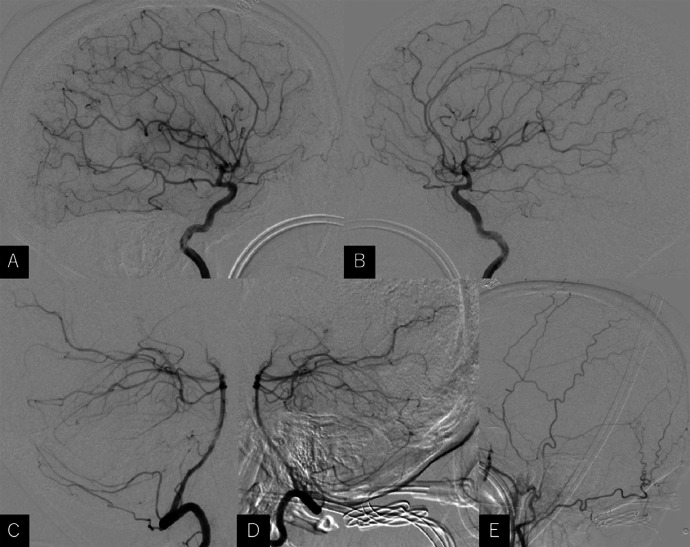
A 53-year-old woman presented with a 2-week history of headache and vertigo. Computed tomography revealed a hyperdense tumor, measuring 30 × 31 × 36 mm in diameter, in the anteromedial parts of the cerebellar hemispheres. Cerebral magnetic resonance imaging 10 days later revealed an apparent extra-axial tumor with broad attachment to the medial tentorium cerebelli and rapid growth to a diameter of 40 × 41 × 46 mm. Cerebral angiography revealed no obvious feeding vessels or tumor stains. The patient underwent biopsy through the left occipital transtentorial route. The histological appearance was consistent with diffuse large B-cell lymphoma. Intracranial lymphoma may present as a dural tumor that mimics a meningioma. Rapid tumor growth incongruous with benign meningiomas should be assumed to be possible lymphoma, and prompt biopsy should be performed.
Keywords: Dural-based; Lymphoma; Mimic meningioma; Occipital transtentorial approach.
© 2024 The Authors. Published by Elsevier Inc. on behalf of University of Washington.
Figures


References
-
- Bassiouni H, Hunold A, Asgari S, Stolke D. Tentorial meningiomas: clinical results in 81 patients treated microsurgically. Neurosurgery. 2004;55(1):116–118. - PubMed
-
- Samii M, Carvalho GA, Tatagiba M, Matthies C, Vorkapic P. Meningiomas of the tentorial notch: surgical anatomy and management. J Neurosurg. 1996;84(3):375–381. - PubMed
-
- Qiu B, Wang Y, Ou S, Guo Z, Wang Y. The unilateral occipital transtentorial approach for pineal region meningioma: a report of 15 cases. Int J Neurosci. 2014;124(10):741–747. - PubMed
-
- Johnson MD, Powell SZ, Boyer PJ, Weil RJ, Moots PL. Dural lesions mimicking meningiomas. Hum Pathol. 2002;33(12):1211–1226. - PubMed
-
- Joshi SS, Joshi S, Muzumdar G, Turel KE, Shah RM, Ammbulkar I, et al. Cranio-spinal Rosai Dorfman disease: case series and literature review. Br J Neurosurg. 2019;33(2):176–183. - PubMed
Publication types
LinkOut - more resources
Full Text Sources
